Sunday, July 16, 2023

Cell Junctions

Cell Junctions

Introduction

If you were building a building, what kinds of connections might you want to put between the rooms? In some cases, you’d want people to be able to walk from one room to another, in which case you’d put in a door. In other cases, you’d want to hold two adjacent walls firmly together, in which case you might put in some strong bolts. And in still other cases, you might need to ensure that the walls were sealed very tightly together – for instance, to prevent water from dripping between them.

As it turns out, cells face the same questions when they’re arranged in a tissue next to other cells. Should they put in doors that connect them directly to their neighbors? Do they need to spot-weld themselves to their neighbors to make a strong layer, or perhaps even form tight seals to prevent water from passing through the tissue? Junctions serving all of these functions can be found in cells of different types, and here, we’ll look at each of them in turn.

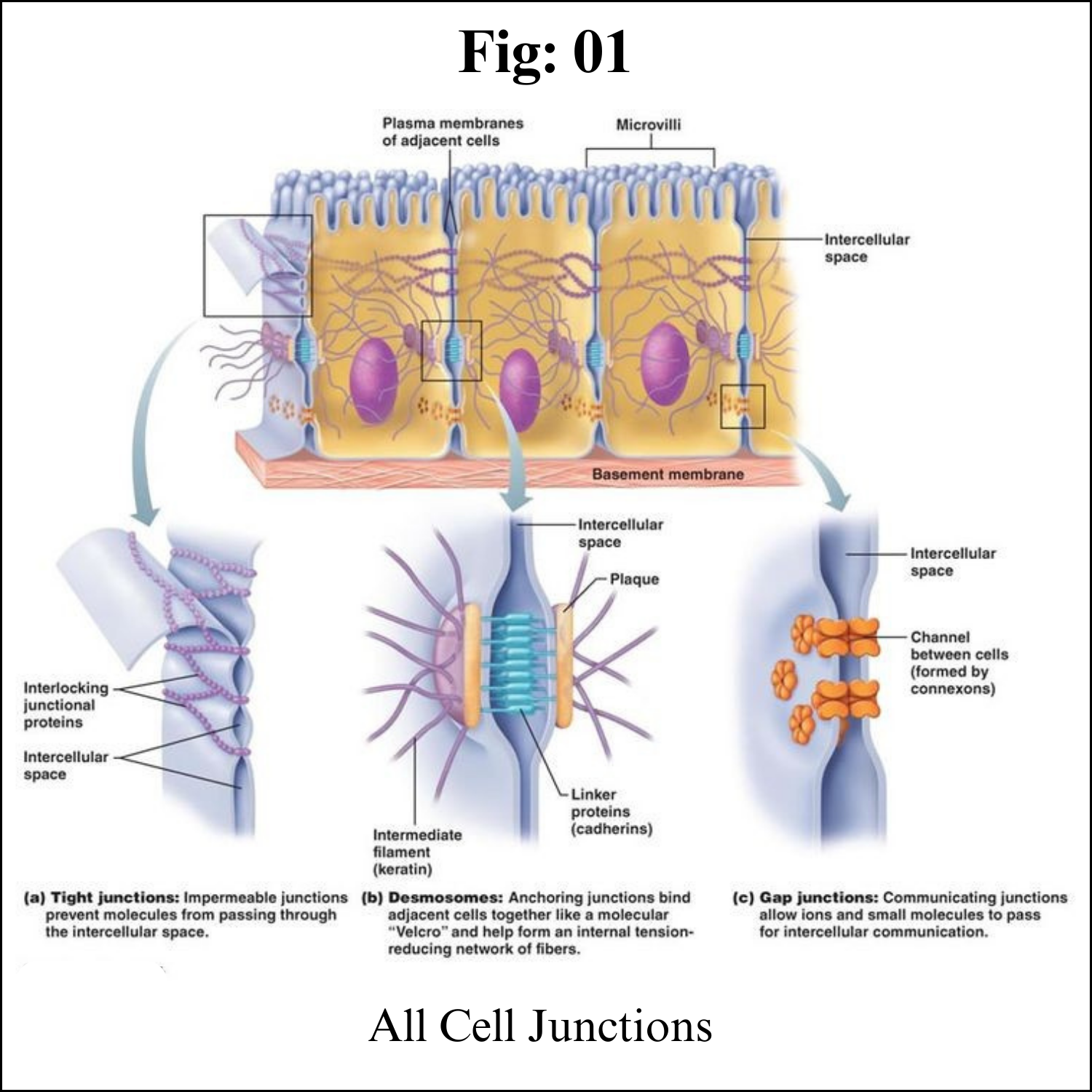
In many animal tissues (e.g., connective tissue), each cell is separated from the next by an extracellular coating or matrix. However, in some tissues (e.g., epithelia), the plasma membranes of adjacent cells are pressed together. Four kinds of junctions occur in vertebrates:

1. Tight junctions
2. Adherens junctions
3. Gap junctions
4. Desmosomes

In many plant tissues, it turns out that the plasma membrane of each cell is continuous with that of the adjacent cells. The membranes contact each other through openings in the cell wall called Plasmodesmata.

Plasmodesmata

Plant cells, surrounded as they are by cell walls, don’t contact one another through wide stretches of plasma membrane the way animal cells can. However, they do have specialized junctions called plasmodesmata (singular, plasmodesma), places where a hole is punched in the cell wall to allow direct cytoplasmic exchange between two cells.
Plasmodesmata are lined with plasma membrane that is continuous with the membranes of the two cells. Each plasmodesma has a thread of cytoplasm extending through it, containing an even thinner thread of endoplasmic reticulum (not shown in the diagram above).

Molecules below a certain size (the size exclusion limit) move freely through the plasmodesmal channel by passive diffusion. The size exclusion limit varies among plants, and even among cell types within a plant. Plasmodesmata may selectively dilate (expand) to allow the passage of certain large molecules, such as proteins, although this process is poorly understood.

Although each plant cell is encased in a boxlike cell wall, it turns out that communication between cells is just as easy, if not easier, than between animal cells. Fine strands of cytoplasm, called plasmodesmata, extend through pores in the cell wall connecting the cytoplasm of each cell with that of its neighbors.

Plasmodesmata provide an easy route for the movement of ions, small molecules like sugars and amino acids, and even macromolecules like RNA and proteins, between cells. The larger molecules pass through with the aid of actin filaments. Plasmodesmata are sheathed by a plasma membrane that is simply an extension of the plasma membrane of the adjoining cells. This raises the intriguing question of whether a plant tissue is really made up of separate cells or is, instead, a syncytium: a single, multinucleated cell distributed throughout hundreds of tiny compartments.


Gap junctions

Functionally, gap junctions in animal cells are a lot like plasmodesmata in plant cells: they are channels between neighboring cells that allow for the transport of ions, water, and other substances cubed. Structurally, however, gap junctions and plasmodesmata are quite different.

In vertebrates, gap junctions develop when a set of six membrane proteins called connexins form an elongated, donut-like structure called a connexon. When the pores, or “doughnut holes,” of connexons in adjacent animal cells align, a channel forms between the cells. (Invertebrates also form gap junctions in a similar way, but use a different set of proteins called innexins.)

Gap junctions are particularly important in cardiac muscle: the electrical signal to contract spreads rapidly between heart muscle cells as ions pass through gap junctions, allowing the cells to contract in tandem.

Gap junctions are intercellular channels some 1.5–2 nm in diameter. These permit the free passage between the cells of ions and small molecules (up to a molecular weight of about 1000 daltons). They are cylinders constructed from 6 copies of transmembrane proteins called connexins. Because ions can flow through them, gap junctions permit changes in membrane potential to pass from cell to cell.


Examples of gap junctions include:

a. The action potential in heart (cardiac) muscle flows from cell to cell through the heart providing the rhythmic contraction of the heartbeat.

b. At some so-called electrical synapses in the brain, gap junctions permit the arrival of an action potential at the synaptic terminals to be transmitted across to the postsynaptic cell without the delay needed for release of a neurotransmitter.

c. As the time of birth approaches, gap junctions between the smooth muscle cells of the uterus enable coordinated, powerful contractions to begin.

d. Several inherited disorders of humans such as certain congenital heart defects and certain cases of congenital deafness have been found to be caused by mutant genes encoding connexins.

Tight junctions

Not all junctions between cells produce cytoplasmic connections. Instead, tight junctions create a watertight seal between two adjacent animal cells.

At the site of a tight junction, cells are held tightly against each other by many individual groups of tight junction proteins called claudins, each of which interacts with a partner group on the opposite cell membrane. The groups are arranged into strands that form a branching network, with larger numbers of strands making for a tighter seal.

The purpose of tight junctions is to keep liquid from escaping between cells, allowing a layer of cells (for instance, those lining an organ) to act as an impermeable barrier. For example, the tight junctions between the epithelial cells lining your bladder prevent urine from leaking out into the extracellular space.

Epithelia are sheets of cells that provide the interface between masses of cells and a cavity or space (a lumen). The portion of the cell exposed to the lumen is called its apical surface. The rest of the cell (i.e., its sides and base) make up the basolateral surface. Tight junctions seal adjacent epithelial cells in a narrow band just beneath their apical surface. They consist of a network of claudins and other proteins. Tight junctions perform two vital functions:

1. They limit the passage of molecules and ions through the space between cells. So most materials must actually enter the cells (by diffusion or active transport) in order to pass through the tissue. This pathway provides tighter control over what substances are allowed through.

2. They block the movement of integral membrane proteins (red and green ovals) between the apical and basolateral surfaces of the cell. Thus the special functions of each surface, for example receptor-mediated endocytosis at the apical surface and exocytosis at the basolateral surface can be preserved.


Desmosomes

Animal cells may also contain junctions called desmosomes, which act like spot welds between adjacent epithelial cells. A desmosome involves a complex of proteins. Some of these proteins extend across the membrane, while others anchor the junction within the cell.

Cadherins, specialized adhesion proteins, are found on the membranes of both cells and interact in the space between them, holding the membranes together. Inside the cell, the cadherins attach to a structure called the cytoplasmic plaque (red in the image at right), which connects to the intermediate filaments and helps anchor the junction.

Desmosomes pin adjacent cells together, ensuring that cells in organs and tissues that stretch, such as skin and cardiac muscle, remain connected in an unbroken sheet.
Desmosomes are localized patches that hold two cells tightly together. They are common in epithelia (e.g., the skin). 

Desmosomes are attached to intermediate filaments of keratin in the cytoplasm. Pemphigus is an autoimmune disease in which the patient has developed antibodies against proteins (cadherins) in desmosomes. The loosening of the adhesion between adjacent epithelial cells causes blistering. 

Carcinomas are cancers of epithelia. However, the cells of carcinomas no longer have desmosomes. This may partially account for their ability to metastasize.

Hemidesmosomes 

These are similar to desmosomes but attach epithelial cells to the basal lamina ("basement membrane") instead of to each other. Pemphigoid is an autoimmune disease in which the patient develops antibodies against proteins (integrins) in hemidesmosomes. This, too, causes severe blistering of epithelia.


The function of cell junctions

The main function of some junctions is to connect the cytoplasm between adjacent cells and allow intercellular transportation and communication. Other junctions mostly function as adherence sites that maintain tissue structure and integrity. All types of tissues in animals have junctions, however, junctions that serve for adherence are more abundant in epithelial tissues (tissues that line internal organs and cavities).

Cell Junctions - Key takeaways

The two main functions of intercellular junctions are to allow intercellular transportation and communication or to maintain tissue structure and integrity through adherence of adjacent cells.

Connections between cell plants are called plasmodesmata, while animal cells can connect through tight junctions, gap junctions, and desmosomes.

Plasmodesmata connect the cytoplasm of adjacent plant cells for intercellular communication and movement of material. Gap junctions allow the intercellular movement of ions and small molecules and are essential for the transmission of electrical signals throughout a tissue. 

Tight junctions and desmosomes are for adherence rather than connection, maintaining tissue integrity. Tight junctions provide a waterproof seal, while desmosomes give resistance to mechanical stress.


Friday, July 14, 2023

Major Histocompatibility Complex (MHC)


Major Histocompatibility Complex (MHC)

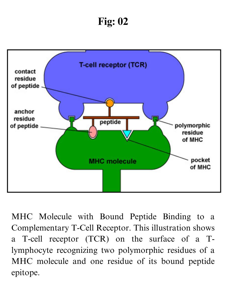
MHC molecules enable T-lymphocytes to recognize epitopes of antigens and discriminate self from non-self. Unlike B-cell receptors on B-lymphocytes that are able to directly bind epitopes on antigens, the T-cell receptors (TCRs) of T-lymphocytes can only recognize epitopes - typically short chains of amino acids called peptides - after they are bound to MHC molecules. 

The MHC genes are the most polymorphic genes in the human genome, possessing many alleles for each gene. 

The MHC genes are codominantly expressed so that an individual expresses the alleles inherited from each parent. 

In this way, the number of MHC molecules that bind peptides for presentation to T-lymphocytes is maximized. 

In addition, each MHC molecule is able to bind a wide variety of different peptides, both self-peptides and foreign peptides. There are two classes of MHC molecules: MHC-I and MHC-II.

MHC-I molecules present epitopes to T8-lymphocytes.

MHC-II molecules present epitopes to T4-lymphocytes.


The expression of MHC molecules is increased by cytokines produced during both innate immune responses and adaptive immune responses. Cytokines such as interferon-alpha, interferon-beta, interferon-gamma, tumor necrosis factor increase the expression of MHC-I molecules, while interferon-gamma is the main cytokine to increase the expression of MHC-II molecules.


MHC-I molecules

MHC-I molecules are designed to enable the body to recognize infected cells and tumor cells and destroy them with cytotoxic T-lymphocytes or CTLs. 

CTLs are effector defense cells derived from naive T8-lymphocytes. MHC-I molecules are:

1. Made by all nucleated cells in the body.

2. Possess a deep groove that can bind peptide epitopes, typically 8-11 amino acids long, typically from endogenous antigens .

3. Present MHC-I/peptide complexes to naive T8-lymphocytes and cytotoxic T-lymphocytes possessing a complementary-shaped T-cell receptor or TCR.

4. Through the process of cross-presentation, some antigen-presenting dendritic cells can cross-present epitopes of exogenous antigens to MHC-I molecules for eventual presentation to naive T8-lymphocytes.

Endogenous antigens are proteins found within the cytosol of human cells. Examples of endogenous antigens include:

a. Viral proteins produced during viral replication;

b. Proteins produced by intracellular bacteria such as Rickettsias and Chlamydias during their replication;

c. Proteins that have escaped into the cytosol from the phagosome of phagocytes such as antigen-presenting cells;

d. Tumor antigens produced by cancer cells; and

e. Self-peptides from host cellular proteins.

During the replication of viruses and intracellular bacteria within their host cell, as well as during the replication of tumor cells, viral, bacterial, or tumor proteins are degraded into a variety of peptide epitopes by cylindrical organelles called proteasomes. The body's own cytosolic proteins are also degraded into peptides by proteasomes.

These peptide epitopes are then attached to a groove of MHC-I molecules that are then transported to the surface of that cell where they can be recognized by a complementary-shaped T-cell receptor (TCR) and a CD8 molecule, a co-receptor, on the surface of either a naive T8-lymphocyte or a cytotoxic T-lymphocyte (CTL). 

The TCRs recognize both the foreign peptide antigen and the MHC molecule. TCRs, however, will not recognize self-peptides bound to MHC-I. As a result, normal cells are not attacked and killed.

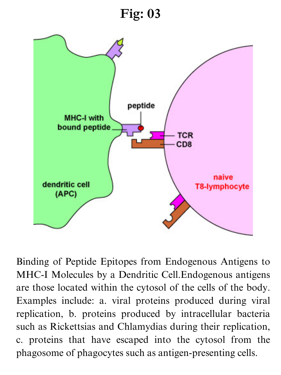
Dendritic cells bind epitopes from endogenous antigens to MHC-I molecules and present them to naive T8-lymphocytes in order to activate these naive T8-lymphocytes.

a. Antigens are engulfed by dendritic cells and placed in a phagosome. Some of the proteins escape from the phagosome into the cytosol of the dendritic cell where they become endogenous antigens.

b. These endogenous antigens pass through proteasomes where they are degraded into a series of peptides.

c. The peptides are transported into the rough endoplasmic reticulum (ER) by a transporter protein called TAP.

d. The peptides then bind to the grooves of newly synthesized MHC-I molecules.

e. The endoplasmic reticulum transports the MHC-I molecules with bound peptides to the Golgi complex.

f. The Golgi complex, in turn, transports the MHC-I/peptide complexes by way of an exocytic vesicle to the cytoplasmic membrane where they become anchored. Here, the peptide and MHC-I/peptide complexes can be recognized by naive T8-lymphocytes by way of TCRs and CD8 molecules having a complementary shape.


Through the process of cross-presentation, some antigen-presenting dendritic cells can cross-present epitopes of exogenous antigens to MHC-I molecules for eventual presentation to naive T8-lymphocytes.

MHC-I molecules with bound peptides on the surface of antigen-presenting dendritic cells can be recognized by a complementary-shaped TCR/CD8 on the surface of a naive T8-lymphocyte to initiate cell-mediated immunity. (Certain dendritic cells, as discussed later, can also cross-present exogenous antigens to MHC-I molecules).

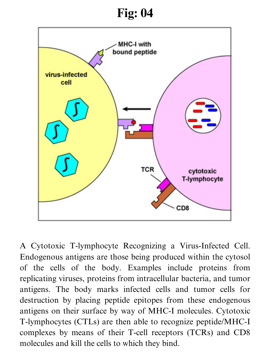
MHC-I molecules with bound peptides on the surface of infected cells and tumor cells can be recognized by a complementary-shaped TCR/CD8 on the surface of a cytotoxic T-lymphocyte or CTL to initiate destruction of the cell containing the endogenous antigen. (CTLs are effector cells derived from naive T8-lymphocytes.) 

Cytotoxic T-lymphocytes (CTLs) are then able to recognize peptide/MHC-I complexes by means of their T-cell receptors (TCRs) and CD8 molecules and kill the cells to which they bind.

a. During viral replication within the host cell, endogenous antigens, such as viral proteins, pass through proteasomes where they are degraded into a series of peptides.

b. The peptides are transported into the rough endoplasmic reticulum (ER) by a transporter protein called TAP.

c. The peptides then bind to the grooves of newly synthesized MHC-I molecules.

d. The endoplasmic reticulum transports the MHC-I molecules with bound peptides to the Golgi complex.

e. The Golgi complex, in turn, transports the MHC-I/peptide complexes by way of an exocytic vesicle to the cytoplasmic membrane where they become anchored. Here, the peptide and MHC-I/peptide complexes can be recognized by CTLs by way of TCRs and CD8 molecules having a complementary shape.


MHC-I molecules are coded for by three MHC-I genes, HLA-A, HLA-B, and HLA-C. As mentioned above, however, there are many different alleles for each gene that a person inherits. In this way, the number of MHC-I molecules that bind peptides for presentation to T-8 lymphocytes is maximized. The expression of MHC-I molecules on all cell types is increased by the cytokines interferon-alpha (IFN-a) and interferon-beta (IFN-ß).

MHC-II molecules

MHC-II molecules are designed to enable T4-lymphocytes to recognize epitopes of exogenous antigens and discriminate self from non-self. MHC-II molecules are:

Made by antigen-presenting cells or APCs, such as dendritic cells, macrophages, and B-lymphocytes.

Possess a deep groove that can bind peptide epitopes, often 10-30 amino acids long but with an optimum length of 12-16 amino acids, typically from exogenous antigens. The peptides interact along their entire length with the groove.

Present MHC-II/peptide complexes to naive T4-lymphocytes or effector T4-lymphocytes that have a complementary shaped T-cell receptor or TCR.

Through the process of cross-presentation, some antigen-presenting dendritic cells can cross-present epitopes of endogenous antigens to MHC-II molecules for eventual presentation to naive T4-lymphocytes.


Exogenous antigens are antigens that enter from outside the body, such as bacteria, fungi, protozoa, and free viruses. These exogenous antigens enter macrophages, dendritic cells, and B-lymphocytes through phagocytosis. 

The microbes are engulfed and placed in a phagosome which then fuses with lysosomes. Following this fusion, the phagolysosome becomes acidified. 

Acidification, in turn, activates the proteases within the phagolysosome enabling protein antigens from the microbe to be degraded into a series of short peptides. 

These peptide epitopes are then attached to MHC-II molecules and are then transported to the surface of the antigen-presenting cell (APC). (Certain dendritic cells, as discussed later, can also cross-present endogenous antigens to MHC-II molecules.)

Some pathogens, such as Mycobacterium tuberculosis, Mycobacterium leprae, and Leishmania, are able to grow in the endocytic vesicles of macrophages without being killed by lysosomes. These macrophages can, however, become activated by T4-effector lymphocytes called TH1 cells and subsequently use intravesicular proteases to degrade the proteins from these pathogens into peptides for presentation to MHC-II molecules that pass through on their way to the cell surface.

Here the MHC-II molecules with bound peptides can be recognized by a complementary-shaped T-cell receptor and a CD4 molecule, a co-receptor, on the surface of a T4-lymphocyte. T4-lymphocytes are the cells the body uses to regulate both humoral immunity and cell-mediated immunity.

MHC-II molecules are coded for by three MHC-II genes, HLA-DR, HLA-DP, and HLA-DQ. Interferon-gamma (IFN- ?) increases the expression of both MHC-I and MHC-II molecules.


Thursday, July 13, 2023

Jean Piaget's - Cognitive Development


Jean Piaget's - Cognitive Development
 

Jean Piaget’s theory of cognitive development suggests that intelligence changes as children grow.

A child’s cognitive development is not just about acquiring knowledge, the child has to develop or construct a mental model of the world.

Cognitive development occurs through the interaction of innate capacities (nature) and environmental events (nurture), and children pass through a series of stages.

About Author


Jean Piaget, (born August 9, 1896, Neuchâtel, Switzerland—died September 16, 1980, Geneva), Swiss psychologist who was the first to make a systematic study of the acquisition of understanding in children. He is thought by many to have been the major figure in 20th-century developmental psychology.

Piaget’s early interests were in zoology; as a youth he published an article on his observations of an albino sparrow, and by 15 his several publications on mollusks had gained him a reputation among European zoologists. 

At the University of Neuchâtel, he studied zoology and philosophy, receiving his doctorate in the former in 1918. Soon afterward, however, he became interested in psychology, combining his biological training with his interest in epistemology. 

He first went to Zürich, where he studied under Carl Jung and Eugen Bleuler, and he then began two years of study at the Sorbonne in Paris in 1919.

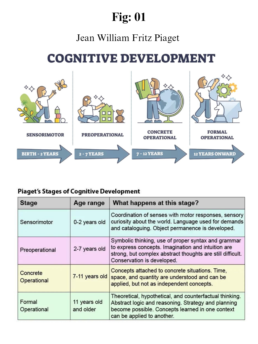
Piaget’s theory of cognitive development proposes 4 stages of development.

1. Sensorimotor stage: birth to 2 years
2. Preoperational stage: 2 to 7 years
3. Concrete operational stage: 7 to 11 years
4. Formal operational stage: ages 12 and up

The sequence of the stages is universal across cultures and follows the same invariant (unchanging) order.

All children go through the same stages in the same order (but not all at the same rate).

Theory Development

Piaget was employed at the Binet Institute in the 1920s, where his job was to develop French versions of questions on English intelligence tests. He became intrigued with the reasons children gave for their wrong answers to the questions that required logical thinking.

He believed that these incorrect answers revealed important differences between the thinking of adults and children.

Piaget branched out on his own with a new set of assumptions about children’s intelligence:

Children’s intelligence differs from an adult’s in quality rather than in quantity. This means that children reason (think) differently from adults and see the world in different ways.

Children actively build up their knowledge about the world. They are not passive creatures waiting for someone to fill their heads with knowledge.

The best way to understand children’s reasoning is to see things from their point of view.

Piaget did not want to measure how well children could count, spell or solve problems as a way of grading their I.Q. 

What he was more interested in was the way in which fundamental concepts like the very idea of number, time, quantity, causality, justice, and so on emerged.

Piaget studied children from infancy to adolescence using naturalistic observation of his own three babies and sometimes controlled observation too. From these, he wrote diary descriptions charting their development.

He also used clinical interviews and observations of older children who were able to understand questions and hold conversations.

Stages of Development

Each child goes through the stages in the same order, and child development is determined by biological maturation and interaction with the environment.

At each stage of development, the child’s thinking is qualitatively different from the other stages, that is, each stage involves a different type of intelligence.

Although no stage can be missed out, there are individual differences in the rate at which children progress through stages, and some individuals may never attain the later stages.

Piaget did not claim that a particular stage was reached at a certain age – although descriptions of the stages often include an indication of the age at which the average child would reach each stage.

The Sensorimotor Stage (Ages: Birth to 2 Years)

The first stage is the sensorimotor stage, and during this stage, the infant focuses on physical sensations and on learning to coordinate their body.

Major Characteristics and Developmental Changes:

The infant learns about the world through their senses and through their actions (moving around and exploring their environment).

During the sensorimotor stage, a range of cognitive abilities develop. These include: object permanence; self-recognition (the child realizes that other people are separate from them); deferred imitation; and representational play.

They relate to the emergence of the general symbolic function, which is the capacity to represent the world mentally

At about 8 months, the infant will understand the permanence of objects and that they will still exist even if they can’t see them and the infant will search for them when they disappear.

During the beginning of this stage, the infant lives in the present. It does not yet have a mental picture of the world stored in its memory therefore it does not have a sense of object permanence.

If it cannot see something, then it does not exist. This is why you can hide a toy from an infant, while it watches, but it will not search for the object once it has gone out of sight.

The main achievement during this stage is object permanence – knowing that an object still exists, even if it is hidden. It requires the ability to form a mental representation (i.e., a schema) of the object.

Towards the end of this stage the general symbolic function begins to appear where children show in their play that they can use one object to stand for another. Language starts to appear because they realize that words can be used to represent objects and feelings.

The child begins to be able to store information that it knows about the world, recall it and label it.

The Preoperational Stage (Ages: 2 – 7 Years)

Piaget’s second stage of intellectual development is the preoperational stage. It takes place between 2 and 7 years. At the beginning of this stage the child does not use operations, so the thinking is influenced by the way things appear rather than logical reasoning.

A child cannot conserve which means that the child does not understand that quantity remains the same even if the appearance changes.

Furthermore, the child is egocentric; he assumes that other people see the world as he does. This has been shown in the three mountains study.

As the preoperational stage develops egocentrism declines and children begin to enjoy the participation of another child in their games and “lets pretend “ play becomes more important.

Toddlers often pretend to be people they are not (e.g. superheroes, policemen), and may play these roles with props that symbolize real life objects. Children may also invent an imaginary playmate.

Major Characteristics and Developmental Changes:

Toddlers and young children acquire the ability to internally represent the world through language and mental imagery.

During this stage, young children can think about things symbolically. This is the ability to make one thing, such as a word or an object, stand for something other than itself.

A child’s thinking is dominated by how the world looks, not how the world is. It is not yet capable of logical (problem solving) type of thought.

Moreover, the child has difficulties with class inclusion; he can classify objects but cannot include objects in subsets, which involves classifying objects as belonging to two or more categories simultaneously.

Infants at this stage also demonstrate animism. This is the tendency for the child to think that non-living objects (such as toys) have life and feelings like a person’s.

By 2 years, children have made some progress toward detaching their thoughts from the physical world. However, have not yet developed logical (or “operational”) thought characteristics of later stages.

Thinking is still intuitive (based on subjective judgements about situations) and egocentric (centered on the child’s own view of the world).

The Concrete Operational Stage (Ages: 7 – 11 Years)

By the beginning of the concrete operational stage, the child can use operations (a set of logical rules) so she can conserve quantities, she realizes that people see the world in a different way than he does (decentring) and he has improved in inclusion tasks. Children still have difficulties with abstract thinking.

Major Characteristics and Developmental Changes:

During this stage, children begin to think logically about concrete events.

Children begin to understand the concept of conservation; understanding that, although things may change in appearance, certain properties remain the same.

During this stage, children can mentally reverse things (e.g. picture a ball of plasticine returning to its original shape).

During this stage, children also become less egocentric and begin to think about how other people might think and feel.

The stage is called concrete because children can think logically much more successfully if they can manipulate real (concrete) materials or pictures of them.

Piaget considered the concrete stage a major turning point in the child’s cognitive development because it marks the beginning of logical or operational thought. This means the child can work things out internally in their head (rather than physically try things out in the real world).

Children can conserve number (age 6), mass (age 7), and weight (age 9). Conservation is the understanding that something stays the same in quantity even though its appearance changes.

But operational thought is only effective here if the child is asked to reason about materials that are physically present. Children at this stage will tend to make mistakes or be overwhelmed when asked to reason about abstract or hypothetical problems.

The Formal Operational Stage (Ages: 12 and Over…)

The formal operational period begins at about age 11. As adolescents enter this stage, they gain the ability to think in an abstract manner, the ability to combine and classify items in a more sophisticated way, and the capacity for higher-order reasoning.

Adolescents can think systematically and reason about what might be as well as what is (not everyone achieves this stage).. This allows them to understand politics, ethics, and science fiction, as well as to engage in scientific reasoning.

Adolescents can deal with abstract ideas: e.g. they can understand division and fractions without having to actually divide things up, and solve hypothetical (imaginary) problems.

Major Characteristics and Developmental Changes:

Concrete operations are carried out on things whereas formal operations are carried out on ideas. Formal operational thought is entirely freed from physical and perceptual constraints.

During this stage, adolescents can deal with abstract ideas (e.g. no longer needing to think about slicing up cakes or sharing sweets to understand division and fractions).

They can follow the form of an argument without having to think in terms of specific examples.

Adolescents can deal with hypothetical problems with many possible solutions. E.g. if asked ‘What would happen if money were abolished in one hour’s time? they could speculate about many possible consequences.

From about 12 years children can follow the form of a logical argument without reference to its content. During this time, people develop the ability to think about abstract concepts, and logically test hypotheses.

This stage sees the emergence of scientific thinking, formulating abstract theories and hypotheses when faced with a problem.


Schemas

Piaget claimed that knowledge cannot simply emerge from sensory experience; some initial structure is necessary to make sense of the world.

According to Piaget, children are born with a very basic mental structure (genetically inherited and evolved) on which all subsequent learning and knowledge are based.

Schemas are the basic building blocks of such cognitive models, and enable us to form a mental representation of the world.

Piaget (1952, p. 7) defined a schema as: “a cohesive, repeatable action sequence possessing component actions that are tightly interconnected and governed by a core meaning.”

In more simple terms, Piaget called the schema the basic building block of intelligent behavior – a way of organizing knowledge. Indeed, it is useful to think of schemas as “units” of knowledge, each relating to one aspect of the world, including objects, actions, and abstract (i.e., theoretical) concepts.

Wadsworth (2004) suggests that schemata (the plural of schema) be thought of as “index cards” filed in the brain, each one telling an individual how to react to incoming stimuli or information.

When Piaget talked about the development of a person’s mental processes, he was referring to increases in the number and complexity of the schemata that a person had learned.

When a child’s existing schemas are capable of explaining what it can perceive around it, it is said to be in a state of equilibrium, i.e., a state of cognitive (i.e., mental) balance.

Operations are more sophisticated mental structures which allow us to combine schemas in a logical (reasonable) way.

As children grow they can carry out more complex operations and begin to imagine hypothetical (imaginary) situations.

Apart from the schemas we are born with schemas and operations are learned through interaction with other people and the environment.

Piaget emphasized the importance of schemas in cognitive development and described how they were developed or acquired.

A schema can be defined as a set of linked mental representations of the world, which we use both to understand and to respond to situations. The assumption is that we store these mental representations and apply them when needed.


Applying Piaget’s Theory to the Classroom

Think of old black and white films that you’ve seen in which children sat in rows at desks, with ink wells, would learn by rote, all chanting in unison in response to questions set by an authoritarian old biddy like Matilda.

Children who were unable to keep up were seen as slacking and would be punished by variations on the theme of corporal punishment. Yes, it really did happen and in some parts of the world still does today.

Piaget is partly responsible for the change that occurred in the 1960s and for your relatively pleasurable and pain free school days.

How to Teach

Within the classroom learning should be student-centered and accomplished through active discovery learning. The role of the teacher is to facilitate learning, rather than direct tuition.

Because Piaget’s theory is based upon biological maturation and stages, the notion of “readiness” is important. Readiness concerns when certain information or concepts should be taught.

According to Piaget’s theory children should not be taught certain concepts until they have reached the appropriate stage of cognitive development.

According to Piaget (1958), assimilation and accommodation require an active learner, not a passive one, because problem-solving skills cannot be taught, they must be discovered.

Therefore, teachers should encourage the following within the classroom:

Educational programs should be designed to correspond to Piaget’s stages of development. Children in the concrete operational stage should be given concrete means to learn new concepts e.g. tokens for counting.

Devising situations that present useful problems, and create disequilibrium in the child.

Focus on the process of learning, rather than the end product of it. Instead of checking if children have the right answer, the teacher should focus on the student’s understanding and the processes they used to get to the answer.

Child-centered approach. Learning must be active (discovery learning).

Children should be encouraged to discover for themselves and to interact with the material instead of being given ready-made knowledge.

Accepting that children develop at different rates so arrange activities for individual children or small groups rather than assume that all the children can cope with a particular activity.

Using active methods that require rediscovering or reconstructing “truths.”

Using collaborative, as well as individual activities (so children can learn from each other).

Role of the Teacher

Evaluate the level of the child’s development so suitable tasks can be set.

Adapt lessons to suit the needs of the individual child (i.e. differentiated teaching).

Be aware of the child’s stage of development (testing).
Teach only when the child is ready. i.e. has the child reached the appropriate stage.

Providing support for the “spontaneous research” of the child.

Using collaborative, as well as individual activities.

Devising situations that present useful problems, and create disequilibrium in the child.

Educational Implications of Piaget’s Theory

The application of Piaget’s theory of cognitive development theory in classroom should be done as follows:

Based on the developmental level of children, the curriculum should provide the required educational experience.

Classroom activities that encourage and assist self learning must be incorporated.

Practical learning situations must be included in the class.
Co-curricular activities that enhance children’s cognitive development must be given equal importance as curricular activities.

The teaching method must be simple to complex and the inclusion of the project teaching method is recommended.

Children learn and think differently from adults therefore, they should be taught accordingly.

The discovery approach to learning must be emphasized.

Monday, July 10, 2023

Prokaryotic Protein Production


Prokaryotic Protein Production 

Cell Signaling + Central dogma 

Ligand - Receptor - Cascade pathway - Transcription (Pre mRNA, tRNA & rRNA) - Post Transcriptional Modification (Mature mRNA) - Translation (Amino acid Sequence) - Post Translational Modification - Protein Production - Cellular functions. 

Prokaryotic Transcription 

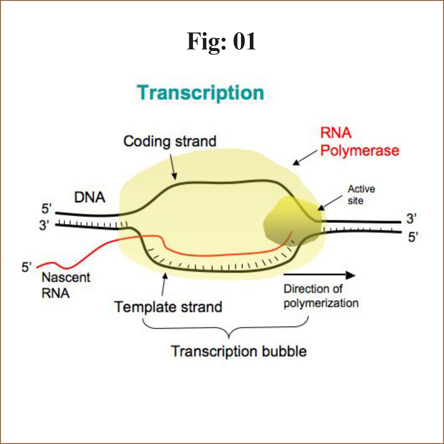
The process of synthesis of RNA by copying the template strand of DNA is called transcription.

During replication the entire genome is copied but in transcription only the selected portion of the genome is copied.

The enzyme involved in transcription is RNA polymerase. Unlike DNA polymerase it can initiate transcription by itself, it does not require primase. More exactly it is a DNA dependent RNA polymerase.

The steps of transcription

Transcription is an enzymatic process. the mechanism of transcription completes in three major steps

1. Initiation:

a) Closed complex formation
b) Open complex formation
c) Tertiary complex formation

2. Elongation

3. Termination:

a) Rho- dependent
b) Rho-independent


1. Initiation:

The transcription is initiated by RNA polymerase holoenzyme from a specific point called promotor sequence.

Bacterial RNA polymerase is the principle enzyme involved in transcription.

Single RNA polymerase is found in a bacteria which is called core polymerase and it consists of α, β, β’ and ω sub units.

The core enzyme bind to specific sequence on template DNA strand called promotor. The binding of core polymerase to promotor is facilitates and specified by sigma (σ) factor. (σ70 in case of E. coli).


The core polymerase along with σ-factor is called Holo-enzyme ie. RNA polymerase holoenzyme.

In case of e. coli, promotor consists of two conserved sequences 5’-TTGACA-3’ at -35 element and 5’-TATAAT-3’ at -10 element. These sequence are upstream to the site from which transcription begins. Binding of holoenzyme to two conserved sequence of promotor form close complex.

In some bacteria, the altered promotor may exist which contain UP-element and some may contain extended -10 elements rather than -35 elements.


Region1: it includes 1.2 and 1.1 region. Region 1.1 acts as molecular mimic of DNA

Region2: it recognizes -10 element in promotor. α-helix recognizes -10 element.

Region 3: it recognizes extended -10 element.

Region 4: it recognizes -35 element in promotor by a structure called helix-turn-helix.

The UP-element is recognized by a carboxyl terminal domain of α-sub unit called αCTD (carboxyl terminal domain) which is connected to αNTD (Amino terminal domain) by flexible linker.

Closed complex: Binding of RNA polymerase holoenzyme to the promotor sequence form closed comolex

Open complex: After formation of closed complex, the RNA polymerase holoenzyme separates 10-14 bases exztending from -11 to +3 called melting. So that open complex is formed. This changing from closed complex to open complex is called isomerization.

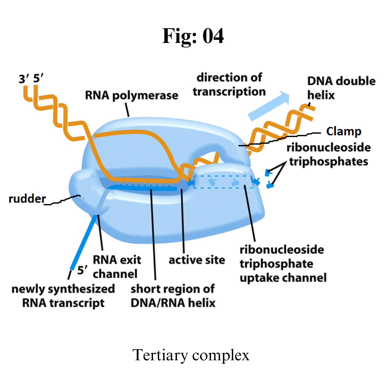
Tertiary complex: RNA polymerase starts synthesizing nucleotide. It doesnot require the help of primase.


If the enzyme synthesize short RNA molecules of less than 10 bp, it does not further elongates which is called abortive initiation. This is because σ 3.2 acting as mimic of RNA and it lies at middle of RNA exit channel in open complex. 

When the RNA polymerase manage to synthesize RNA more than 10 bp long, it eject the σ 3.2 region and RNA further elongates and exit from RNA exit channel. This is the formation of tertiary complex.

2. Elongation:

After synthesis of RNA more than 10 bp long, the σ-factor is ejected and the enzyme move along 5’-3’ direction continuously synthesizing RNA.

The synthesized RNA exit from RNA exit channel.

The synthesized RNA is proof reads by Hydrolytic editing. For this the polymerase back track by one or more nucleotide and cleave the RNA removing the error and synthesize the correct one. The Gre factor enhance this proof reading process.
Pyrophospholytic editing another mechanism of removing altered nucleotide.
 
3. Termination:

There are two mechanism of termination.

i. Rho independent:

In this mechanism, transcription is terminated due to specific sequence in terminator DNA.
The terminator DNA contains invert repeat which cause complimentary pairing as transcript RNA form hair pin structure.
This invert repeat is followed by larger number of TTTTTTTT(~8 bp) on template DNA. The uracil appear in RNA. The load of hair pin structure is not tolerated by A=U base pair so the RNA get separated from RNA-DNA heteroduplex.

ii. Rho dependent:

In this mechanism, transcription is terminated by rho (ρ) protein.
It is ring shaped single strand binding ATpase protein.
The rho protein bind the single stranded RNA as it exit from polymerase enzyme complex and hydrolyse the RNA from enzyme complex.
The rho protein does not bind to those RNA whose protein is being translated. Rather it bind to RNA after translation.
In bacteria transcription and translation occur simultaneously so the rho protein bind the RNA after translation has completed but transcription is still ON.

Post-Transcriptional Modification 

Prokaryotic post-transcriptional regulators typically modulate RNA decay, translation initiation efficiency or transcript elongation. Different types of prokaryotic post-transcriptional regulators have been identified, including small RNAs (sRNAs) and RNA-binding proteins (RBPs). sRNAs are typically defined as non-coding RNA molecules that bind with limited complementarity near the ribosome binding site (RBS) of their target mRNA, causing competition with the ribosome for binding to this region. 

However, the number of sRNAs that deviate from this general definition is increasing. The new insights into the post-transcriptional mechanisms of sRNAs and their role in gene expression regulation were reviewed recently. 
Here, RBPs involved in post-transcriptional regulation are discussed.

 For some of these proteins, the mechanism of action and the targets are well described, as for CsrA and Hfq. Their post-transcriptional function in Escherichia coli was already reported almost 20 years ago. Lately, more insight was gained into the diverse mechanisms these two well-studied proteins use to regulate the expression of their target genes and how they regulate their own expression or activity in E. coli and in other bacteria. 

Additional RBPs involved in post-transcriptional regulation have been identified only recently and not much is known about their post-transcriptional function. In this review, the general working mechanisms of RBPs are discussed first. Afterward, examples of well-known and recently identified proteins, from E. coli and from other bacteria, are described.

General Mechanisms of Regulatory Proteins that Act Post-Transcriptionally

Bacterial post-transcriptionally active regulatory proteins typically bind RNA molecules and regulate translation initiation, stability, and transcript elongation of their RNA targets, using different regulatory mechanisms. 

These mechanisms include

(i) adaptation of the susceptibility of the target RNAs to RNases,

(ii) modulation of the accessibility of the RBS of mRNA targets for ribosome binding, 

(iii) acting as a chaperone for the interaction of the RNA target with other effector molecules, and 

(iv) modulation of transcription terminator/antiterminator structure formation, and will be described hereafter.

Prokaryotic Translation

As with mRNA synthesis, protein synthesis can be divided into three phases: initiation, elongation, and termination.


Initiation

Protein synthesis begins with the formation of an initiation complex. In E. coli, this complex involves the small 30S ribosome, the mRNA template, three initiation factors (IFs; IF-1, IF-2, and IF-3), and a special initiator tRNA, called tRNAMetf.

In E. coli mRNA, a sequence upstream of the first AUG codon, called the Shine-Dalgarno sequence (AGGAGG), interacts with the rRNA molecules that compose the ribosome. 
This interaction anchors the 30S ribosomal subunit at the correct location on the mRNA template. Guanosine triphosphate (GTP), which is a purine nucleotide triphosphate, acts as an energy source during translation—both at the start of elongation and during the ribosome’s translocation. Binding of the mRNA to the 30S ribosome also requires IF-III.

The initiator tRNA then interacts with the start codon AUG (or rarely, GUG). This tRNA carries the amino acid methionine, which is formulated after its attachment to the tRNA. The formylation creates a “faux” peptide bond between the formyl carboxyl group and the amino group of the methionine. Binding of the fMet-tRNA-Metf is mediated by the initiation factor IF-2. 

The fMet begins every polypeptide chain synthesized by E. coli, but it is usually removed after translation is complete. When an in-frame AUG is encountered during translation elongation, a non-formylated methionine is inserted by a regular Met-tRNAMet. 
After the formation of the initiation complex, the 30S ribosomal subunit is joined by the 50S subunit to form the translation complex. 

Elongation

In prokaryotes, we will review elongation from the perspective of E. coli. When the translation complex is formed, the tRNA binding region of the ribosome consists of three compartments. The A (aminoacyl) site binds incoming charged aminoacyl tRNAs. 

The P (peptidyl) site binds charged tRNAs carrying amino acids that have formed peptide bonds with the growing polypeptide chain but have not yet dissociated from their corresponding tRNA. The E (exit) site releases dissociated tRNAs so that they can be recharged with free amino acids. The initiating methionyl-tRNA, however, occupies the P site at the beginning of the elongation phase of translation in both prokaryotes and eukaryotes.

During translation elongation, the mRNA template provides tRNA binding specificity. As the ribosome moves along the mRNA, each mRNA codon comes into register, and specific binding with the corresponding charged tRNA anticodon is ensured. If mRNA were not present in the elongation complex, the ribosome would bind tRNAs nonspecifically and randomly.

Elongation proceeds with charged tRNAs sequentially entering and leaving the ribosome as each new amino acid is added to the polypeptide chain. Movement of a tRNA from A to P to E site is induced by conformational changes that advance the ribosome by three bases in the 3′ direction. The energy for each step along the ribosome is donated by elongation factors that hydrolyze GTP. 
GTP energy is required both for the binding of a new aminoacyl-tRNA to the A site and for its translocation to the P site after formation of the peptide bond. Peptide bonds form between the amino group of the amino acid attached to the A-site tRNA and the carboxyl group of the amino acid attached to the P-site tRNA. 

The formation of each peptide bond is catalyzed by peptidyl transferase, an RNA-based enzyme that is integrated into the 50S ribosomal subunit. The energy for each peptide bond formation is derived from the high-energy bond linking each amino acid to its tRNA. 
After peptide bond formation, the A-site tRNA that now holds the growing peptide chain moves to the P site, and the P-site tRNA that is now empty moves to the E site and is expelled from the ribosome. Amazingly, the E. coli translation apparatus takes only 0.05 seconds to add each amino acid, meaning that a 200-amino-acid protein can be translated in just 10 seconds.


Termination

Termination of translation occurs when a nonsense codon (UAA, UAG, or UGA) is encountered. Upon aligning with the A site, these nonsense codons are recognized by protein release factors that resemble tRNAs. 
The releasing factors in both prokaryotes and eukaryotes instruct peptidyl transferase to add a water molecule to the carboxyl end of the P-site amino acid. This reaction forces the P-site amino acid to detach from its tRNA, and the newly made protein is released. 

The small and large ribosomal subunits dissociate from the mRNA and from each other; they are recruited almost immediately into another translation initiation complex. After many ribosomes have completed translation, the mRNA is degraded so the nucleotides can be reused in another transcription reaction.

Post-Translational Modification


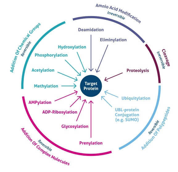
The increase in complexity from the level of the genome to the proteome is further facilitated by protein post-translational modifications (PTMs). 

PTMs are chemical modifications that play a key role in functional proteomic because they regulate activity, localization, and interaction with other cellular molecules such as proteins, nucleic acids, lipids and cofactors.

PTMs estimations demonstrate that single genes encode multiple proteins. Genomic recombination, transcription initiation at alternative promoters, differential transcription termination, and alternative splicing of the transcript are mechanisms that generate different mRNA transcripts from a single gene.

Post-translational modifications are key mechanisms to increase proteomic diversity. While the genome comprises 20,000 to 25,000 genes, the proteome is estimated to encompass over 1 million proteins. 
Changes at the transcriptional and mRNA levels increase the size of the transcriptome relative to the genome, and the myriad of different post-translational modifications exponentially increases the complexity of the proteome relative to both the transcriptome and genome.

Post-translational modification can occur at any step in the "life cycle" of a protein. For example, many proteins are modified shortly after translation is completed to mediate proper protein folding or stability or to direct the nascent protein to distinct cellular compartments (e.g., nucleus, membrane). 
Other modifications occur after folding and localization are completed to activate or inactivate catalytic activity or to otherwise influence the biological activity of the protein. Proteins are also covalently linked to tags that target a protein for degradation. 

Besides single modifications, proteins are often modified through a combination of post-translational cleavage and the addition of functional groups through a step-wise mechanism of protein maturation or activation.
Protein PTMs can also be reversible depending on the nature of the modification. For example, kinases phosphorylate proteins at specific amino acid side chains, which is a common method of catalytic activation or inactivation. 
Conversely, phosphatases hydrolyze the phosphate group to remove it from the protein and reverse the biological activity. Proteolytic cleavage of peptide bonds is a thermodynamically favorable reaction and therefore permanently removes peptide sequences or regulatory domains.

Consequently, the analysis of proteins and their post-translational modifications is particularly important for the study of heart disease, cancer, neurodegenerative diseases and diabetes. 

The characterization of PTMs, although challenging, provides invaluable insight into the cellular functions underlying etiological processes. Technically, the main challenges to studying post-translationally modified proteins are the development of specific detection and purification methods. 

Fortunately, these technical obstacles are being overcome with a variety of new and refined proteomics technologies.


Phosphorylation

Reversible protein phosphorylation, principally on serine, threonine or tyrosine residues, is one of the most important and well-studied post-translational modifications. Phosphorylation plays critical roles in the regulation of many cellular processes, including cell cycle, growth, apoptosis and signal transduction pathways. In the following example, western blot analysis was used to evaluate phosphoprotein specificity in lysates obtained from serum-starved HeLa and NIH 3T3 cancer cell lines stimulated with epidermal growth factor (EGF) and platelet derived growth factor (PDGF), respectively.

Glycosylation

Protein glycosylation is acknowledged as one of the major post-translational modifications, with significant effects on protein folding, conformation, distribution, stability and activity. Glycosylation encompasses a diverse selection of sugar-moiety additions to proteins that ranges from simple monosaccharide modifications of nuclear transcription factors to highly complex branched polysaccharide changes of cell surface receptors. Carbohydrates in the form of asparagine-linked (N-linked) or serine/threonine-linked (O-linked) oligosaccharides are major structural components of many cell surface and secreted proteins.


Methylation

The transfer of one-carbon methyl groups to nitrogen or oxygen (N- and O-methylation, respectively) to amino acid side chains increases the hydrophobicity of the protein and can neutralize a negative amino acid charge when bound to carboxylic acids. Methylation is mediated by methyltransferases, and S-adenosylmethionine (SAM) is the primary methyl group donor.

N-acetylation

N-acetylation, or the transfer of an acetyl group to nitrogen, occurs in almost all eukaryotic proteins through both irreversible and reversible mechanisms. N-terminal acetylation requires the cleavage of the N-terminal methionine by methionine aminopeptidase (MAP) before replacing the amino acid with an acetyl group from acetyl-CoA by N-acetyltransferase (NAT) enzymes. This type of acetylation is co-translational, in that N-terminus is acetylated on growing polypeptide chains that are still attached to the ribosome. While 80 to 90% of eukaryotic proteins are acetylated in this manner, the exact biological significance is still unclear.

Acetylation at the ε-NH2 of lysine (termed lysine acetylation) on histone N-termini is a common method of regulating gene transcription. Histone acetylation is a reversible event that reduces chromosomal condensation to promote transcription, and the acetylation of these lysine residues is regulated by transcription factors that contain histone acetyltransferase (HAT) activity. While transcription factors with HAT activity act as transcription co-activators, histone deacetylase (HDAC) enzymes are co-repressors that reverse the effects of acetylation by reducing the level of lysine acetylation and increasing chromosomal condensation.

Sirtuins (silent information regulator) are a group of NAD-dependent deacetylases that target histones. As their name implies, they maintain gene silencing by hypoacetylating histones and have been reported to aid in maintaining genomic stability.

While acetylation was first detected in histones, cytoplasmic proteins have been reported to also be acetylated, and therefore acetylation seems to play a greater role in cell biology than simply transcriptional regulation. Furthermore, crosstalk between acetylation and other post-translational modifications, including phosphorylation, ubiquitination and methylation, can modify the biological function of the acetylated protein.

Protein acetylation can be detected by chromatin immunoprecipitation (ChIP) using acetyllysine-specific antibodies or by mass spectrometry, where an increase in histone by 42 mass units represents a single acetylation.

Saturday, July 8, 2023

Reflex action

Reflex action 

Introduction 

Every time you accidentally touch a hot surface, your hand withdraws itself immediately. It is like an automated action that happens without you having to think about it. In scientific terms, this action is called Reflex Action. Such Actions occur because of numerous neural pathways that are known as reflex arcs that act on an impulse before it reaches the brain. These actions can be inherited from parents or are learned throughout their lifetime. Read the article to explore more about the physiology and significance of reflex action.

Reflex action

Reflex action refers to the simplest form of response in the nervous system, which may be defined as the spontaneous, automatic and mechanical response to a stimulus acting on a specific receptor without conscious effort or thought and requires the involvement of a part of the central nervous system.

Physiology of Reflex Action

i. Reflex action is a rapid, simple, automatic response to a stimulus that is not under the voluntary control of the brain.

ii. It is an involuntary action.

iii. It takes very less time, about a few milliseconds, to show response during reflex action.

iv. Reflex action generally involves the spinal cord for quick response to a specific stimulus. Such reflexes are termed spinal reflexes. Then information input goes on to the brain, where the thinking process occurs later.

v. Reflexes maintain balance & posture. E.g. Spinal reflexes – control trunk and limb muscles.

vi. Reflex actions that involve smooth muscles are called visceral reflexes (e.g. beating of the heart), while reflex actions that involve skeletal muscles are called somatic reflexes (e.g. Knee-jerk).

vii. Some reflexes, though performed without our will but, are in our knowledge, like watering of the mouth, blinking of eyes, etc.

viii. But some reflexes are there which are performed without our knowledge, like the flow of bile from the gallbladder.

Types of Reflex Action

Natural reflex– It refers to the reflexes in which no previous learning is required, i.e., are inherited from parents or inborn or unconditioned reflexes, like salivation, swallowing, peristalsis, blinking of eyes, etc.

Conditioned reflex– It refers to the reflexes that are acquired or learned during its lifetime. It is developed by experience and is not transmitted through heredity—E.g. Salivation in a hungry dog in response to a dinner bell.

Reflex Action Examples

Examples of Natural Reflexes:

1. Withdrawal of hand suddenly from a hot object or if it gets pricked.
2. Coughing
3. Sneezing
4. Closing of eyelids when some particles enter the eye.
5. Shivering when it is too cold.
6. Knee-jerk reflex- This can be experienced when the area below the kneecap is tapped.

a. Message received by the receptors sent to the muscles of the thigh. Then, muscles in the front of our thigh contract and the muscles in its back relax. Resulting in foot jerking.

Examples of Conditioned or acquired Reflexes

1. Playing of musical instrument
2. Tying shoelace
3. Applying brakes when someone suddenly comes in front.
4. Typing on the computer.

Reflex Arc

Reflexes are motor responses to specific sensory stimuli, and this process takes place in so less time that it can hardly be estimated when the stimulus was generated and when the action took place. These reflex actions are mediated via the reflex arc.

The reflex arc is the neural pathway that begins with a sensory neuron at a receptor (e.g., a pain receptor in the fingertip) and ends with a motor neuron at an effector (e.g., a skeletal muscle), and as mentioned above, it has following parts:

1. A receptor, which receives stimulus from the surroundings.
2. Afferent or sensory nerve, which takes a sensory impulse to the spinal cord or brain.
3. The central nervous system (brain or spinal cord) interprets the impulse and sets a motor impulse accordingly.
4. Efferent or Motor nerve, which carries motor impulse to the specific effector organs.
5. Effector organ, which acts according to the information received from the motor neuron.
6. Interneurons may be present between sensory and motor neurons.


Importance of Reflex Action

As it is a fast action, it enables the animal to respond immediately to the harmful stimuli so that no harm is caused to it.
It relieves the brain of too much work, thus preventing it from being exhausted.

Conclusion

From the above discussion, we can conclude that there are some actions that can be controlled by the spinal cord itself. These types of actions are involuntary, i.e., not in our control. 

For example, if we touch a hot object, we immediately remove our hands from it. Had this been taken care of by the brain, then some severe damage would have been caused to the hand.

So, we can say that these types of actions need immediate reaction to prevent damage to the organs of the body. Such types of actions are reflex actions. 

It can be either natural or conditioned. Natural reflex actions are inherited from parents, while conditioned reflexes are learned throughout their lifetime.

Thursday, July 6, 2023

CRISPR-Cas


CRISPR - Cas 

CRISPR (Clustered Regularly Interspaced Short Palindromic Repeats)- Cas (CRISPR-associated) nucleases system.

Introduction 

Genome editing is the modification of genomic DNA at a specific target site in a wide variety of cell types and organisms, including insertion, deletion and replacement of DNA, resulting in inactivation of target genes, acquisition of novel genetic traits and correction of pathogenic gene mutations. In recent years, with the rapid development of life sciences, genome editing technology has become the most efficient method to study gene function, explore the pathogenesis of hereditary diseases, develop novel targets for gene therapy, breed crop varieties, and so on. 

At present, there are three mainstream genome editing tools in the world, zinc finger nucleases (ZFNs), transcription activator-like effector nucleases (TALENs) and the RNA-guided CRISPR (clustered regularly interspaced short palindromic repeats)-Cas (CRISPR-associated) nucleases systems. 

Due to the advantages of simple design, low cost, high efficiency, good repeatability and short-cycle, CRISPR-Cas systems have become the most widely used genome editing technology in molecular biology laboratories all around the world.

Overview 

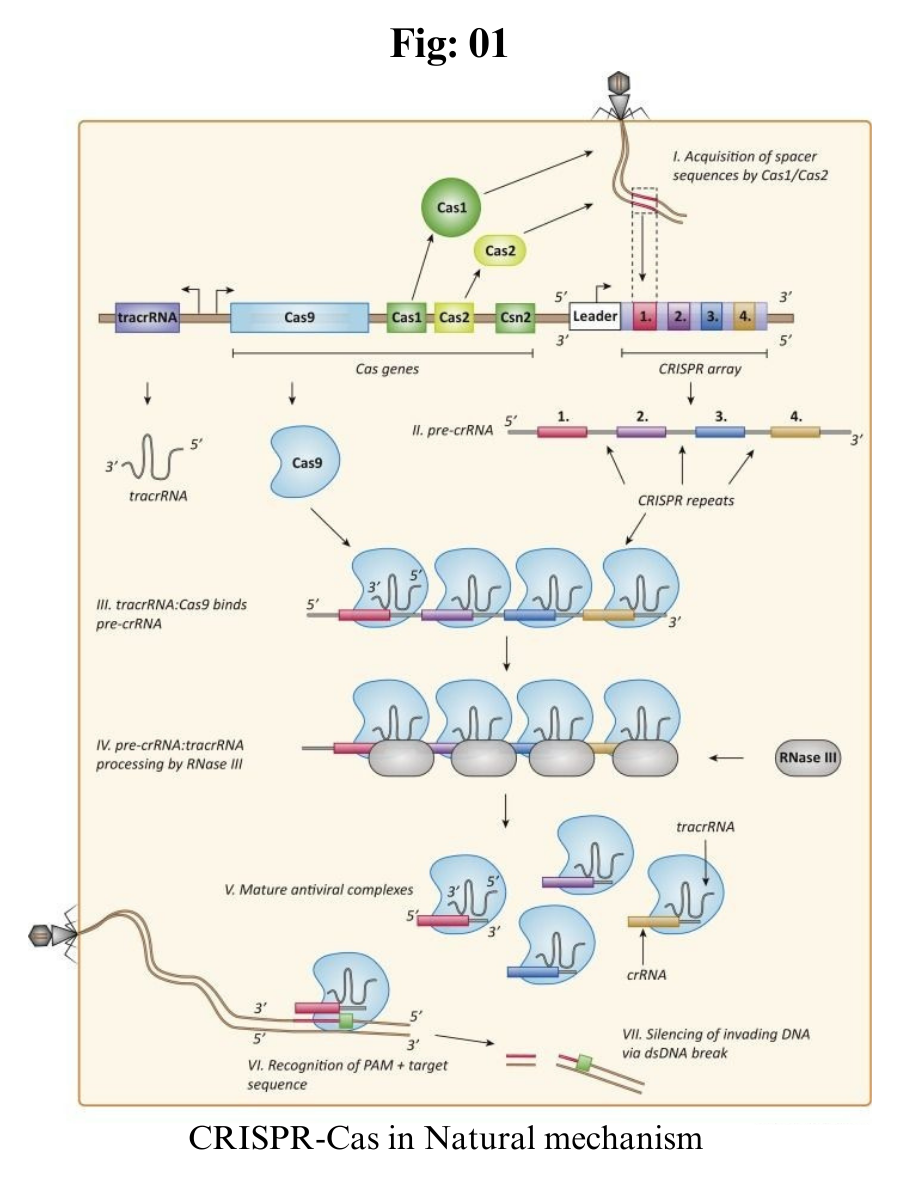
CRISPR-Cas is an adaptive immune system existing in most bacteria and archaea, preventing them from being infected by phages, viruses and other foreign genetic elements. 

It is composed of CRISPR repeat-spacer arrays, which can be further transcribed into CRISPR RNA (crRNA) and trans-activating CRISPR RNA (tracrRNA), and a set of CRISPR-associated (cas) genes which encode Cas proteins with endonuclease activity. 

When the prokaryotes are invaded by foreign genetic elements, the foreign DNA can be cut into short fragments by Cas proteins, then the DNA fragments will be integrated into the CRISPR array as new spacers. 

Once the same invader invades again, crRNA will quickly recognize and pair with the foreign DNA, which guides Cas protein to cleave target sequences of foreign DNA, thereby protecting the host.


CRISPR Components 

CRISPR: Clustered Regularly Interspaced Short Palindromic Repeats of genetic information that some bacterial species use as part of an antiviral system. A group of scientists, including our co-founder Dr. Emmanuelle Charpentier, discovered how to use this system as a gene-editing tool. 

Cas9: a CRISPR-associated (Cas) endonuclease, or enzyme, that acts as “molecular scissors” to cut DNA at a location specified by a guide RNA

Deoxyribonucleic acid (DNA): the molecule that most organisms use to store genetic information, which contains the “instructions for life”

Ribonucleic acid (RNA): a molecule related to DNA that living things use for a number of purposes, including transporting and reading the DNA “instructions”

Guide RNA (gRNA): a type of RNA molecule that binds to Cas9 and specifies, based on the sequence of the gRNA, the location at which Cas9 will cut DNA

PAM: An NGG PAM at the 3′ end of the target DNA site is essential for the recognition and cleavage of the target gene by Cas9 protein. Besides classical NGG PAM sites, other PAM sites such as NGA and NAG also exist, but their efficiency of genome editing is not high. However, such PAM sites only exist in about one-sixteenth of the human genome, thereby largely restricting the targetable genomic loci. For this purpose, several Cas9 variants have been developed to expand PAM compatibility.

Types of CRISPR-Cas

CRISPR-Cas systems can be classified into 2 classes (Class 1 and Class 2), 6 types (I to VI) and several subtypes, with multi-Cas protein effector complexes in Class 1 systems (Type I, III, and IV) and a single effector protein in Class 2 systems (Type II, V, and VI).

The Type II CRISPR-Cas9 system derived from Streptococcus pyogenes (SpCas9) is one of the best characterized and most commonly used categories in numerous CRISPR-Cas systems. The main components of the CRISPR-Cas9 system are RNA-guided Cas9 endonuclease and a single-guide RNA (sgRNA). 

The Cas9 protein possesses two nuclease domains, named HNH and RuvC, and each cleaves one strand of the target double-stranded DNA. A single-guide RNA (sgRNA) is a simplified combination of crRNA and tracrRNA. 

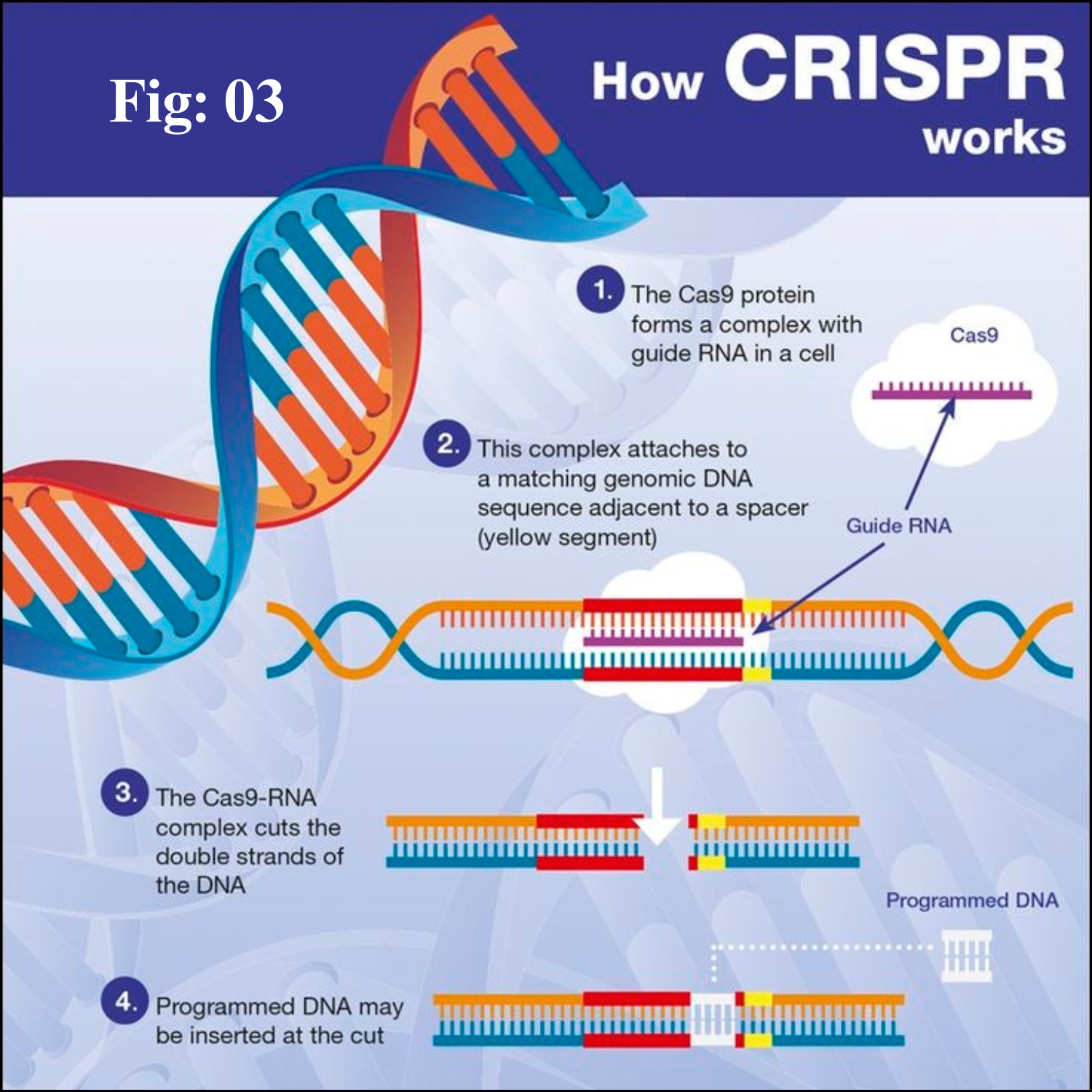
The Cas9 nuclease and sgRNA form a Cas9 ribonucleoprotein (RNP), which can bind and cleave the specific DNA target. Furthermore, a protospacer adjacent motif (PAM) sequence is required for Cas9 protein binding to the target DNA. 

Process

During genome editing process, sgRNA recruits Cas9 endonuclease to a specific site in the genome to generate a double-stranded break (DSB), which can be repaired by two endogenous self-repair mechanisms, the error-prone non-homologous end joining (NHEJ) pathway or the homology-directed repair (HDR) pathway. 

Under most conditions, NHEJ is more efficient than HDR, for it is active in about 90% of the cell cycle and not dependent on nearby homology donors. NHEJ can introduce random insertions or deletions (indels) into the cleavage sites, leading to the generation of frameshift mutations or premature stop codons within the open reading frame (ORF) of the target genes, finally inactivating the target genes. 

Alternatively, HDR can introduce precise genomic modifications at the target site by using a homologous DNA repair template. Furthermore, large fragment deletions and simultaneous knockout of multiple genes could be achieved by using multiple sgRNAs targeting one single gene or more.


Applications of CRISPR-Cas systems in gene therapy

Gene therapy refers to the introduction of foreign genes into target cells to treat specific diseases caused by mutated or defective genes. 

Target cells of gene therapy are mainly divided into two categories: somatic cells and germ line cells. However, since germ line gene therapy is complicated in technique as well as involves ethical and security issues, today gene therapy is limited to somatic cell gene therapy. 

Traditional gene therapy is usually carried out by homologous recombination or lentiviral delivery. Nevertheless, the efficiency of homologous recombination is low, and lentiviral vectors are randomly inserted into the recipient genome, which may bring potential security risks to clinical applications. 

Currently, with the rapid development of CRISPR-Cas systems, they have been widely applied in gene therapy for treating various human diseases, monogenic diseases, infectious diseases, cancer, etc. 

Furthermore, some CRISPR-mediated genome-editing therapies have already reached the stage of clinical testing. Table 4 briefly summarizes the ongoing clinical trials of gene therapy using genome-editing technology, including ZFN, TALEN and CRISPR-Cas systems.

β-Thalassaemia

β-Thalassaemia, a hereditary hemolytic anemia disease, is one of the most common and health-threatening monogenic diseases in the world. It is characterized by mutations in the β-globin (HBB) gene, leading to severe anemia caused by decreased hemoglobin (Hb) level. 

For the moment, the only way to cure β-thalassemia is hematopoietic stem cell transplantation (HSCT). Yet, high cost of treatment and shortage of donors limit its clinical application. Other therapy, for example, blood transfusion, can only sustain the life of patients but can’t cure the disease. 

To better treat β-thalassemia, researchers have turned their attention to gene therapy. A major technical idea is to repair the defective β-globin gene of iPSCs from patients with β-thalassemia by CRISPR-Cas9 technology, then red blood cells can be produced normally and the disease could be cured. 

Besides, reactivating fetal hemoglobin (HbF) expression has also been proposed to be an effective method to treat β-thalassemia through knockout of BCL11A gene, which suppresses the expression of fetal hemoglobin.

Sickle Cell Disease

Additionally, CRISPR-Cas systems have also been used for the treatment of other hematologic diseases, such as sickle cell disease (SCD) and hemophilia B (HB). 

SCD is a monogenic disease caused by a single-nucleotide mutation in human β-globin gene, leading to a substitution of glutamic acid by valine and the production of an abnormal version of β-globin, which is known as hemoglobin S (HbS). 

CRISPR-Cas9 system has been used to treat SCD by repairing the β-globin gene mutation or reactivating HbF expression. 

HB is an X-linked hereditary bleeding disorder caused by deficiency of coagulation factor IX, and the most common treatment for hemophilia B is supplement blood coagulation factor. 

Huai et al. injected naked Cas9-sgRNA plasmid and donor DNA into the adult mice of F9 mutation HB mouse model for gene correction. 

Meanwhile, Cas9/sgRNA were also microinjected into germline cells of this HB mouse model for gene correction. Both in vivo and ex vivo experiments were sufficient to resolve the coagulation deficiency. 

Guan et al. corrected the F9 Y371D mutation in HB mice using CRISPR-Cas9 mediated in situ genome editing, which greatly improved the hemostatic efficiency and increased the survival of HB mice.


HIV (Human immunodeficiency virus)

Human immunodeficiency virus (HIV), a kind of retrovirus, mainly attacks the human immune system, especially the CD4+ T lymphocytes. When human cells are invaded by HIV, the viral sequences can be integrated into the host genome, blocking cellular and humoral immunity while causing acquired immunodeficiency syndrome (AIDS). 

There is still no known cure for AIDS but it could be treated. Although antiretroviral therapy can inhibit HIV-1 replication, the viral sequences still exist in the host genome, and they could be reactivated at any time. 

CRISPR-Cas9 system can target long terminal repeat (LTR) and destruct HIV-1 proviruses, thus it is possible to completely eliminate HIV-1 from the genome of infected host cells. In addition, resistance to HIV-1 infection could be induced by knockout of the HIV co-receptor CCR5 gene in CD4+ T cells.


HPV (Human Papilloma Virus)

Cervical cancer is the second most common gynecologic malignant tumor. The incidence is increasing year by year and young people are especially prone to this disease. It was found that the occurrence of cervical cancer is closely related to HPV (human papillomavirus) infection. 

HPV is a double-stranded cyclic DNA virus, E6 and E7 genes located in HPV16 early regions are carcinogenic genes. Researchers designed sgRNAs targeting E6 and E7 genes to block the expression of E6 and E7 protein, subsequently the expression of p53 and pRb was restored to normal, finally increasing tumor cells apoptosis and suppressing subcutaneous tumor growth in in vivo experiments. Moreover, HPV virus proliferation was blocked through cutting off E6/E7 genes, and the virus in the bodies could be eliminated.

Cancer

Cancer is the second leading cause of death worldwide after cardiovascular diseases, and it is also a medical problem that needs to be solved urgently. A variety of genetic or epigenetic mutations have been accumulated in the cancer genome, which can activate proto-oncogenes, inactivate tumor suppressors and produce drug resistance. 

So far, CRISPR-Cas systems have been used to correct the oncogenic genome/epigenome mutations in tumor cells and animal models, resulting in inhibition of tumor cell growth and promotion of cell apoptosis, thereby inhibiting tumor growth.

Delivery methods

Nowadays, how to effectively deliver CRISPR-Cas components to specific cells, tissues and organs for precisely directed genome editing is still a major problem in gene therapy. Ideal delivery vectors should have the advantages of non-toxicity, well targeting property, high efficiency, low cost, and biodegradability. 

At present, three main delivery methods have been employed in delivering CRISPR-Cas components, including physical, viral and non-viral methods. 

Physical methods are the simplest way to deliver CRISPR-Cas components, including electroporation, microinjection and mechanical cell deformation. They are simple and efficient, which can also improve the expression of genes, and are widely applied in in vitro experiments. 

In addition, viral vectors, such as adenovirus, adeno-associated virus (AAV) and lentivirus viral vectors, are being widely used for both in vitro/ex vivo and in vivo delivery due to their high delivery efficiency. 

They are commonly used for gene delivery in gene therapy, and some of them have been approved for clinical use. However, the safety issue of viral vectors is still a major problem that needs to be solved in pre-clinical trials. 

Therefore, researchers have turned their attention to non-viral vectors, for instance, liposomes, polymers and nanoparticles. Based on the advantages of safety, availability and cost-effectiveness, they are becoming a hotspot for the delivery of CRISPR-Cas components.


Ethical Issues 

CRISPR-Cas mediated genome editing has attracted much attention since its advent in 2012. In theory, each gene can be edited by CRISPR-Cas systems, even genes in human germ cells. However, germline gene editing is forbidden in many countries including China, for it could have unintended consequences and bring ethical and safety concerns.

However, in March 2015, a Chinese scientist, Junjiu Huang, published a paper about gene editing in human tripronuclear zygotes in the journal Protein & Cell, which brings the ethical controversy of human embryo gene editing to a climax. Since then, genome editing has been challenged by ethics and morality, and legal regulation of genome editing has triggered a heated discussion all around the world.

Then, on Nov. 28, 2018, the day before the opening of the second international human genome editing summit, Jiankui He, a Chinese scientist from the Southern University of Science and Technology, announced that a pair of gene-edited babies, named Lulu and Nana, were born healthy in China this month. They are the world’s first gene-edited babies, whose CCR5 gene has been modified, making them naturally resistant to HIV infection after birth. The announcement has provoked shock, even outrage among scientists around the world, causing widespread controversy in the application of genome editing.

The society was shocked by this breaking news, for it involves genome editing in human embryos and propagating into future generations, triggering a chorus of criticism from the scientific community and bringing concerns about ethics and security in the use of genome editing. 

Therefore, scientists call on the Chinese government to investigate the matter fully and establish strict regulations on human genome editing. Global supervisory system is also needed to ensure genome editing of human embryos moving ahead safely and ethically.

Conclusion

Since CRISPR-Cas mediated genome editing technologies have provided an accessible and adaptable means to alter, regulate, and visualize genomes, they are thought to be a major milestone for molecular biology in the 21st century. 

So far, CRISPR-Cas systems have been broadly applied in gene function analysis, human gene therapy, targeted drug development, animal model construction and livestock breeding, which fully prove their great potential for further development. 

However, there are still some limitations to overcome in the practical applications of CRISPR-Cas systems, and great efforts still need to be made to evaluate their long-term safety and effectiveness.

Reverse Transcription

Reverse Transcription Introduction Reverse transcription is the synthesis of DNA from an RNA template. This process is driven by RNA-depende...