Introduction
These animals are known as ‘sea squirt’. The life-history of urochordates passes through a dramatic change. Their chordate characters are more pronounced during the larval period. While in adults they are more like invertebrates than chordates. Therefore, the characters are described in two heads — larval characters and adult characters.
Larval characters of Urochordates
1. Elongated larva of Urochordata is known as ascidian tadpole larva. Adults emerge from the larva by the process of metamorphosis.
2. Notochord restricted at the caudal end, hence the name Urochordata.
3. Dorsal hollow nerve cord spreads end to end.
4. Pharyngeal gill slits are present.
5. Highly active post anal tail is prominent.
Adult characters of Urochordata
1. The body of the adult is covered by a tunic (hence named Tunicata). The tunic is composed of a protein tunicin and a polysaccharide similar to plant cellulose.
2. Adults are sessile and attached to the substratum of the sea.
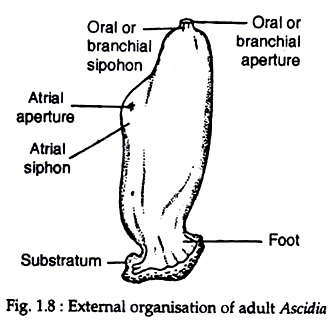
3. Incurrent branchial siphon, and ex-current atrial siphon, form entrance and exit portals for the water that circulates through the body.
4. Branchial siphon opens into a branchial basket, i.e. pharynx.
5. Tiny finger-like sensory tentacles encircle the incurrent siphon to examine the incoming water and prevent large particles from entering.
6. These are hermaphrodite animals; reproduce both sexually and asexually.
Classification of Urochordata
This subphylum is divided into three classes — Ascidiacea, Thaliacea and Larvacea.
The characters and examples of these classes are given here:
A. Class — Ascidiacea:
General characters
1. Comprises mostly brightly coloured marine animals.
2. Some species are solitary, others are colonial.
3. Adults are sessile, but larvae are plank
tonic and do not feed.
4. Adults having sac-like body, covered by tunic.
5. Most of the chordate characters that were present during larval period disappear during metamorphosis into adult.
6. In adult, nervous system transforms into a nerve ganglion.
Examples:
Ascidia, Ciona, Herdmania.
B. Class — Thaliacea:
General Characters
1. These are free-living pelagic urochordates.
2. The tunic is transparent and thin.
3. They possess encircling circumferential bands of muscles within the walls of the test.
4. Incurrent and ex-current siphons are present at opposite end of the body.
5. A few pharyngeal gill slits are present.
6. In the life-cycle polymorphism and clear alternation of generations are evident.
Examples:
Salpa, Doliolum.
C. Class — Larvacea/Appendicularia:
General characters
1. These are tiny marine planktonic urochordates found worldwide.
2. Larvacea received their name because the adults retain larval characteristics similar in some way to the ascidian tadpole with its tail and trunk. The general resemblance of adult larvaceans to ascidian tadpoles suggests that larvaceans may be neotenous form.
3. They produce a remarkable feeding apparatus (house) that consists of three components: screens, filters and expanded gelatinous matrix. Disturbed or actively feeding larvaceans abandon their old house and builds a new one.
4. The trunk holds major body organs.
5. The tail is thin and flat.
6. Muscle bands act on notochord to produce movement.
7. A tubular nerve cord is present.
8. All species, except one, are monoecious, and most of these are protandrous.
Examples:
Oikopleura, Appendicularia.
Ascidiacea (Sea squirts)
Phylum Chordata
Class Ascidiacea
Number of families 24
Physical characteristics
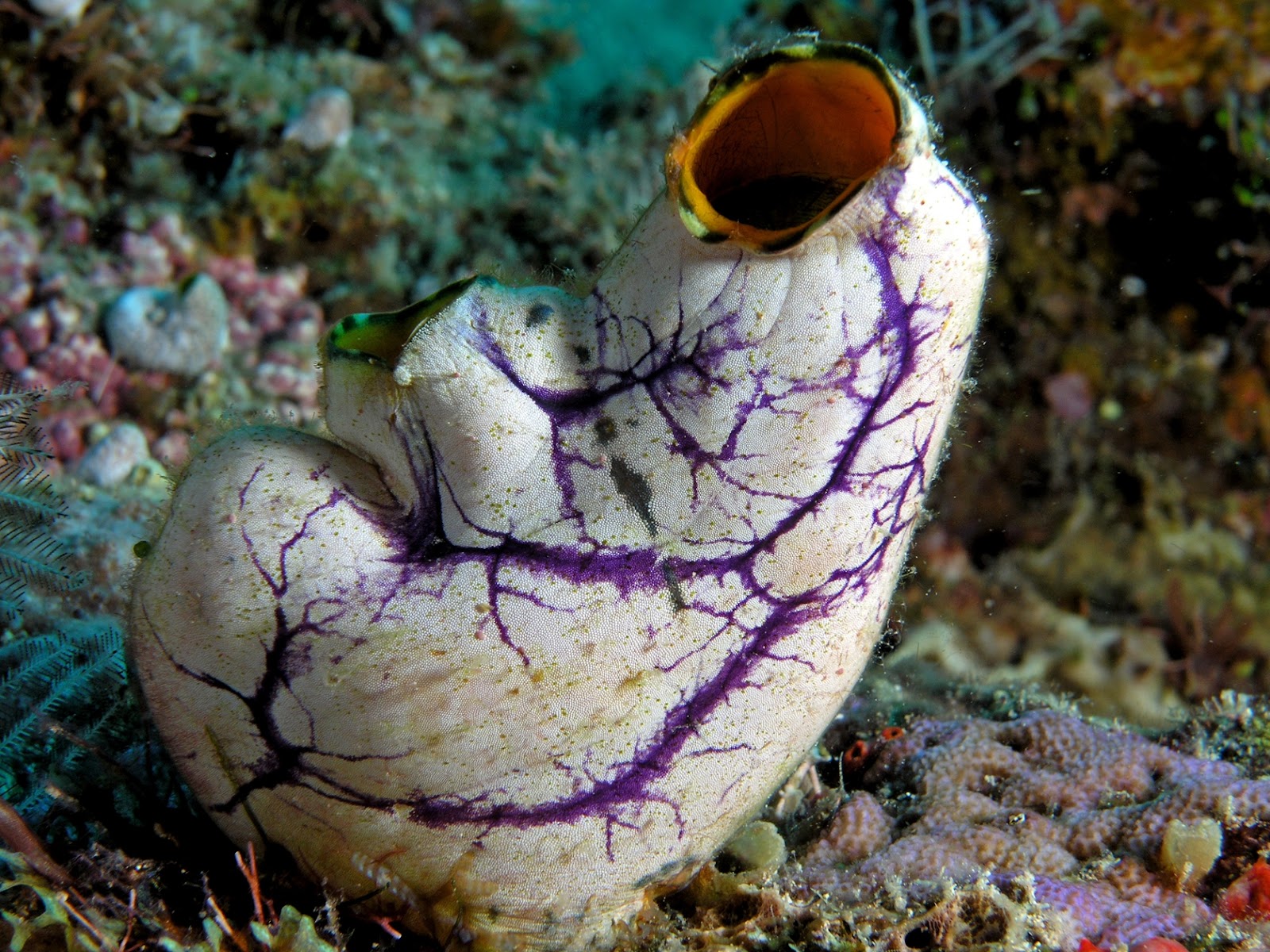
Usually, ascidians are described as sessile organisms with sac-like bodies firmly attached by the posterior end to the substratum. In reality, the shape of different ascidian species may be very diverse, and sometimes it is even difficult to recognize them as belonging to Ascidiacea.
Although they grow side by side, is hard to imagine that the large, upright, cylindrical, solitary Ciona species belong to the same order as the Didemnum, which look like thin, encrusting stones and resemble a sponge colony.
The body is always covered with the test, a protective layer of cellulose-like material secreted by the epithelium. The test may be clear and often brightly colored, as in the spectacular Pacific species Halocynthia aurantium, or it may be covered by various kinds of spines, as in the northern Boltenia echinata; it even may contain calcareous spicules, as in snow-white Bathypera ovoida.
Among species living on a sandy or muddy bottom, the test often has long, thin outgrows, or test hairs, and sometimes is covered by a dense layer of sand grains, making the specimen cryptic, like many species of the family Molgulidae.
The body muscles typically are arranged in transverse and longitudinal bands. Sometimes they are numerous, thick, and strong, forming a solid muscular wall; in many species, however, the muscles are represented by only a few thin fibers.
There are two openings on the body: an oral or branchial opening and an atrial opening. These openings may be sessile or set on the ends of siphons of various lengths, the atrial siphon always dorsal to the branchial siphon.
The oral siphon leads to the voluminous pharynx, called in ascidians the "branchial sac," The branchial sac occupies most the space in the body of solitary ascidians. The space inside the branchial sac is the branchial cavity, and the space between the branchial sac and the body wall is the atrial cavity.
The atrial cavity opens to the exterior through the atrial siphon, and the branchial and atrial cavities are filled with seawater.
The seawater, with food particles and oxygen, is drawn into the branchial sac through the branchial siphon and a circle of oral tentacles and passes from the branchial to the atrial cavity through numerous perforations in the wall of the branchial sac, where food items are sieved; then filtered water moves out from the body through the atrial siphon.
Perforations of the wall of the branchial sac generally are small and have ciliated margins called stigmata, The shape of the stigmata is an important taxonomic character: they may be straight and arranged in transverse or sometimes longitudinal rows, or they may be spiral figures, sometimes forming high funnels protruding into the branchial cavity.
The wall of the branchial sac has transverse and longitudinal branchial vessels crossing each other and forming rectangular meshes. In some deepwater ascidians true stigmata are absent, the wall of the branchial sac is reduced, and the branchial sac is represented only by wide rectangular meshes formed by crossing longitudinal and transverse branchial vessels.
The branchial sac is bilaterally symmetrical, with its mid-dorsal line marked by a fold termed the "dorsal lamina." The midventral line has an endostyle, a groove lined with ciliated glandular epithelium secreting mucus.
The mucus constantly moves from the endostyle to the dorsal lamina and then, with the filtered food particles, to the esophagus, to which the bottom of the branchial sac opens.
The esophagus typically is short and always is much narrower than the branchial sac. It leads to the stomach and then to the intestine. The intestine makes a loop and opens into the atrial cavity.
The position of the gut loop in relation to the branchial sac is an important taxonomic character of the suborder and family levels: the gut loop may be on the left or, in one family, on the right side of the branchial sac; between the branchial sac and the body wall; or, mostly in colonial ascidians, under the branchial sac.
Gonads are hermaphroditic and situated in the gut loop, under the gut, or on the body wall on the sides of the branchial sac. Gonoducts always open into the atrial cavity, and only in one highly specialized genus do they penetrate the test and open directly to the exterior.
The nervous system, as in all sessile animals, is simple and represented by an elongated ganglion situated on the dorsal side between the siphons and a few nerves running from it.
The heart is a thin-walled tubular organ on the ventral side of the body or, in colonial ascidians, in the bottom of elongated zooids.
In the case of colonial ascidians, the test forms the so-called common test, a mass in which individuals, called "zooids," are completely or partly embedded. The general plan of the structure of the zooids is the same as in solitary ascidians, but details may differ significantly.
The body may be undivided or divided into two (thorax and abdomen) or three (thorax, abdomen, and post-abdomen) regions. The thorax contains the branchial sac, the gut loop is in the abdomen, and the gonads are either in the abdomen or the post-abdomen.
Branchial siphons of all zooids in a colony open directly to the exterior, but atrial apertures in many species open into the cloacal cavity within the common test. The colony may contain single or several isolated cloacal cavities, each exposed to the exterior through one or several openings.
Zooids connected with one cloacal cavity form a system. The shape of the systems varies; zooids may be arranged in circular systems around the single cloacal opening in the center, or they may form long double rows along cloacal canals.
The form of the systems usually can be recognized easily on living colonies; the systems make a characteristic pattern on the surface of the colony, which, in certain cases, helps to identify the species.
The size of most ascidians varies from 0.04 to 0.4 in (1–10 mm), rarely 0.6 in (15 cm). Certain species, however, are much larger; in favorable conditions some solitary species may reach 19.7 in (50 cm) in height, and thin, encrusting colonies of certain species of didemnids grow to 9.8 sq ft (3 sq m).
The largest species is the colonial Antarctic Distaplia cylindrica; its long, sausage-shaped colonies may be up to 23 ft (7 m) in length and 3 in (8 cm) in diameter. The smallest species is the deepwater Minipera pedunculata, whose diameter is only 0.02 in (0.5 mm).
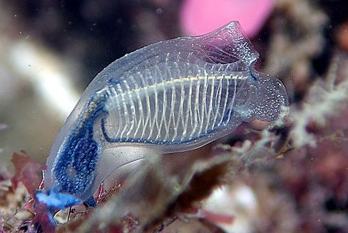
Many ascidians are brightly colored, usually red, brown, and yellow or, rarely, blue. The coloring of colonial ascidians is especially diverse: that of the zooids and the common test may be different, resulting in complex, often very beautiful patterns on the colony surface. Some species, such as Clavelina, have a transparent body, often marked with variously colored spots and lines.
Many ascidians have a peculiar feature: they are able to accumulate vanadium. More primitive species tend to have higher levels of vanadium in their tissues.
Behavior
Ascidians are sessile, usually firmly fixed to the substratum. In response to an external stimulus, they can only slowly contract the body and close the siphons. Some of the very few interstitial species can actively move between gravel particles.
Very slow movement of the colony also has been recorded in one or two tropical colonial species. Abyssal Situla and Megalodicopia species can quickly close their enormously large, bilobed branchial siphons in attempt to catch small swimming invertebrates, such as copepods.
Feeding Ecology And Diet
Almost all ascidians are filter feeders. The cilia lining the stigmata on the pharyngeal wall constantly pump water with suspended organic particles through the branchial sac.
This feeding method is called active filtration: an ascidian expends energy to create a steady flow of water through the branchial sac. With the filtration membrane, a constantly moving mucous sheet on the inner surface of the branchial sac, ascidians are able to capture very fine organic particles including bacteria and phytoplankton.
The species living in water with higher concentrations of suspended particles usually have more complex and dense branchial sacs; the stigmata in these species typically are small and numerous, and high branchial folds increase the branchial surface.
At great depths, water contains very little organic matter. Ascidians having the usual type of branchial sac, with ciliated stigmata, are very small in size there. Some deepwater species, such as Culeolus, have lost true ciliated stigmata; the branchial sac in such species resembles a loose net made by the crossing vessels, without tissue between them.
These branchial sacs have less resistance to water flow than do the dense filters of shallow-water ascidians. These species typically have very large, widely opened branchial siphons oriented to the water current, allowing so-called passive filtration: water passes through the branchial sac without any muscular action or beating of the cilia, thus saving energy.
Such highly adapted abyssal species often are large. Ascidians of the family Octacnemidae have a mixed diet. Some of them apparently can catch small swimming invertebrates with their large, bilobed branchial siphons.
Reproductive biology
All ascidians are hermaphroditic. All colonial species of the suborder Aplousobranchia are viviparous; in these species ova are fertilized internally and developed within the parent colony, in the atrial cavity of zooids or in special outgrowths of the zooid body wall, termed brood pooches, or in the common test of the colony.
Viviparous species release swimming larvae. Solitary species are either viviparous or oviparous, releasing ova. In viviparous solitary species, larvae are incubated in the atrial cavity.
Ascidian larvae are called tadpole larvae. Larvae have an oval trunk, usually less than 0.04 in (1 mm) long, and a tail that is longer than the trunk. The larval trunk contains the larval organs, including the cerebral vesicle with a light receptor and statocyte and adhesive papillae on the anterior end, and a rudiment of the future adult.
In many colonial species this rudimentary ascidian within the larval trunk is rather well developed and resembles a zooid of the adult ascidian. Sometimes, as in colonial Diplosoma, the larval trunk may contain two or more future zooids.
Ascidian larvae never feed; they swim for a short time and then attach to the substratum and start metamorphosis. During metamorphosis all larval organs degenerate, and an ascidian develops from the rudiment located in the larval trunk.
In the case of solitary ascidians only one specimen develops from each larva, whereas in colonial ascidians individuals (zooids) replicate themselves by asexual reproduction (budding) and form a colony.
Retrogressive Metamorphosis
Some of the urochordates like herdmania possess eyespot, statocyst, a long tail and a well developed noto-chord in the larval stage. But as they grow into the adult form the eyespot and statocyst disappear and the tail and notochord get reduced.
It is a biological process by which an animal physically develops after birth through cell growth and differentiation. The transformation of a maggot into an adult fly and of a tadpole into an adult frog are examples of metamorphosis.
'Retrogressive metamorphosis' means degenerative changes wherein an active larva transforms into a sedentary adult. For example, in Urochordata, the larva bears all advanced characters of Chordata but after metamorphosis, the adult loses its chordate characters.
Thus, here the larva exhibits advanced characters and during metamorphosis, retrogression of characters occurs, such type of metamorphosis is called retrogressive metamorphosis. Let us consider an example of Herdmania, a Urochordate.
Changes during Metamorphosis in Herdmania (Urochordata) are:
-The notochord, nerve cord muscles, and tail will be reduced. All these structures help the larva to swim freely in the water. But they are not useful to adults.
-The alimentary canal becomes complicated. The pharynx enlarges in size. The number of gill slits will increase by divisions.
-The stomach, gonads, and intestine will grow.
-The nervous system is reduced.
-The atrial cavity enlarges into a sac-like structure.
-The eyespot and statocyst will completely disappear.


