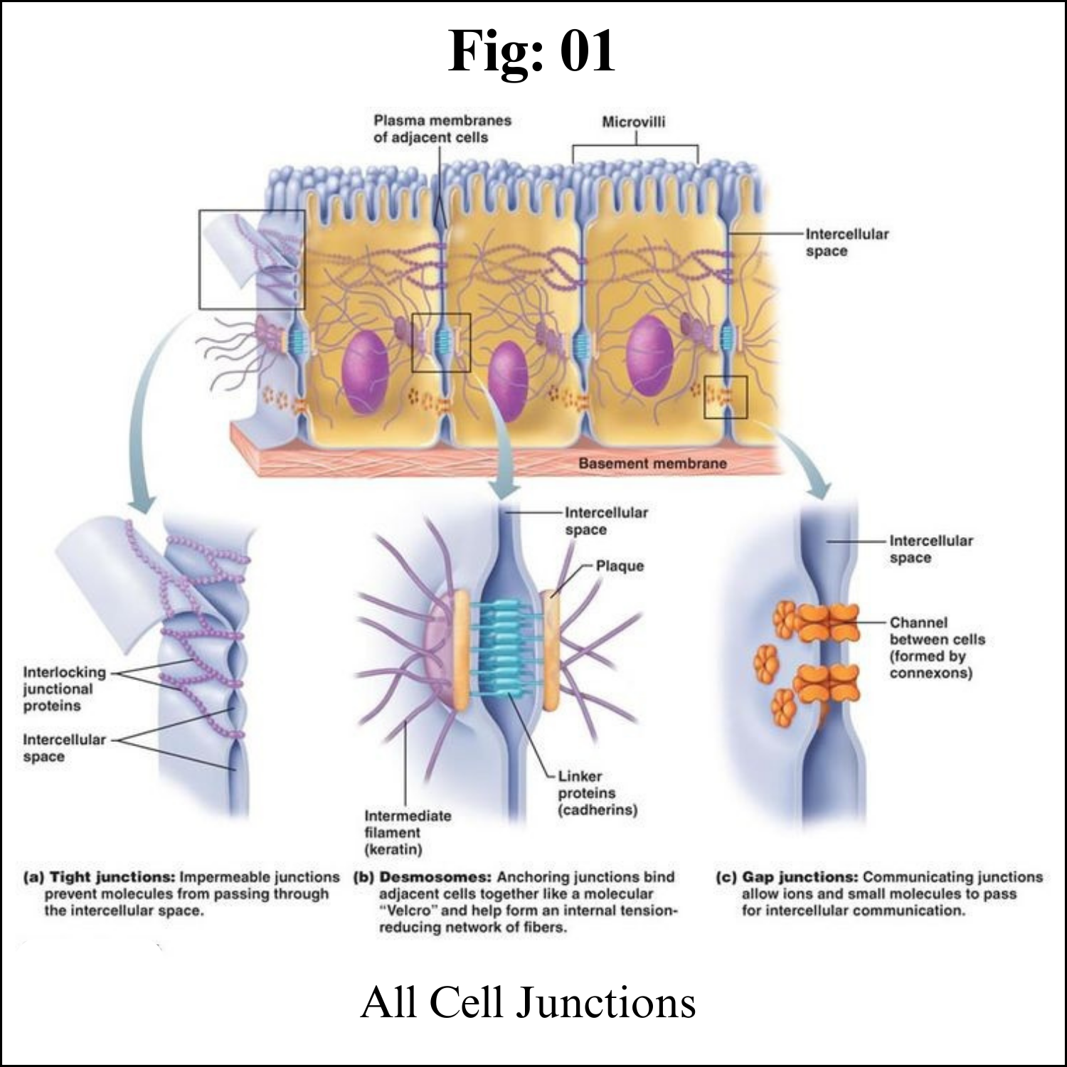
Cell Junctions
Introduction
If you were building a building, what kinds of connections might you want to put between the rooms? In some cases, you’d want people to be able to walk from one room to another, in which case you’d put in a door. In other cases, you’d want to hold two adjacent walls firmly together, in which case you might put in some strong bolts. And in still other cases, you might need to ensure that the walls were sealed very tightly together – for instance, to prevent water from dripping between them.
As it turns out, cells face the same questions when they’re arranged in a tissue next to other cells. Should they put in doors that connect them directly to their neighbors? Do they need to spot-weld themselves to their neighbors to make a strong layer, or perhaps even form tight seals to prevent water from passing through the tissue? Junctions serving all of these functions can be found in cells of different types, and here, we’ll look at each of them in turn.
In many animal tissues (e.g., connective tissue), each cell is separated from the next by an extracellular coating or matrix. However, in some tissues (e.g., epithelia), the plasma membranes of adjacent cells are pressed together. Four kinds of junctions occur in vertebrates:
1. Tight junctions
2. Adherens junctions
3. Gap junctions
4. Desmosomes
In many plant tissues, it turns out that the plasma membrane of each cell is continuous with that of the adjacent cells. The membranes contact each other through openings in the cell wall called Plasmodesmata.
Plasmodesmata
Plant cells, surrounded as they are by cell walls, don’t contact one another through wide stretches of plasma membrane the way animal cells can. However, they do have specialized junctions called plasmodesmata (singular, plasmodesma), places where a hole is punched in the cell wall to allow direct cytoplasmic exchange between two cells.
Plasmodesmata are lined with plasma membrane that is continuous with the membranes of the two cells. Each plasmodesma has a thread of cytoplasm extending through it, containing an even thinner thread of endoplasmic reticulum (not shown in the diagram above).
Molecules below a certain size (the size exclusion limit) move freely through the plasmodesmal channel by passive diffusion. The size exclusion limit varies among plants, and even among cell types within a plant. Plasmodesmata may selectively dilate (expand) to allow the passage of certain large molecules, such as proteins, although this process is poorly understood.
Although each plant cell is encased in a boxlike cell wall, it turns out that communication between cells is just as easy, if not easier, than between animal cells. Fine strands of cytoplasm, called plasmodesmata, extend through pores in the cell wall connecting the cytoplasm of each cell with that of its neighbors.
Plasmodesmata provide an easy route for the movement of ions, small molecules like sugars and amino acids, and even macromolecules like RNA and proteins, between cells. The larger molecules pass through with the aid of actin filaments. Plasmodesmata are sheathed by a plasma membrane that is simply an extension of the plasma membrane of the adjoining cells. This raises the intriguing question of whether a plant tissue is really made up of separate cells or is, instead, a syncytium: a single, multinucleated cell distributed throughout hundreds of tiny compartments.
Gap junctions
Functionally, gap junctions in animal cells are a lot like plasmodesmata in plant cells: they are channels between neighboring cells that allow for the transport of ions, water, and other substances cubed. Structurally, however, gap junctions and plasmodesmata are quite different.
In vertebrates, gap junctions develop when a set of six membrane proteins called connexins form an elongated, donut-like structure called a connexon. When the pores, or “doughnut holes,” of connexons in adjacent animal cells align, a channel forms between the cells. (Invertebrates also form gap junctions in a similar way, but use a different set of proteins called innexins.)
Gap junctions are particularly important in cardiac muscle: the electrical signal to contract spreads rapidly between heart muscle cells as ions pass through gap junctions, allowing the cells to contract in tandem.
Gap junctions are intercellular channels some 1.5–2 nm in diameter. These permit the free passage between the cells of ions and small molecules (up to a molecular weight of about 1000 daltons). They are cylinders constructed from 6 copies of transmembrane proteins called connexins. Because ions can flow through them, gap junctions permit changes in membrane potential to pass from cell to cell.
Examples of gap junctions include:
a. The action potential in heart (cardiac) muscle flows from cell to cell through the heart providing the rhythmic contraction of the heartbeat.
b. At some so-called electrical synapses in the brain, gap junctions permit the arrival of an action potential at the synaptic terminals to be transmitted across to the postsynaptic cell without the delay needed for release of a neurotransmitter.
c. As the time of birth approaches, gap junctions between the smooth muscle cells of the uterus enable coordinated, powerful contractions to begin.
d. Several inherited disorders of humans such as certain congenital heart defects and certain cases of congenital deafness have been found to be caused by mutant genes encoding connexins.
Tight junctions
Not all junctions between cells produce cytoplasmic connections. Instead, tight junctions create a watertight seal between two adjacent animal cells.
At the site of a tight junction, cells are held tightly against each other by many individual groups of tight junction proteins called claudins, each of which interacts with a partner group on the opposite cell membrane. The groups are arranged into strands that form a branching network, with larger numbers of strands making for a tighter seal.
The purpose of tight junctions is to keep liquid from escaping between cells, allowing a layer of cells (for instance, those lining an organ) to act as an impermeable barrier. For example, the tight junctions between the epithelial cells lining your bladder prevent urine from leaking out into the extracellular space.
Epithelia are sheets of cells that provide the interface between masses of cells and a cavity or space (a lumen). The portion of the cell exposed to the lumen is called its apical surface. The rest of the cell (i.e., its sides and base) make up the basolateral surface. Tight junctions seal adjacent epithelial cells in a narrow band just beneath their apical surface. They consist of a network of claudins and other proteins. Tight junctions perform two vital functions:
1. They limit the passage of molecules and ions through the space between cells. So most materials must actually enter the cells (by diffusion or active transport) in order to pass through the tissue. This pathway provides tighter control over what substances are allowed through.
2. They block the movement of integral membrane proteins (red and green ovals) between the apical and basolateral surfaces of the cell. Thus the special functions of each surface, for example receptor-mediated endocytosis at the apical surface and exocytosis at the basolateral surface can be preserved.
Desmosomes
Animal cells may also contain junctions called desmosomes, which act like spot welds between adjacent epithelial cells. A desmosome involves a complex of proteins. Some of these proteins extend across the membrane, while others anchor the junction within the cell.
Cadherins, specialized adhesion proteins, are found on the membranes of both cells and interact in the space between them, holding the membranes together. Inside the cell, the cadherins attach to a structure called the cytoplasmic plaque (red in the image at right), which connects to the intermediate filaments and helps anchor the junction.
Desmosomes pin adjacent cells together, ensuring that cells in organs and tissues that stretch, such as skin and cardiac muscle, remain connected in an unbroken sheet.
Desmosomes are localized patches that hold two cells tightly together. They are common in epithelia (e.g., the skin).
Desmosomes are attached to intermediate filaments of keratin in the cytoplasm. Pemphigus is an autoimmune disease in which the patient has developed antibodies against proteins (cadherins) in desmosomes. The loosening of the adhesion between adjacent epithelial cells causes blistering.
Carcinomas are cancers of epithelia. However, the cells of carcinomas no longer have desmosomes. This may partially account for their ability to metastasize.
Hemidesmosomes
These are similar to desmosomes but attach epithelial cells to the basal lamina ("basement membrane") instead of to each other. Pemphigoid is an autoimmune disease in which the patient develops antibodies against proteins (integrins) in hemidesmosomes. This, too, causes severe blistering of epithelia.
The function of cell junctions
The main function of some junctions is to connect the cytoplasm between adjacent cells and allow intercellular transportation and communication. Other junctions mostly function as adherence sites that maintain tissue structure and integrity. All types of tissues in animals have junctions, however, junctions that serve for adherence are more abundant in epithelial tissues (tissues that line internal organs and cavities).
Cell Junctions - Key takeaways
The two main functions of intercellular junctions are to allow intercellular transportation and communication or to maintain tissue structure and integrity through adherence of adjacent cells.
Connections between cell plants are called plasmodesmata, while animal cells can connect through tight junctions, gap junctions, and desmosomes.
Plasmodesmata connect the cytoplasm of adjacent plant cells for intercellular communication and movement of material. Gap junctions allow the intercellular movement of ions and small molecules and are essential for the transmission of electrical signals throughout a tissue.
Tight junctions and desmosomes are for adherence rather than connection, maintaining tissue integrity. Tight junctions provide a waterproof seal, while desmosomes give resistance to mechanical stress.


