Sunday, August 20, 2023

Coelom (Body Cavity)


Coelom 

Introduction:

The coelom is one of the characteristic features of metazoans. The true coelom is a body cavity formed during embryo development from the three germinal layers. The body cavity meaning a fluid filled space that can accomodate organs. The coelom is lined by mesodermal epithelium cells. Presence or absence of coelom is one of the criteria for classifying animals.

Definition:

" The coelom is the fluid-filled body cavity present between the alimentary canal and the body wall." 

The true coelom has a mesodermal origin. It is lined by mesoderm. The peritoneal cavity present in the abdomen and similar spaces around other organs such as lungs, heart are parts of the coelom. Coelom differs in its structure and formation process. 

Structure, Formation and Types of Coelom:

There are three types of structural body formation present in animals related to coelom:

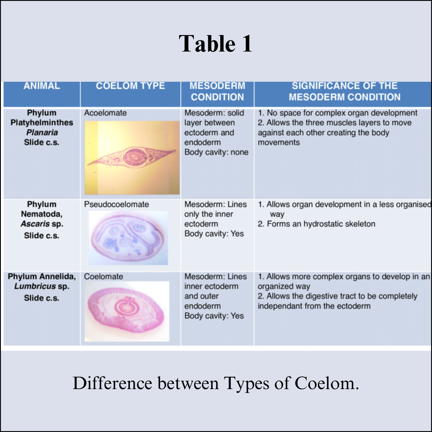
1. Acoelomate:

Coelom is absent. The blastocoel is completely occupied by mesoderm. E.g. Porifera, Coelenterata and Flatworms (Platyhelminthes). There is only spongocoel or coelenteron present.

2. Pseudocoelomate:

True coelom is not present. The blastocoel is partly filled by mesodermal cells. The body cavity is lined by mesoderm only towards the body wall and mesoderm is not present towards the gut. E.g. Roundworms (Aschelminthes)

3. Eucoelomate:

Animals that have a true coelom. The coelom is lined by mesoderm on both the sides, towards the body wall and towards the gut. The blastocoel present in the gastrula gets completely replaced by a true coelom. The body organs are suspended in the coelom by mesenteries. E.g. from the phylum Annelida to Chordata.


Eucoelomates are further divided into Protostomes and Deuterostomes on the basis of different embryonic development. The process of coelom formation in protostomes and deuterostomes is different. 

The coelom is categorized into two types on the basis of formation, namely, Schizocoelom and Enterocoelom.

a. Schizocoelom: It is present in the protostomes. The body cavity or coelom originates from the splitting of the mesoderm. One part attaches to ectoderm and the other surrounds the endoderm. The space between them develops into the coelom. The blastopore forms the mouth. Examples of schizocoelous animals are Annelida, Arthropoda and Mollusca. In Arthropoda and Mollusca the coelom is filled by blood and is known as Haemocoel.

b. Enterocoelom: It is present in the deuterostomes. The coelom is formed from the fusion of the internal outgrowths of the archenteron, that pinches off and fuses together to form coelom lined by mesoderm. Examples of enterocoelous animals are Echinodermates and Chordates.


Coelom Function

Coelom works as a shock absorber and protects from any kind of mechanical shock. It gives more flexibility to the body organs to move and protects from any damage on minor bends by cushioning the internal organs.

The coelomic fluid acts as a hydrostatic skeleton, which helps in the locomotion of soft-bodied animals and gives the body a definite shape. Contracting muscles can push against the coelomic fluid because of the fluid pressure.

The coelomocyte cells, that either float freely in the coelom or attached to the wall, support the immune system. They support the immune system by initiating humoral immune response and phagocytosis.

The coelomic fluid also helps in gaseous transport and transport of nutrients and waste products.

Coelom gives the extra space required by organs to develop and function. E.g. pumping action of the heart, carrying a child in the womb, etc. is possible due to coelom.

Saturday, August 19, 2023

Mitotic Cell Division


Mitotic Cell Division 

Introduction 

Perhaps the most amazing thing about mitosis is its precision, a feature that has intrigued biologists since Walther Flemming first described chromosomes in the late 1800s. Although Flemming was able to correctly deduce the sequence of events in mitosis, this sequence could not be experimentally verified for several decades, until advances in light microscopy made it possible to observe chromosome movements in living cells. Researchers now know that mitosis is a highly regulated process involving hundreds of different cellular proteins. The dynamic nature of mitosis is best appreciated when this process is viewed in living cells.

Mitosis Occupies a Portion of the Cell Cycle

In his pioneering studies of mitosis, Flemming noted that the nuclear material, which he named "chromatin" for its ability to take up stains, did not have the same appearance in all cells. (We still use the word "chromatin" today, albeit in a more biochemical sense to refer to complexes of nuclear DNA and protein.) 

Specifically, in some cells, chromatin appeared as an amorphous network, although in other cells, it appeared as threadlike bodies that Flemming named "mitosen." Based on his observations, Flemming had the insight to propose that chromatin could undergo reversible transformations in cells. 

Today, scientists know that Flemming had successfully distinguished chromosomes in the interphase portion of the cell cycle from chromosomes undergoing mitosis, or the portion of the cell cycle during which the nucleus divides. With very few exceptions, mitosis occupies a much smaller fraction of the cell cycle than interphase.

The difference in DNA compaction between interphase and mitosis is dramatic. A precise estimate of the difference is not possible, but during interphase, chromatin may be hundreds or even thousands of times less condensed than it is during mitosis. 

For this reason, the enzyme complexes that copy DNA have the greatest access to chromosomal DNA during interphase, at which time the vast majority of gene transcription occurs. 

In addition, chromosomal DNA is duplicated during a sub portion of interphase known as the S, or synthesis, phase. As the two daughter DNA strands are produced from the chromosomal DNA during S phase, these daughter strands recruit additional histones and other proteins to form the structures known as sister chromatids. 

The sister chromatids, in turn, become "glued" together by a protein complex named cohesin. Cohesin is a member of the SMC, or structural maintenance of chromosomes, a family of proteins. SMC proteins are DNA-binding proteins that affect chromosome architectures; indeed, cells that lack SMC proteins show a variety of defects in chromosome stability or chromosome behavior. 

Current data suggest that cohesin complexes may literally form circles that encompass the two sister chromatids. At the end of S phase, cells are able to sense whether their DNA has been successfully copied, using a complicated set of checkpoint controls that are still not fully understood. For the most part, only cells that have successfully copied their DNA will proceed into mitosis.


Chromatin Is Extensively Condensed as Cells Enter Mitosis

The most obvious difference between interphase and mitosis involves the appearance of a cell's chromosomes. During interphase, individual chromosomes are not visible, and the chromatin appears diffuse and unorganized. Recent research suggests, however, that this is an oversimplification and that chromosomes may actually occupy specific territories within the nucleus. 

In any case, as mitosis begins, a remarkable condensation process takes place, mediated in part by another member of the SMC family, condensin. 

Like cohesin, condensin is an elongated complex of several proteins that binds and encircles DNA. In contrast to cohesin, which binds two sister chromatids together, condensin is thought to bind a single chromatid at multiple spots, twisting the chromatin into a variety of coils and loops.


The Mitotic Spindle Aids in Chromosome Separation

During mitosis, chromosomes become attached to the structure known as the mitotic spindle. In the late 1800s, Theodor Boveri created the earliest detailed drawings of the spindle based on his observations of cell division in early Ascaris embryos. 

Boveri's drawings, which are amazingly accurate, show chromosomes attached to a bipolar network of fibers. Boveri observed that the spindle fibers radiate from structures at each pole that we now recognize as centrosomes, and he also noted that each centrosome contains two small, rodlike bodies, which are now known as centrioles. 

Boveri observed that the centrioles duplicate before the chromosomes become visible and that the two pairs of centrioles move to separate poles before the spindle assembles. We now know that centrioles duplicate during S phase, although many details of this duplication process are still under investigation.

The composition of the spindle fibers remained unknown until the 1960s, when tubulin was discovered and techniques were developed for visualizing spindles using electron microscopes. 

It is now well-established that spindles are bipolar arrays of microtubules composed of tubulin and that the centrosomes nucleate the growth of the spindle microtubules. During mitosis, many of the spindle fibers attach to chromosomes at their kinetochores, which are specialized structures in the most constricted regions of the chromosomes. 

The length of these kinetochore-attached microtubules then decreases during mitosis, pulling sister chromatids to opposite poles of the spindle. Other spindle fibers do not attach to chromosomes, but instead form a scaffold that provides mechanical force to separate the daughter nuclei at the end of mitosis.


Mitosis Is Divided into Well-Defined Phases

From his many detailed drawings of mitosen, Walther Flemming correctly deduced, but could not prove, the sequence of chromosome movements during mitosis. 

Flemming divided mitosis into two broad parts: a progressive phase, during which the chromosomes condensed and aligned at the center of the spindle, and a regressive phase, during which the sister chromatids separated. 

Our modern understanding of mitosis has benefited from advances in light microscopy that have allowed investigators to follow the process of mitosis in living cells. Such live cell imaging not only confirms Flemming's observations, but it also reveals an extremely dynamic process that can only be partially appreciated in still images.

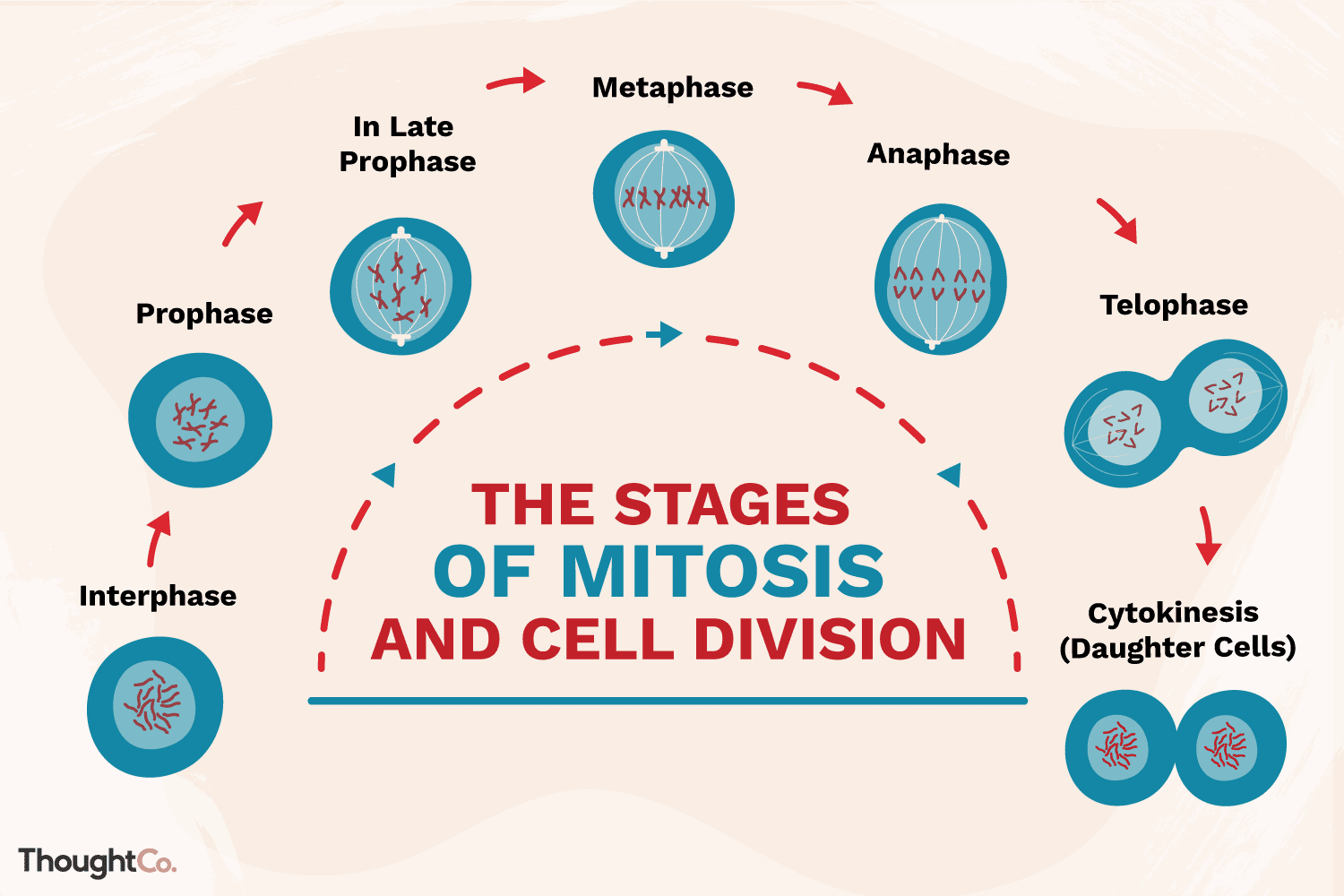
Today, mitosis is understood to involve five phases, based on the physical state of the chromosomes and spindle. These phases are prophase, prometaphase, metaphase, anaphase, and telophase. 

Cytokinesis is the final physical cell division that follows telophase, and is therefore sometimes considered a sixth phase of mitosis. All phases of mitosis, as well as the flanking periods of interphase and cytokinesis before and after. 

Researchers' biochemical understanding of mitotic phases has greatly increased in recent years, revealing that this highly orchestrated process involves hundreds, if not thousands, of cellular proteins.

Prophase

Mitosis begins with prophase, during which chromosomes recruit condensin and begin to undergo a condensation process that will continue until metaphase. In most species, cohesin is largely removed from the arms of the sister chromatids during prophase, allowing the individual sister chromatids to be resolved. Cohesin is retained, however, at the most constricted part of the chromosome, the centromere. During prophase, the spindle also begins to form as the two pairs of centrioles move to opposite poles and microtubules begin to polymerize from the duplicated centrosomes.

Prometaphase

Prometaphase begins with the abrupt fragmentation of the nuclear envelope into many small vesicles that will eventually be divided between the future daughter cells. The breakdown of the nuclear membrane is an essential step for spindle assembly. Because the centrosomes are located outside the nucleus in animal cells, the microtubules of the developing spindle do not have access to the chromosomes until the nuclear membrane breaks apart.

Prometaphase is an extremely dynamic part of the cell cycle. Microtubules rapidly assemble and disassemble as they grow out of the centrosomes, seeking out attachment sites at chromosome kinetochores, which are complex platelike structures that assemble during prometaphase on one face of each sister chromatid at its centromere. As prometaphase ensues, chromosomes are pulled and tugged in opposite directions by microtubules growing out from both poles of the spindle, until the pole-directed forces are finally balanced. Sister chromatids do not break apart during this tug-of-war because they are firmly attached to each other by the cohesin remaining at their centromeres. At the end of prometaphase, chromosomes have a bi-orientation, meaning that the kinetochores on sister chromatids are connected by microtubules to opposite poles of the spindle.

Metaphase

Next, chromosomes assume their most compacted state during metaphase, when the centromeres of all the cell's chromosomes line up at the equator of the spindle. Metaphase is particularly useful in cytogenetics, because chromosomes can be most easily visualized at this stage. Furthermore, cells can be experimentally arrested at metaphase with mitotic poisons such as colchicine. Video microscopy shows that chromosomes temporarily stop moving during metaphase. A complex checkpoint mechanism determines whether the spindle is properly assembled, and for the most part, only cells with correctly assembled spindles enter anaphase.

Anaphase 

The progression of cells from metaphase into anaphase is marked by the abrupt separation of sister chromatids. A major reason for chromatid separation is the precipitous degradation of the cohesin molecules joining the sister chromatids by the protease separase.

Two separate classes of movements occur during anaphase. During the first part of anaphase, the kinetochore microtubules shorten, and the chromosomes move toward the spindle poles. During the second part of anaphase, the spindle poles separate as the non-kinetochore microtubules move past each other. These latter movements are currently thought to be catalyzed by motor proteins that connect microtubules with opposite polarity and then "walk" toward the end of the microtubules.

Telophase 

Mitosis ends with telophase, or the stage at which the chromosomes reach the poles. The nuclear membrane then reforms, and the chromosomes begin to decondense into their interphase conformations. Telophase is followed by cytokinesis, or the division of the cytoplasm into two daughter cells. The daughter cells that result from this process have identical genetic compositions.

Cytokinesis

Cytokinesis is the physical process of cell division, which divides the cytoplasm of a parental cell into two daughter cells. It occurs concurrently with two types of nuclear division called mitosis and meiosis, which occur in animal cells. Mitosis and each of the two meiotic divisions result in two separate nuclei contained within a single cell. 

Cytokinesis performs an essential process to separate the cell in half and ensure that one nucleus ends up in each daughter cell. Cytokinesis starts during the nuclear division phase called anaphase and continues through telophase. 

A ring of protein filaments called the contractile ring forms around the equator of the cell just beneath the plasma membrane. The contractile ring shrinks at the equator of the cell, pinching the plasma membrane inward, and forming what is called a cleavage furrow. 

Eventually, the contractile ring shrinks to the point that there are two separate cells each bound by its own plasma membrane.


Cell Cycle Checkpoints

Cell cycle checkpoints are surveillance mechanisms that monitor the order, integrity, and fidelity of the major events of the cell cycle. These include growth to the appropriate cell size, the replication and integrity of the chromosomes, and their accurate segregation at mitosis. 

Many of these mechanisms are ancient in origin and highly conserved, and hence have been heavily informed by studies in simple organisms such as the yeasts. 

Others have evolved in higher organisms, and control alternative cell fates with significant impact on tumor suppression. Here, we consider these different checkpoint pathways and the consequences of their dysfunction on cell fate.

The central machines that drive cell cycle progression are the cyclin-dependent kinases (CDKs). These are serine/threonine protein kinases that phosphorylate key substrates to promote DNA synthesis and mitotic progression. 

The catalytic subunits are in molar excess, but lack activity until bound by their cognate cyclin subunits, which are tightly regulated at both the levels of synthesis and ubiquitin-dependent proteolysis. 

Cyclin-binding allows inactive CDKs to adopt an active configuration akin to monomeric and active kinases. Layered on top of this regulation, CDK activity can also be negatively regulated by the binding of small inhibitory proteins, the CKIs, or by inhibitory tyrosine phosphorylation which blocks phosphate transfer to substrates.


The Mitotic Spindle Checkpoint

The segregation of sister chromatids at anaphase is under the mechanical control of the mitotic spindle. The spindle is composed of microtubules and several motor proteins at both the centrosomal and kinetochore ends, plus additional motors that provide force between overlapping microtubules that do not attach to kinetochores. 

It is essential that spindle attachment occurs in a bi-oriented fashion such that sister chromatids are under tension at metaphase, and attached to both poles of the spindle. Once all kinetochores are attached and aligned at the metaphase plate, anaphase can proceed as is promoted by the activity of a large E3 ubiquitin ligase known as the Anaphase-Promoting Complex or Cyclosome (APC/C). 

This ligase targets a number of proteins, but most essential are the mitotic cyclins, which abolishes CDK activity, and securin, the degradation of which allows separase to be released and cleave cohesin complexes at the kinetochores. 

APC activity is controlled by two accessory proteins: Cdc20, which functions up to the metaphase–anaphase, and Cdh1, which continues to facilitate APC-mediated ubiquitination once cyclin and separase degradation has begun. Once sister chromatid cohesion is released, spindle tension and the associated motor proteins enable sister chromatids to move apart and form identical daughter nuclei.

The spindle checkpoint functions to prevent activation of APC Cdc20 under conditions where kinetochores are not occupied by spindle microtubules, or are attached but not under tension (for example, when attached to the same pole, known as a merotelic attachment). 

Under these conditions, the spindle checkpoint protein Mad2 (Mitotic Arrest Deficient) inhibits Cdc20 activity both in the context of Cdc20 at unattached kinetochores, where it forms a mitotic checkpoint complex, and at APC-bound molecules. 

Cdc20 is also regulated by the mitotic checkpoint kinase Bub1 in yeast (Budding uninhibited by benomyl) and its cousin Bub1R in mammals. As Cdc2 is inactive, so is APC, and hence, cells cannot enter anaphase.

The spindle checkpoint includes a number of other proteins, with the list growing with evolutionary complexity. In addition, the formation of the spindle and the detection and correction of spindle defects are under the control of the Polo, Aurora, and NIMA-related (Nek) kinases. 

In this regard, the spindle checkpoint shares the same basic premise as those controlling DNA integrity discussed above—prevent a cell cycle transition while other effectors correct a genome-altering defect. However, the mitotic checkpoint is unique in that it functions to maintain CDK activity, whereas those functioning in interphase aim to maintain CDK inactivity.

Cell Size Control

In order to maintain cell size and ensure that each daughter cell is endowed with the appropriate amount of genetic and biosynthetic material, cells must, on average, exactly double their contents before division. 

Control of cell size is critical for regulating nutrient distribution for the cell and for regulating organ size and function in multicellular organisms. 

The existence of cell size checkpoints has been proposed for allowing cells to coordinate cell size with cell cycle progression. Cell size checkpoints have been observed in G1 and G2. 

Early evidence for these checkpoints came from observations that the size of new daughter cells after mitosis affects cell cycle progression: large daughter cells speed up progression through G1 and/or G2, and small daughter cells delay exit from these growth phases. 

However, different species and cell types vary widely in the location of these checkpoints within the cell cycle, and thus in how the cell cycle is affected in response to change in cell size.

Not surprisingly, much of what is known about size checkpoints at the molecular level is based on regulation of the proteins involved in G1 and G2/M progression. 

Control of the G1 cell size checkpoint has been studied most extensively in budding yeast, where the cyclin Cln3, which activates Start, regulates cell size. 

Control of the G2/M cell size checkpoint has been studied most extensively in fission yeast, where Cdc25 and Wee1 respond to cell size and nutritional status in their control of the Cdc2-cyclin B complex. 

One proposed mechanism for control of cell size is via the monitoring of protein translation. Ribosomal mass, and thus translational activity, should correlate with the size of the cell, so it is thought that there is some product of translation called a “translational sizer” that increases in abundance with cell size and that exerts control over the cell cycle after a certain amount has accumulated. 

Cln3 and Cdc25 are both proposed translational sizers. This hypothesis also offers an explanation for how cell size and the cell cycle respond to nutritional status. 

In yeast, several signaling pathways, including the PKA and TOR pathways, are proposed to mediate nutrient control of the cell cycle, and the unifying characteristic of these pathways is that they control ribosome biogenesis, such that translational activity serves as a cellular indicator of nutritional status.

DNA Damage Responses

Throughout interphase, DNA damage elicits a cell cycle arrest that allows time for repair pathways to operate prior to commitment to subsequent phases of the cell cycle. 

The source of DNA damage may be intrinsic, such as intermediates of metabolism, attrition of telomeres, oncogene overexpression, and DNA replication errors. 

Alternatively, there are many extrinsic sources of DNA damage ranging from sunlight, to carcinogens, ionizing radiation or other anticancer therapeutics. While there are many lesion-specific responses for DNA repair, different lesions in genomic DNA activate common checkpoint pathways whose goal is to maintain CDKs in an inactive state until the lesion is removed. 

Broadly speaking, DNA damage checkpoints can be separated into those controlled by the tumor suppressor and transcription factor p53, and those ultimately under the control of the checkpoint kinase Chk1, and we will consider the latter first.

In higher organisms, the transcription factor p53 is a critical component of DNA damage checkpoints, particularly in G1 phase. p53 is regulated by a plethora of posttranslational modifications, including N-terminal phosphorylation on serine-15, which is catalyzed by ATR and its cousins ATM (Ataxia Telangiectasia Mutated) and DNA-PKcs (DNA-dependent protein kinase, catalytic subunit). 

Similar to ATR, these kinases are targeted to double-strand DNA breaks by interacting proteins: the MRN (Mre11-Rad50-Nbs1) complex for ATM, and the Ku70–Ku80 complex for DNA-PKcs. 

Activated p53 is stabilized through protection from its E3 ubiquitin ligase Mdm2, and as a tetramer transactivates the expression of a large number of genes, including the cyclin-dependent kinase inhibitor (CKI) p21. 

Through this mechanism, G1 CDKs are inhibited, and DNA damage is repaired prior to DNA replication. However, p53 can also repress the expression of genes, and is required for prolonged G2 arrest in the face of persistent DNA damage. 

Moreover, p53 can direct the alternative cell fates of apoptosis or senescence. Indeed, the cell cycle arrest function of p53 seems to be a later adopted function, as Drosophila p53 regulates apoptosis, but not cell cycle progression.

Monitoring DNA Replication

S phase marks a particularly vulnerable time for cells to cope with DNA damage. Not only must lesions be repaired as in G1 and G2 cells, but they also act as a physical impediment to the replicative polymerases. 

DNA replication is initiated at specific sites, the replication origins. These are epigenetically defined by a number of proteins that ensure they fire (start replicating) once and only once per cell cycle. 

Replication origin firing is controlled by the phosphorylation of two proteins, Cdt1 and Cdc6, which is catalyzed by both CDKs and the Dbf4-dependent protein kinase (DDK) Cdc7. Such phosphorylation not only initiates replication but also leads to degradation of these proteins, and hence the origin cannot be refire.

When the polymerase and its associated proteins (the replisome) encounter a blockade to progression, it is imperative that the replisome remains stably associated with the replicating chromatid so that replication can resume once the blockade is removed. Such blockades can be modified dNTPs, abasic sites, protein–DNA complexes, or result from the depletion of dNTPs. This replisome stabilization is the function of the intra-S-phase checkpoint.

The effector kinase of the intra-S-phase checkpoint is known as Cds1 in fission yeast or Chk2 in humans. Despite its related name, Chk2 is not biochemically or functionally related to Chk1. 

Cds1/Chk2 has an N-terminal phospho-S/T-binding Forkhead-Associated (FHA) domain followed by a kinase domain. Upon replication stalling, the replisome component Mrc1 (Mediator of the replication checkpoint) is phosphorylated by ATR. 

This creates a binding site for Cds1/Chk2, which is then phosphorylated by ATR, and then fully activated by autophosphorylation. Activated Cds1/Chk2 then stabilizes the stalled replisome by phosphorylation of several subunits, notably the MCM helicase.

In budding yeast, the Rad53 kinase serves the function of Cds1/Chk2. Like Cds1/Chk2, Rad53 has an N-terminal FHA domain followed by a kinase domain. However, Rad53 has an additional C-terminal FHA domain not seen in Cds1 that is important in its activation by DNA damage.

Summary

Mitosis is a part of cell division during which the nucleus of a cell is divided into two daughter nuclei having an identical number of chromosomes.

The cell cycle between the two subsequent cell divisions is called interphase, and consists of the following phase;

G1 phase, during which a cell grows and decides whether to divide or not

S phase, during which the DNA of a cell is replicated resulting in duplication of chromosomes

G2 phase, during which the proteins necessary for mitosis are formed by the cell

The cells that do not receive a signal for cell division enter the quiescent G0 phase. 

The process of mitosis consists of the following phases;

Prophase, during which the nuclear envelope degenerates, chromosomes become prominent, and the mitotic apparatus starts forming.

Metaphase: In this phase, mitotic spindles are formed, attaching to the chromatids, pulling them and aligning along the mitotic plate.

Anaphase; During this phase, chromosomes are segregated, and sister chromatids are pulled towards the opposite poles. Cells also begin to elongate.

Telophase, it results in the formation of the nuclear envelope around the sister chromatids and two nuclei are formed

Mitosis is followed by cytokinesis that divides the cell into two daughter cells. 

Animal cell is divided by cleavage furrow

Plant cell is divided by formation of fusion plate via Golgi-derived vesicles

The process of mitosis have the following significance in the life of organisms;

It is involved in both intrauterine and extrauterine growth and development

It is involved in the replacement of the cells that are continuously sloughed off

The process of regeneration is dependent on mitosis

Asexual reproduction takes place via mitosis.

Thursday, August 3, 2023

Drug Delivery System


Drug delivery systems

Introduction:

Drug delivery systems describe technologies that carry drugs into or throughout the body. These technologies include the method of delivery, such as a pill that you swallow or a vaccine that is injected. Drug delivery systems can also describe the way that drugs are ‘packaged’—like a micelle or a nanoparticle—that protects the drug from degradation and allows it to travel wherever it needs to go in the body. The field of drug delivery has advanced dramatically in the past few decades, and even greater innovations are anticipated in the coming years. Biomedical engineers have contributed substantially to our understanding of the physiological barriers to efficient drug delivery and have also contributed to the development of several new modes of drug delivery that have entered clinical practice.

Yet, with all of this progress, many disease treatments still have unacceptable side effects. Side effects occur because drugs interact with healthy organs or tissues, and this can limit our ability to treat many diseases such as cancer, neurodegenerative diseases, and infectious diseases. Continuing advances in this space will help to facilitate the targeted delivery of drugs while also mitigating their side effects.

Drug delivery methods

Drugs can be introduced into the body via several different routes. These routes are generally classified by their “starting point” – the location at which the drug is administered. Each route has its own advantages and disadvantages.


Buccal drug delivery

Buccal drug delivery describes the administration of a drug through the buccal mucosa (lining of the cheek). Whilst this route of delivery avoids first-pass effects (rapid drug uptake and metabolism into inactive compounds by the liver), it presents as a “challenging” barrier to drug absorption, especially for larger biopharmaceuticals – delivery is currently limited to small molecule drugs with lipophilic properties as they can readily cross the membrane. 

As the buccal route is often used for extended-release drug delivery (whereby the drug is released in a controlled way over an extended time period), formulations that can attach to the mucosa are typically preferred. A variety of formulations have been developed for buccal delivery including; tablets, gels, lozenges and patches.

Nasal drug delivery 

Nasal drug delivery involves the delivery of a drug via the nasal cavity of the nose. Nasal spray medications are usually used for the treatment of local diseases affecting the upper respiratory tract (e.g., nasal congestion, allergic rhinitis). However, in some circumstances (where for example rapid onset is required) this delivery approach can be exploited for systemic delivery of small molecule drugs – e.g., the migraine medication olmatriptan. 

The thin nasal mucosa is heavily vascularized meaning transfer to the systemic blood circulation is rapid, and similarly to buccal administration, first-pass metabolism can be avoided. Liquid and (less frequently) powder formulations can be used for nasal drug delivery.

Ocular drug delivery 

Ocular drug delivery has been somewhat of a difficult task for drug delivery scientists, due to the eye’s unique anatomy and physiology – both static, dynamic and metabolic ocular barriers impede the absorption of drugs through the eye. 

There are several different administration routes for the delivery of drugs to specific parts of the eye. Researchers have been able to partially combat the challenges of delivering drugs to ocular tissues through identifying specific efflux and influx transporters in the eye and modifying drugs to target these transporters.

Oral drug delivery

Oral drug delivery is by far the most well-known and often the preferred route of drug administration due to its non-invasive nature, ease-of-use, cost-effectiveness, and the highly absorptive properties of the gastrointestinal (GI) tract. 

For oral drug delivery to be successful, the aqueous solubility of the drug compound in the GI system should be assessed to determine if modifications are required to improve bioavailability. 

Oral drug delivery does however have drawbacks when it comes to its suitability for certain patient populations including; pediatric, geriatric and those with cognitive impairment.

Pulmonary drug delivery

Pulmonary drug delivery describes the administration of a drug via inhalation through the mouth and into the airways. Inhaled medications are an effective means for treating local disease of the lungs. 

More recently, pulmonary drug delivery has also been studied as a potential route of administration for systemic diseases due to the vast absorptive surface area and highly permeable membrane of the alveolar region. Pulmonary delivery is unaffected by dietary complications and interpatient metabolic variation, two additional advantages.

Sublingual drug delivery

Sublingual drug delivery is the term used for administration of a drug under the tongue, which is then absorbed into the bloodstream via the tongue’s ventral surface and the floor of the mouth. 

Sublingual absorption is rapid, therefore onset of action can be achieved quickly. This route of delivery also avoids hepatic first‐pass metabolism. It does, however, disrupt talking, eating and drinking which is disadvantageous. 

In addition, absorption and consequently efficacy of the drug is reduced by smoking due to vasoconstriction of the vessels, therefore use in smokers is not recommended.

Transdermal drug delivery

Transdermal drug delivery is a method of delivering a drug systemically by applying a formulation onto intact skin. Initially the drug penetrates the stratum corneum and then progresses through the deeper epidermis and dermis where it is finally systemically absorbed via the dermal microcirculation. The fact that it is non-invasive and suitable for unconscious or vomiting patients are two key advantages.

Vaginal/anal drug delivery

Vaginal/anal drug delivery routes have a faster onset of action compared to the oral route and a higher bioavailability. Rectal medications can be used to exhibit local effects (e.g., laxative) or systemic effects (e.g., analgesics when other routes are contraindicated). 

Vaginal drug administration avoids first-pass metabolism and is unaffected by gastrointestinal disturbances. The vaginal route is often considered for administration of hormones and to address women’s health issues. Numerous vaginal formulation options are available including; hydrogels, tablets, pessaries or suppositories.


Drug delivery vehicles

Drug molecules that exhibit low bioavailability require protection from degradation (enzymatic and acid-catalyzed) once inside the body15 – ~40 % of novel active pharmaceutical ingredients (APIs) are rejected by the pharmaceutical industry due to low bioavailability. To address this, carrier systems have been developed to increase bioavailability and defend drug molecules against degradation. Here, we look at three types of carrier-based delivery.


Nanoparticles

Nanoparticles are typically 100–500 nm in size. By modifying their properties, it is possible to optimize bioavailability, decrease clearance, and increase stability, making them ideal “carriers” for delivering a particular drug to its specific target-tissue. 

Nanoparticles have good solubility and consequently increased bioavailability due to their small size and larger surface area. Their appeal as drug carriers is enhanced by their ability to:

1. cross the blood–brain barrier (BBB)
2. enter the pulmonary system 
3. pass through the tight junctions of endothelial cells

Nanoparticles can enter the body via three main routes; injection, inhalation, or taken orally. To develop the “ideal” nanoparticle-based delivery system you must first determine how the body will handle exogenous particulate matter. If the body detects a nanoparticle as “foreign”, the body’s natural immune response will result in it being cleared from the body – this issue can be addressed by changing the surface properties of the particle. By incorporating polymer complexes into the surface, binding of plasma proteins (which typically increase clearance) is prevented.

Nanoparticles have been explored as carriers for drugs to treat numerous conditions including cancer, neurological disorders and acquired immune deficiency syndrome (AIDS).

Liposomes

Liposomes are spherical vesicles consisting of one (or more) phospholipid bilayers. These vesicles present themselves as an attractive delivery system due to them possessing flexible biochemical and physiochemical properties allowing them to be easily manipulated. 

Liposomes have a unique ability to encase lipophilic and hydrophilic compounds, making them suitable carriers for a range of drugs. Other advantages include their capacity to self-assemble, ability to carry large drug-loads and their biocompatibility. Being composed of natural phospholipids makes them “pharmacologically inactive”, meaning they display minimal toxicity. Liposomes can be categorized into four main types:

a. Conventional
b. Theranostic
c. PEGylated
d. Ligand-targeted

It is worth noting that whilst liposomes can be easily manipulated and modified – with modification comes increased chances of detection and clearance.

Microspheres

Microspheres are small (1–1000 µm) spherical particles,19 that are typically prepared from linear polymers and are described as “free flowing powdered drug delivery systems”. 

When it comes to their use as a drug delivery system, they possess numerous advantageous properties including being; biodegradable, biocompatible and easily administered. 

They can be used for both localized and targeted delivery of drugs. The drug is dispersed throughout the polymer matrix of the microsphere. Microspheres can be prepared using either natural polymers or synthetic polymers.

Synthetic polymers can be further classified depending on whether they are biodegradable or non-biodegradable. The persistence of non-biodegradable microspheres in the body can increase the risk of toxicity over longer time periods. Biodegradable polymers, however, do not pose the same risk, making them better suited to parenteral applications.

Other carrier-based methods include resealed erythrocytes, niosomes and monoclonal antibodies.

Current Scenario:

Clinicians historically have attempted to administer interventions to areas of the body directly affected by disease. Instead of delivering drugs systemically, which affects the whole body, some drugs can be administered locally, which can decrease side effects and drug toxicity while maximizing a treatment’s impact. 

A topical (used on the skin) antibacterial ointment for the treatment of a localized infection or a cortisone injection to relieve pain in a joint can avoid some of the systemic side effects of these medications. There are other ways to achieve targeted drug delivery, but some medications can only be given systemically.

Another example of a drug delivery system includes the components of a vaccine that helps it to travel inside the body. Vaccines work by providing our immune system with instructions to recognize and attack a pathogen. 

These ‘instructions’—such as mRNA, in the case of some COVID-19 vaccines—must be packaged so that it is not degraded by the body and can reach its target. The packaging used for COVID-19 mRNA vaccines are lipid nanoparticles, which protects the fragile mRNA cargo and facilitates its delivery into cells.

Technologies to deliver the drugs: 

Current research on drug delivery systems can be described in two broad categories: routes of delivery and delivery vehicles.

Routes of Delivery

Medications can be taken in a variety of ways—by swallowing, by inhalation, by absorption through the skin, or by injection. Each method has advantages and disadvantages, and not all methods can be used for every medication. Improving current delivery methods or designing new ones can enhance the use of existing medications.

Microneedle patch for painless vaccinations: 

Microneedle arrays are one example of a new method to deliver medications through the skin. In these arrays, dozens of microscopic needles, each far thinner than a strand of human hair, can be fabricated to contain a medicine. The needles are so small that, although they penetrate the skin, they don’t reach the nerves and can deliver medications painlessly.

Scientists are developing such a patch with an array of dissolvable microneedles for vaccine delivery. These patches are easy to use and do not require refrigeration or special disposal methods, so they could be used by patients at home. This technology could be especially helpful in low-resource communities that may not have many health care providers or adequate storage facilities for traditional, refrigerated medicines.

Robotic pill for oral drug delivery of complex drugs: 

Self-injections are used to manage some diseases, such as diabetes and Crohn’s disease. Medications for these conditions may require an injection because the drugs used are often complex and easily degradable, and therefore can’t be taken orally. However, self-injection can represent burdens for patients, including the frequency of the injections and the potential for needle stick injuries.

Scientists are developing an alternative method for self-injection: a robotic pill that can be loaded with complex, liquid drugs. Once swallowed, this pill makes its way to the stomach, where the drug is injected into the stomach tissue. The pill is then excreted through the gastrointestinal tract. While these robotic pills have only been evaluated in animal models so far, they could potentially offer an alternative to self-injection across a range of conditions.

Delivery Vehicles

Drug delivery vehicles represent different ways that medications can be packaged so that the drug can safely travel within the body. Some common examples of drug delivery vehicles include micelles, liposomes, or nanoparticles. 

Different drug delivery vehicles can improve the targeting of the drug by helping the medication travel exactly where it needs to go. Additionally, research in this space allows for the development of new ways to package hard-to-use drugs, for reasons like size or fragility. 

These improvements in biotechnology are leading to improved medications that can target diseases more effectively and precisely. 

Nanoparticle carriers for the treatment of eye disorders: 

Gene transfer therapies—where genetic material that codes for therapeutic proteins is introduced into cells—represent a promising way to treat a variety of eye disorders, including macular degeneration. Current delivery methods have limitations, such as the size of the genetic material that they can hold, and their tendency to initiate an immune response.

Scientists are developing a nanoparticle to carry genetic material that overcomes these limitations. These nanoparticles are not readily detected by the immune system and can hold larger genes than current methods. In a mouse model of macular degeneration, the researchers found that injection with their gene-loaded nanoparticles resulted in a 60% reduction in abnormal blood vessels (a characteristic of the disease that causes vision impairments) compared with controls. While still in the early stages, this research could lead to better treatments for eye disorders.

Mimicking immune cells to combat inflammation: 

While inflammation is an essential part of our immune response to harmful substances, excessive inflammation in the vascular system can ultimately cause tissue injury, notably in the lungs. Acute respiratory distress syndrome (ARDS) is a life-threatening condition characterized by sudden and severe damage in the lungs which can cause low blood oxygen levels. Treatments typically include mechanical ventilation, but there are no recommended pharmacological interventions. Mortality rates for ARDS are high, ranging from 35-46%.

Part of the vascular inflammation process includes the migration and adhesion of neutrophils, a type of immune cell, into the lungs, where they bind to endothelial cells. Taking advantage of this behavior, NIBIB-funded researchers are designing nanovesicles that mimic neutrophils, which could be loaded with anti-inflammatory drugs that then deposit their therapeutic cargo in the lungs. This new drug delivery system, inspired by biology, is still early in development.

Wednesday, July 26, 2023

Physiology of Learning and Memory


Physiology of Learning and Memory  

Learning and the development of memory are processes that cannot be separated from psychology and sociology. This blog deals with the physiology of learning and memory. Topics include understanding the relationship between experience and the storage of acquired knowledge, how the brain deals with “useless” knowledge, and how infants start to understand their surroundings.

Learning

Learning is the process of acquiring new knowledge or skills by study, instruction, or experience. Learning is the precondition for the brain to store experiences and use those experiences in our actions to gain benefits and prevent damage.

Humans adapt to their environment by learning. When an individual learns to expect and prepare for a significant event such as the arrival of food or pain, it is called learning by classical conditioning. When a person learns to repeat a behavior that brings a reward and understands how to avoid doing something that brings unwanted results, the process is termed learning by operant conditioning. When an individual learns about things they have neither experienced nor observed merely based on observation and through language, the process is termed cognitive learning. This chapter will discuss how a person learns.


Formation of synapses

Learning processes during the first 6 months of an infant’s life hold great significance in the development of the nervous system. Environmental stimuli and experiences also play a role as they lead to the formation of new synapses and the improvement of already existing synaptic connections. The ability of the brain to form and improve these connections are referred to as neuroplasticity, which is based on physiological or neuroanatomical conditions.

Infants learning

Most brain cells are already formed in utero. The 1st neurons and synapses begin to develop in the spinal cord approximately 7 weeks after conception. However, most nerve cells of infants are incapable of communication at the time of birth as they are not yet connected. These connections develop during the first 3 years of life by the formation of dendrites, which enable cells to absorb information. Moreover, synaptic connections are formed, which are responsible for relaying information. The extent of these connections is far greater than what is required, making it easy to adjust later if needed.

Learning capacity of infants

Babies react strongly to stimuli, which is an important indication that the learning process is being carried out. Besides, these stimuli are necessary for the brain to develop. Thus, an environment with few stimuli hinders the development of infants.

Infants can recognize faces and vocal sounds during their developmental stage, enabling them to differentiate between familiar faces and strangers. After imprinting their personal surroundings, infants lose a certain amount of flexibility related to their mental abilities. However, learning processes become more specific. This development occurs during the first 6 months of life.

Brain Work

The brain uses neurons to communicate and, to a large extent, manage itself. However, the development of neurons and the resulting brain capacities depend on environmental and sensory stimuli. The brain works in conjunction with the spinal cord to send and receive information via neurons. The brain processes perceptions, which are also connected; accordingly, it uses experiences that are already stored. However, most perceptions are suppressed. The brain differentiates perceptions that must be processed during the learning process based on the following aspects:

1. Relevance
2. Value of new knowledge
3. Significance
4. Meaningfulness

Importance of Emotions During the Learning Process

Cognition and emotions play a major role in the learning process. Sensations are used as somatic markers, which influence processing, storage, and memory. Learning also includes the strengthening of the most used neuronal pathways such that they can be used longer and, above all, faster.

Learning as Formation and Deformation in the Brain

The learning process starts with the processing of external influences. Learning leads to changes in the brain, which can be classified under 4 categories, namely, expanding, tuning, reconstructing, and pruning.

Expanding refers to improving the number and strength of neuronal connections by developing a network of already existing information. Tuning describes the process of creating new connections. Relearning occurs during the process of reconstructing. In this time-consuming and exhaustive process, pre-existing learning achievements (motor patterns and routine processes) are replaced by new ones that are better suited for the respective tasks. Pruning refers to regression of the neuronal pathways that are seldom or never used. During this process, connections can be changed such that they can no longer be activated.

Learning can be categorized as follows:

a. Intentional learning
b. Individual learning
c. Collective learning
d. Physical learning
e. Social learning

Significance of social interaction and physical activity

Humans need social interactions, and this also applies to the brain. Mirror neurons in the brain are responsible for development of the required cognitive orientation patterns. A mirror neuron is a type of sensory-motor cell located in the brain that is activated when an individual performs an action or observes another individual performing the same action. Thus, the neuron “mirrors” others’ actions. Physical activity is also important for brain performance, and this particularly applies in the 1st few years of life.

Localization of Learning Processes and Memory

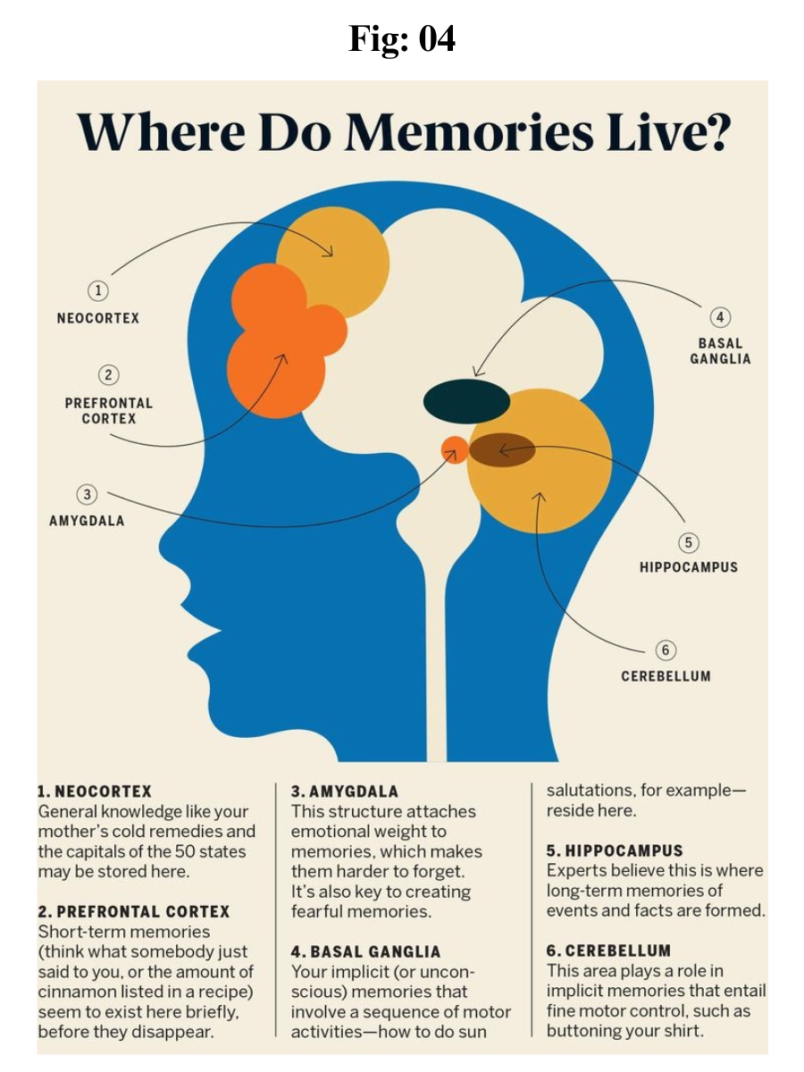
Neurons of the cerebellum and basal ganglia are responsible for motor learning. Declarative memory is located in the medial temporal lobe. Hippocampal lesions lead to anterograde amnesia, a condition in which new information cannot be stored.

Process of Learning in the Brain

The hippocampus plays an important role in learning. The physiologic substrate of learning consists of continuous electrophysiologic, morphologic, and molecular changes in nerve cells. Long-term potentiation is necessary for the long-term availability of information. It facilitates the stimulation of afferent axons over a period of weeks and also leads to greater calcium influx.

Thoughts stored in the long-term memory are available during the entire lifespan of that individual. The process of creating long-term memories is mediated by neurotransmitters such as glutamate (glutamic acid).

Types of Learning

1. Motor Learning: Our day to day activities like walking, running, driving, etc, must be learnt for ensuring a good life. These activities to a great extent involve muscular coordination.

2. Verbal Learning: It is related with the language which we use to communicate and various other forms of verbal communication such as symbols, words, languages, sounds, figures and signs.

3. Concept Learning: This form of learning is associated with higher order cognitive processes like intelligence, thinking, reasoning, etc, which we learn right from our childhood. Concept learning involves the processes of abstraction and generalization, which is very useful for identifying or recognizing things.

4. Discrimination Learning: Learning which distinguishes between various stimuli with its appropriate and different responses is regarded as discrimination stimuli.

5. Learning of Principles: Learning which is based on principles helps in managing the work most effectively. Principles based learning explains the relationship between various concepts.

6. Attitude Learning: Attitude shapes our behavior to a very great extent, as our positive or negative behavior is based on our attitudinal predisposition.


Memory Systems

The striatum is associated with procedural memory and uses the pathway of the neocortex. Associative learning for emotional and motor processes occurs in the amygdala and cerebellum, respectively. Nonassociative learning occurs in the form of habituation and sensitization (both via reflex circuits).

Hebbian theory

The Hebbian theory explains how connections between certain neurons can be strengthened. This theory asserts that “neurons that fire together, wire together,” which means that when activity in a cell repeatedly elicits action potentials in a 2nd cell, the synaptic strength is potentiated.

If an axon of neuron A is located close enough to neuron B such that neuron B can be stimulated by neuron A repeatedly or continuously, the efficiency of neuron A for the stimulation by neuron B is increased by growth processes or changes in metabolism in 1 or both neurons. This suggests that experience-related changes in the nervous systems depend on certain conditions.

Development of Memory and the Papez Circuit

The Papez circuit exists in all mammals and is important for memory development. It is located in the center of the 
limbic system, which is present above the brain stem. The Papez circuit plays a vital role in social behavior, solicitude, love, fear, and learning by imitation.

Papez circuit

The Papez circuit is a chain of neurons named after its discoverer James Papez. Research on the tasks of the Papez circuit for memory performance is ongoing. However, the assumption that the circuit controls anger and rage is already outdated, as it has been discovered that the circuit is even more complex than Papez had thought.

It is currently believed that the Papez circuit plays a role in memory storage by transferring information from the primary memory (short-term memory) to the secondary (long-term memory) or tertiary memory (an independent component of long-term memory).

The Papez circuit proceeds as follows:

hippocampus → fornix → mammillary body in the hypothalamus (corpora mammillaria) → cingulate cortex → hippocampus. 

Different Types of Memory

The specialist term for mind and memory is called the mnestic function. Some things are easier to remember than others. For example, important events are easier to remember than those that hold no meaning, and positive experiences are easier to remember than neutral experiences. Moreover, the process of remembering is easier in a prevailing positive mood, which also means that remembering things is more difficult in a state of 
fatigue or grief.

Process of encoding information

Encoding is the process of transferring sensory information into a construct, which is then stored in our memory system. Working memory stores information for immediate use as part of mental activity (i.e., learning or problem solving).

It is believed to include a phonological loop (i.e., a component of the working memory model that deals with spoken and written material), a visuospatial sketchpad, a central executive, and an episodic buffer.

It allows for the manipulation and organization of information as opposed to short-term memory.

The primary effect is a cognitive bias that results in a subject recalling the 1st items on a list.

Items that have been encoded for the longest duration are transferred to the long-term memory.

The recency effect is a cognitive bias that results in a subject recalling the last items on a list. 

Items in the phonological loop are highly accessible.

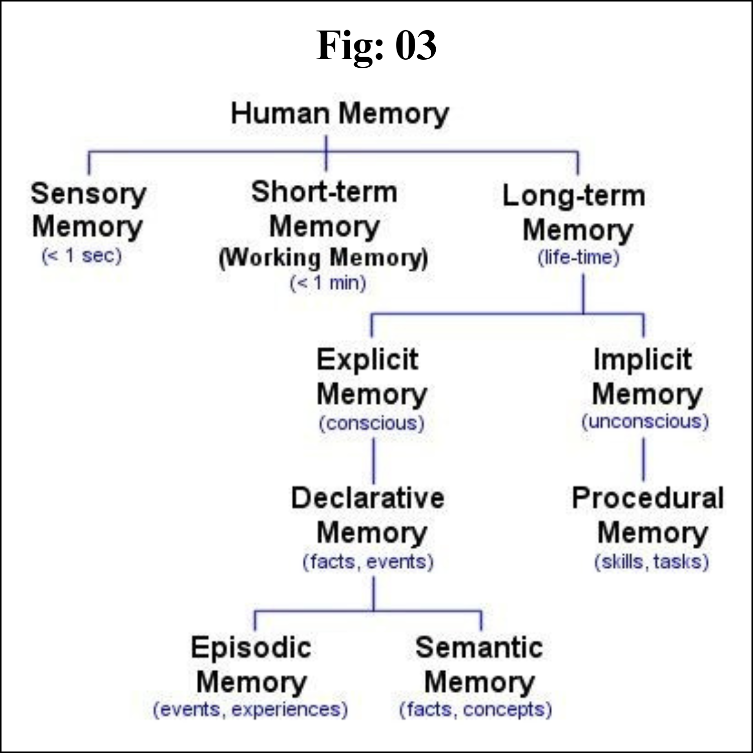
Humans have declarative memory (explicit memory) and 
procedural memory (implicit memory). 

Declarative memory stores information that can be reproduced because humans are conscious of the experience. In contrast, procedural memory stores experiences for which one has no direct memory of the learning process. Nevertheless, this type of memory influences our behavior. A classic example of procedural memory is the learning of a new language.


Sensory (ultra-short-term) memory

The ultra-short-term memory receives stimuli from sensory organs in the form of neuronal excitation. This process has a duration of < 1 second, and perception is via the eyes or ears. Ultra-short-term memory via the eye is referred to as iconic memory, whereas that via the ears is known as echoic memory (it perishes just as fast). Only the stimuli that reach the short-term memory remain, as ultra-short-term memory has no storage capabilities.

Short-term/working memory (primary memory)

Memories in the primary (short-term/working) memory are available for as long as we occupy ourselves with them. If that process is interrupted, the memory is lost too. Memories that begin in the primary memory can be available permanently, but only if they are transferred to the long-term memory. Short-term memory is the usual transitory path for experiences to pass into long-term memory; however, this is occasionally short-circuited so that information can pass directly from the sensory memory to the long-term memory.

The hippocampus, located in the cerebral cortex, is involved in transmitting information from the primary memory to the long-term memory. The hippocampus is thought to be involved in this process because when lesions appear in the hippocampus, only the short-term memory remains intact. Another term for primary memory is labile memory, as it is very unstable. A single distraction is enough to forget the information that has been perceived or heard. Calcium plays a major role in these processes.

Long-term memory

Repetition is particularly important for storing memories in long-term memory. This concept is easily understandable when the high amount of repetition required to learn new movement patterns is considered, e.g., when learning a new sport.

Semantic networks and spreading networks

Information is stored in our long-term memory as an organized network.

Individual ideas or hubs are called nodes (e.g., cities on a map).

Nodes are connected by links or associations (e.g., roads between cities).

The strength of the association depends on how frequently and deeply the connection is made.

Processing material in different ways leads to the establishment of multiple connections.

Nodes are activated only when they reach a response threshold.

The response threshold is reached by the summation of input signals from multiple nodes.

Activation of a node leads to stimulation of neighboring connecting nodes.

Activation of a few nodes can lead to a pattern of activation within the network that spreads inward (spreading activation).

It explains contextual cues, priming, and associations.


Pathology of Memory Performance

The highly complex system of learning and memory is susceptible to malfunctions. If anomalies occur, the differential diagnosis must be made with the greatest care, because even changes in the mineral balance of the body can lead to disorders that give the impression of a disease (e.g., calcium deficiency). Furthermore, cases of 
dementia are increasing, which is partly due to the increase in life expectancy.

Amnesia

Amnesia is a memory disorder in which an individual loses access to stored information. “Amnesia” is derived from the Greek words a (without) and mnémÄ“ (memory). Amnesia is not an independent disease, but the symptom of a disease or the consequence of an influence on the brain. The influence can be internal or external. Individuals with amnesia cannot recall prior experiences or knowledge; this can affect either all or only certain parts and types of information. For example, patients can lose access to memories from certain stages of their lives. In most cases, they can remember events that occurred long ago rather than events that occurred recently. Several forms of amnesia cannot be differentiated from each other; however, the following variants are of importance:

Retrograde amnesia: a type of amnesia in which a person cannot recall memories that were formed before the event that caused the amnesia. Only recently stored memories are affected and not memories from years before. 

Transient global amnesia: a self-limiting clinical syndrome lasting < 24 hours and characterized by the acute onset of anterograde amnesia. Affected individuals are disoriented and ask repetitive questions. There may be an inability to recall general or personal information (retrograde amnesia) while the episode lasts.

Anterograde amnesia: an inability to form new memories/acquire new knowledgePsychogenic amnesia: refers to cases of memory loss presumed to have a psychological rather than a neurological cause Alzheimer disease (AD) 

Alzheimer disease is characterized by variable degrees of cortical atrophy, seen as gyral narrowing and sulcal widening, mostly in the frontal, temporal, and parietal lobes. If there is marked atrophy, there will be compensatory ventricular enlargement (hydrocephalus ex vacuo) secondary to the reduced brain volume. The outcome is the impaired relay of information. Furthermore, AD may make information processing or learning nearly impossible. The basic abnormality in AD is the accumulation of Aβ and tau proteins in specific regions of the brain due to excessive production and defective removal. Amyloid plaques and neurofibrillary tangles are the 2 pathologic hallmarks of AD.

The regions of the brain involved in processing information and memory performance are particularly affected by AD.

Monday, July 24, 2023

Nervous System


Nervous system
 

Introduction 

Although terminology seems to indicate otherwise, there is really only one nervous system in the body. Although each subdivision of the system is also called a "nervous system," all of these smaller systems belong to the single, highly integrated nervous system. Each subdivision has structural and functional characteristics that distinguish it from the others. The nervous system as a whole is divided into two subdivisions: the central nervous system (CNS) and the peripheral nervous system (PNS).

The Central Nervous System (CNS)

The brain and spinal cord are the organs of the central nervous system. Because they are so vitally important, the brain and spinal cord, located in the dorsal body cavity, are encased in bone for protection. The brain is in the cranial vault, and the spinal cord is in the vertebral canal of the vertebral column. Although considered to be two separate organs, the brain and spinal cord are continuous at the foramen magnum.

The Peripheral Nervous System (PNS)

The organs of the peripheral nervous system are the nerves and ganglia. Nerves are bundles of nerve fibers, much like muscles are bundles of muscle fibers. Cranial nerves and spinal nerves extend from the CNS to peripheral organs such as muscles and glands. Ganglia are collections, or small knots, of nerve cell bodies outside the CNS.

The peripheral nervous system is further subdivided into an afferent (sensory) division and an efferent (motor) division. The afferent or sensory division transmits impulses from peripheral organs to the CNS. The efferent or motor division transmits impulses from the CNS out to the peripheral organs to cause an effect or action.

Finally, the efferent or motor division is again subdivided into the somatic nervous system and the autonomic nervous system. The somatic nervous system, also called the somatomotor or somatic efferent nervous system, supplies motor impulses to the skeletal muscles. 

Because these nerves permit conscious control of the skeletal muscles, it is sometimes called the voluntary nervous system. The autonomic nervous system, also called the visceral efferent nervous system, supplies motor impulses to cardiac muscle, to smooth muscle, and to glandular epithelium. 

It is further subdivided into sympathetic and parasympathetic divisions. Because the autonomic nervous system regulates involuntary or automatic functions, it is called the involuntary nervous system.


The Central Nervous System

The CNS consists of the brain and spinal cord, which are located in the dorsal body cavity. The brain is surrounded by the cranium, and the spinal cord is protected by the vertebrae. The brain is continuous with the spinal cord at the foramen magnum. In addition to bone, the CNS is surrounded by connective tissue membranes, called meninges, and by cerebrospinal fluid.

Meninges

There are three layers of meninges around the brain and spinal cord. The outer layer, the dura mater, is tough white fibrous connective tissue. The middle layer of meninges is arachnoid, which resembles a cobweb in appearance, is a thin layer with numerous threadlike strands that attach it to the innermost layer. 

The space under the arachnoid, the subarachnoid space, is filled with cerebrospinal fluid and contains blood vessels. The pia mater is the innermost layer of meninges. This thin, delicate membrane is tightly bound to the surface of the brain and spinal cord and cannot be dissected away without damaging the surface.

Meningiomas are tumors of the nerve tissue covering the brain and spinal cord. Although meningiomas are usually not likely to spread, physicians often treat them as though they were malignant to treat symptoms that may develop when a tumor applies pressure to the brain.


Brain

The brain is divided into the cerebrum, diencephalons, brain stem, and cerebellum.


Cerebrum

The largest and most obvious portion of the brain is the cerebrum, which is divided by a deep longitudinal fissure into two cerebral hemispheres. The two hemispheres are two separate entities but are connected by an arching band of white fibers, called the corpus callosum that provides a communication pathway between the two halves.

Each cerebral hemisphere is divided into five lobes, four of which have the same name as the bone over them: the fontal lobe, the parietal lobe, the occipital lobe, and the temporal lobe. A fifth lobe, the insula or Island of Reil, lies deep within the lateral sulcus.

Diencephalon

The diencephalons is centrally located and is nearly surrounded by the cerebral hemispheres. It includes the thalamus, hypothalamus, and epithalamus. The thalamus, about 80 percent of the diencephalons, consists of two oval masses of gray matter that serve as relay stations for sensory impulses, except for the sense of smell, going to the cerebral cortex. 

The hypothalamus is a small region below the thalamus, which plays a key role in maintaining homeostasis because it regulates many visceral activities. The epithalamus is the most dorsal portion of the diencephalons. This small gland is involved with the onset of puberty and rhythmic cycles in the body. It is like a biological clock.

Brain Stem

The brainstem is the region between the diencephalons and the spinal cord. It consists of three parts: midbrain, pons, and medulla oblongata. The midbrain is the most superior portion of the brain stem. The pons is the bulging middle portion of the brain stem. 

This region primarily consists of nerve fibers that form conduction tracts between the higher brain centers and spinal cord. The medulla oblongata, or simply medulla, extends inferiorly from the pons. It is continuous with the spinal cord at the foramen magnum. 

All the ascending (sensory) and descending (motor) nerve fibers connecting the brain and spinal cord pass through the medulla.

Cerebellum

The cerebellum, the second largest portion of the brain, is located below the occipital lobes of the cerebrum. Three paired bundles of myelinated nerve fibers, called cerebellar peduncles, form communication pathways between the cerebellum and other parts of the central nervous system.

Ventricles and Cerebrospinal Fluid

A series of interconnected, fluid-filled cavities are found within the brain. These cavities are the ventricles of the brain, and the fluid is cerebrospinal fluid (CSF).

Spinal Cord

The spinal cord extends from the foramen magnum at the base of the skull to the level of the first lumbar vertebra. The cord is continuous with the medulla oblongata at the foramen magnum. Like the brain, the spinal cord is surrounded by bone, meninges, and cerebrospinal fluid.

The spinal cord is divided into 31 segments with each segment giving rise to a pair of spinal nerves. At the distal end of the cord, many spinal nerves extend beyond the conus medullaris to form a collection that resembles a horse's tail. This is the cauda equina. In the cross section, the spinal cord appears oval in shape.


The spinal cord has two main functions:

. Serving as a conduction pathway for impulses going to and from the brain. Sensory impulses travel to the brain on ascending tracts in the cord. Motor impulses travel on descending tracts.

. Serving as a reflex center. The reflex arc is the functional unit of the nervous system. Reflexes are responses to stimuli that do not require conscious thought and consequently, they occur more quickly than reactions that require thought processes. For example, with the withdrawal reflex, the reflex action withdraws the affected part before you are aware of the pain. Many reflexes are mediated in the spinal cord without going to the higher brain centers.


The Peripheral Nervous System


The peripheral nervous system consists of the nerves that branch out from the brain and spinal cord. These nerves form the communication network between the CNS and the body parts. The peripheral nervous system is further subdivided into the somatic nervous system and the autonomic nervous system. The somatic nervous system consists of nerves that go to the skin and muscles and is involved in conscious activities. The autonomic nervous system consists of nerves that connect the CNS to the visceral organs such as the heart, stomach, and intestines. It mediates unconscious activities.

Structure of a Nerve

A nerve contains bundles of nerve fibers, either axons or dendrites, surrounded by connective tissue. Sensory nerves contain only afferent fibers, long dendrites of sensory neurons. Motor nerves have only efferent fibers, long axons of motor neurons. Mixed nerves contain both types of fibers.

A connective tissue sheath called the epineurium surrounds each nerve. Each bundle of nerve fibers is called a fasciculus and is surrounded by a layer of connective tissue called the perineurium. Within the fasciculus, each individual nerve fiber, with its myelin and neurilemma, is surrounded by connective tissue called the endoneurium. A nerve may also have blood vessels enclosed in its connective tissue wrappings.

Cranial Nerves

Twelve pairs of cranial nerves emerge from the inferior surface of the brain. All of these nerves, except the vagus nerve, pass through foramina of the skull to innervate structures in the head, neck, and facial region.

The cranial nerves are designated both by name and by Roman numerals, according to the order in which they appear on the inferior surface of the brain. Most of the nerves have both sensory and motor components. Three of the nerves are associated with the special senses of smell, vision, hearing, and equilibrium and have only sensory fibers. Five other nerves are primarily motor in function but do have some sensory fibers for proprioception. The remaining four nerves consist of significant amounts of both sensory and motor fibers.

Acoustic neuromas are benign fibrous growths that arise from the balance nerve, also called the eighth cranial nerve or vestibulocochlear nerve. These tumors are non-malignant, meaning that they do not spread or metastasize to other parts of the body. The location of these tumors is deep inside the skull, adjacent to vital brain centers in the brain stem. As the tumors enlarge, they involve surrounding structures which have to do with vital functions. In the majority of cases, these tumors grow slowly over a period of years. In other cases, the growth rate is more rapid and patients develop symptoms at a faster pace. Usually, the symptoms are mild and many patients are not diagnosed until some time after their tumor has developed. Many patients also exhibit no tumor growth over a number of years when followed by yearly MRI scans.

Spinal Nerves

Thirty-one pairs of spinal nerves emerge laterally from the spinal cord. Each pair of nerves corresponds to a segment of the cord and they are named accordingly. This means there are 8 cervical nerves, 12 thoracic nerves, 5 lumbar nerves, 5 sacral nerves, and 1 coccygeal nerve.

Each spinal nerve is connected to the spinal cord by a dorsal root and a ventral root. The cell bodies of the sensory neurons are in the dorsal root ganglion, but the motor neuron cell bodies are in the gray matter. The two roots join to form the spinal nerve just before the nerve leaves the vertebral column. Because all spinal nerves have both sensory and motor components, they are all mixed nerves.

Autonomic Nervous System

The autonomic nervous system is a visceral efferent system, which means it sends motor impulses to the visceral organs. It functions automatically and continuously, without conscious effort, to innervate smooth muscle, cardiac muscle, and glands. It is concerned with heart rate, breathing rate, blood pressure, body temperature, and other visceral activities that work together to maintain homeostasis.

The autonomic nervous system has two parts, the sympathetic division and the parasympathetic division. Many visceral organs are supplied with fibers from both divisions. In this case, one stimulates and the other inhibits. This antagonistic functional relationship serves as a balance to help maintain homeostasis.

Conclusion

Key notes 

The nervous system is the major controlling, regulatory, and communicating system in the body. It is the center of all mental activity including thought, learning, and memory.

The various activities of the nervous system can be grouped together as three general, overlapping functions: sensory, integrative, and motor.

Neurons are the nerve cells that transmit impulses. Supporting cells are neuroglia.

The three components of a neuron are a cell body or soma, one or more afferent processes called dendrites, and a single efferent process called an axon.

The central nervous system consists of the brain and spinal cord. Cranial nerves, spinal nerves, and ganglia make up the peripheral nervous system.

The afferent division of the peripheral nervous system carries impulses to the CNS; the efferent division carries impulses away from the CNS.

There are three layers of meninges around the brain and spinal cord. The outer layer is dura mater, the middle layer is arachnoid, and the innermost layer is pia mater.

The spinal cord functions as a conduction pathway and as a reflex center. Sensory impulses travel to the brain on ascending tracts in the cord. Motor impulses travel on descending tracts.


# # # # # 

Reverse Transcription

Reverse Transcription Introduction Reverse transcription is the synthesis of DNA from an RNA template. This process is driven by RNA-depende...