Saturday, July 22, 2023

Neurons - An Overview


Neuron - An Overview 

Introduction

Neurons are nerve cells that send messages all over your body to allow you to do everything from breathing to talking, eating, walking, and thinking. Until recently, most neuroscientists (scientists who study the brain) thought we were born with all the neurons we were ever going to have. As children, we might grow some new neurons to help build the pathways—called neural circuits—that act as information highways between different areas of the brain. However, scientists believed that once a neural circuit was in place, adding any new neurons would change the flow of information and break the brain’s communication system

Discovery of Neurogenesis

In 1962, scientist Joseph Altman challenged this belief when he saw evidence of neurogenesis (the birth of neurons) in a region of the adult rat brain called the hippocampus. He later reported that newborn neurons traveled from their birthplace in the hippocampus to other parts of the brain. In 1979, another scientist, Michael Kaplan, confirmed Altman’s findings in the rat brain; and in 1983, he found special kinds of cells—called neural precursor cells—with the ability to become brain cells like neurons, in adult monkeys.

These discoveries about neurogenesis in the adult brain were surprising to other researchers who thought they were not true in humans. Fortunately, in the early 1980s, a scientist trying to understand how birds learn to sing began to see how neurogenesis in the adult brain might make sense. In a series of experiments, Fernando Nottebohm and his research team showed that the numbers of neurons in the forebrains (areas controlling complex behaviors) of male canaries dramatically increased during the mating season, when the birds learn new songs to attract females.

Why did these bird brains add neurons at such an important time in learning? Nottebohm believed it was because newborn neurons helped store new song patterns within the pathways of the forebrain; these new neurons made learning new songs possible! If birds made new neurons to help them remember and learn, Nottebohm thought the brains of mammals—like humans—might too.

Other scientists, like Elizabeth Gould, later found evidence of newborn neurons in a distinct area of the brain in monkeys, and Fred Gage and Peter Eriksson showed that the adult human brain produce new neurons in a similar area.

Neurogenesis in the adult human brain is still tricky for neuroscientists to show, let alone learn about, how it impacts the brain and its functions. Still, scientists are intrigued by current research on neurogenesis and the possible role of new neurons in the adult brain for learning and memory.

Architecture of Neuron 

Neurons, also known as nerve cells, send and receive signals from your brain. While neurons have a lot in common with other types of cells, they’re structurally and functionally unique.

Specialized projections called axons allow neurons to transmit electrical and chemical signals to other cells. Neurons can also receive these signals via rootlike extensions known as dendrites.

A 2009 study estimated that the human brain houses about 86 billion neurons trusted source. The creation of new nerve cells is called neurogenesis. While this process isn’t well understood, we know that it’s much more active when you’re an embryo. However, 2013 evidenceTrusted Source suggests that some neurogenesis occurs in adult brains throughout our lives.

As researchers gain insight into both neurons and neurogenesis, many are also working to uncover links to neurodegenerative diseases such as Alzheimer’s and Parkinson’s.


Parts of a Neuron 

Neurons vary in size, shape, and structure depending on their role and location. However, nearly all neurons have three essential parts: a cell body, an axon, and dendrites.

Cell body

Also known as a soma, the cell body is the core section of the neuron. The cell body contains genetic information, maintains the neuron’s structure, and provides energy to drive activities.

Like other cell bodies, a neuron’s soma contains a nucleus and specialized organelles. It’s enclosed by a membrane that both protects it and allows it to interact with its immediate surroundings.

Axon

An axon is a long, tail-like structure. It joins the cell body at a specialized junction called the axon hillock. Many axons are insulated with a fatty substance called myelin. Myelin helps axons to conduct an electrical signal.

Neurons usually have one main axon.

Dendrites

Dendrites are fibrous roots that branch out from the cell body. Like antennae, dendrites receive and process signals from the axons of other neurons. Neurons can have more than one set of dendrites, known as dendritic trees.

How many they have generally depends on their role. For instance, Purkinje cells are a special type of neuron found in a part of the brain called the cerebellum. These cells have highly developed dendritic trees which allow them to receive thousands of signals.

Types of neurons

Neurons vary in structure, function, and genetic makeup. Given the sheer number of neurons, there are thousands of different types, much like there are thousands of species of living organisms on Earth.

However, there are five major neuron forms. Each combines several elements of the basic neuron shape.

Multipolar neurons. These neurons have a single axon and symmetrical dendrites that extend from it. This is the most common form of neuron in the central nervous system.

Unipolar neurons. Usually only found in invertebrate species, these neurons have a single axon.

Bipolar neurons. Bipolar neurons have two extensions extending from the cell body. At the end of one side is the axon, and the dendrites are on the other side. These types of neurons are mostly found in the retina of the eye. But they can also be found in parts of the nervous system that help the nose and ear function.

Pyramidal neurons. These neurons have one axon but several dendrites to form a pyramid type shape. These are the largest neuron cells and are mostly found in the cortex. The cortex is the part of the brain responsible for conscious thoughts.

Purkinje neurons. Purkinje neurons have multiple dendrites that fan out from the cell body. These neurons are inhibitory neurons, meaning they release neurotransmitters that keep other neurons from firing.

In terms of function, scientists classify neurons into three broad types: sensory, motor, and interneurons.

Sensory neurons

Sensory neurons help you:

. taste
. smell
. hear
. see
. feel things around you

Sensory neurons are triggered by physical and chemical inputs from your environment. Sound, touch, heat, and light are physical inputs. Smell and taste are chemical inputs.

For example, stepping on hot sand activates sensory neurons in the soles of your feet. Those neurons send a message to your brain, which makes you aware of the heat.

Motor neurons

Motor neurons play a role in movement, including voluntary and involuntary movements. These neurons allow the brain and spinal cord to communicate with muscles, organs, and glands all over the body.

There are two types of motor neurons: lower and upper. Lower motor neurons carry signals from the spinal cord to the smooth muscles and skeletal muscles. Upper motor neurons carry signals between your brain and spinal cord.

When you eat, for instance, lower motor neurons in your spinal cord send signals to the smooth muscles in your esophagus, stomach, and intestines. These muscles contract, which allows food to move through your digestive tract.

Interneurons

Interneurons are neural intermediaries found in your brain and spinal cord. They’re the most common type of neuron. They pass signals from sensory neurons and other interneurons to motor neurons and other interneurons. Often, they form complex circuits that help you to react to external stimuli.

For instance, when you touch something sharp like a cactus, sensory neurons in your fingertips send a signal to interneurons in your spinal cord. Some interneurons pass the signal on to motor neurons in your hand, which allows you to move your hand away. Other interneurons send a signal to the pain center in your brain, and you experience pain.


Neuron Birth 

Many neuroscientists disagree about how many and how often new neurons are created in the brain. Most of the brain’s neurons are already created by the time we’re born, but there is evidence to support the theory that neurogenesis is a lifelong process.

Neurons are born in areas of the brain that are full of neural stem cells, or precursor cells. Stem cells have the potential to make most, if not all, of the different types of neurons and glia found in the brain.

Neuroscientists have observed how neural stem cells behave during experiments in the laboratory. Although this may not be exactly how these cells act when they’re in the brain, it gives us information about how they might function when they’re in the brains of humans or other animals.

The science of stem cells is still very new and could change with additional discoveries, but researchers have learned enough to be able to describe how neural stem cells create the other cells of the brain. The way that stem cells can become other types of brain cells is similar to the idea of a family tree.

Neural stem cells increase by dividing in two. Then they can become either two new stem cells, or two early progenitor cells (parent cells to new neurons or glia), or one of each.

When a stem cell divides to produce another stem cell, it is said to self-renew. This new cell has the potential to make more stem cells.

When a stem cell divides to produce an early progenitor cell, it is said to differentiate. Differentiation means that the new cell is more specialized in how it’s formed and what it can do.

Early progenitor cells can make other progenitor cells, self-renew like stem cells, or can change in either of two ways. One way will make new astrocytes. The other way will make neurons or oligodendrocytes.


Neuron - A Road trip 

Once a neuron is born, it must travel to the place in the brain where it will do its work.

How does a neuron know where to go? What helps it get there?

Scientists have seen that neurons use at least two different methods to travel:

Some neurons travel, or migrate, by following the long fibers of cells called radial glia. These fibers stretch from the inner layers to the outer layers of the brain. Neurons glide along the fibers until they reach their destination.

Neurons also travel by using chemical signals. Scientists have found special molecules on the surface of neurons—adhesion molecules—that attach to similar molecules on nearby glial cells or nerve axons. These chemical signals guide the neurons to their final location.

Not all neurons are successful in their journey. Scientists think that only a third reach their destination. Some cells die in development or while traveling.

Other neurons survive the trip but end up where they should not be. Accidental changes (called mutations) in the genes that control migration create brain areas of misplaced or oddly formed neurons that can cause disorders like childhood epilepsy. Some researchers think that certain disorders, such as schizophrenia and dyslexia, are partly the result of misguided neurons. 


Differentiation - Neuron gain its Structure 

Once a neuron reaches its destination, it must settle into work. There is still a lot that scientists don’t understand about the part of neurogenesis called differentiation.

Neurons are responsible for sending and receiving neurotransmitters—chemicals that carry information between brain cells.

Depending on its location, a neuron can perform the job of a sensory neuron, a motor neuron, or an interneuron, sending and receiving specific neurotransmitters.

In the developing brain, a neuron depends on molecular signals from other cells, such as astrocytes, to determine its shape and location, the kind of transmitter it produces, and the other neurons it will connect to. These freshly born cells create neural circuits—or information pathways connecting neurons to neurons—that will be in place throughout adulthood.

But in the adult brain, neural circuits are already developed, and neurons must find a way to fit in. As new neurons settle in, they start to look like the surrounding cells. They develop axons and dendrites and begin to communicate with their neighbors through synapses.

Synapse 

Synapse, also called Neuronal Junction, the site of transmission of electric nerve impulses between two nerve cells (neurons) or between a neuron and a gland or muscle cell (effector). A synaptic connection between a neuron and a muscle cell is called a neuromuscular junction.

At a chemical synapse each ending, or terminal, of a nerve fiber (presynaptic fiber) swells to form a knoblike structure that is separated from the fiber of an adjacent neuron, called a postsynaptic fiber, by a microscopic space called the synaptic cleft. The typical synaptic cleft is about 0.02 micron wide. 

The arrival of a nerve impulse at the presynaptic terminals causes the movement toward the presynaptic membrane of membrane-bound sacs, or synaptic vesicles, which fuse with the membrane and release a chemical substance called a neurotransmitter. 

This substance transmits the nerve impulse to the postsynaptic fiber by diffusing across the synaptic cleft and binding to receptor molecules on the postsynaptic membrane. The chemical binding action alters the shape of the receptors, initiating a series of reactions that open channel-shaped protein molecules. 

Electrically charged ions then flow through the channels into or out of the neuron. This sudden shift of electric charge across the postsynaptic membrane changes the electric polarization of the membrane, producing the postsynaptic potential, or PSP. 

If the net flow of positively charged ions into the cell is large enough, then the PSP is excitatory; that is, it can lead to the generation of a new nerve impulse, called an action potential.

Once they have been released and have bound to postsynaptic receptors, neurotransmitter molecules are immediately deactivated by enzymes in the synaptic cleft; they are also taken up by receptors in the presynaptic membrane and recycled. This process causes a series of brief transmission events, each one taking place in only 0.5 to 4.0 milliseconds.

A single neurotransmitter may elicit different responses from different receptors. For example, norepinephrine, a common neurotransmitter in the autonomic nervous system, binds to some receptors that excite nervous transmission and to others that inhibit it. 

The membrane of a postsynaptic fiber has many different kinds of receptors, and some presynaptic terminals release more than one type of neurotransmitter. Also, each postsynaptic fiber may form hundreds of competing synapses with many neurons. 

These variables account for the complex responses of the nervous system to any given stimulus. The synapse, with its neurotransmitter, acts as a physiological valve, directing the conduction of nerve impulses in regular circuits and preventing random or chaotic stimulation of nerves.

Electric synapses allow direct communications between neurons whose membranes are fused by permitting ions to flow between the cells through channels called gap junctions. Found in invertebrates and lower vertebrates, gap junctions allow faster synaptic transmission as well as the synchronization of entire groups of neurons. 

Gap junctions are also found in the human body, most often between cells in most organs and between glial cells of the nervous system. Chemical transmission seems to have evolved in large and complex vertebrate nervous systems, where transmission of multiple messages over longer distances is required.


Neurotransmitters

Neurotransmitters are chemical messengers that your body can’t function without. Their job is to carry chemical signals (“messages”) from one neuron (nerve cell) to the next target cell. The next target cell can be another nerve cell, a muscle cell or a gland.

Your body has a vast network of nerves (your nervous system) that send and receive electrical signals from nerve cells and their target cells all over your body. Your nervous system controls everything from your mind to your muscles, as well as organ functions. 

In other words, nerves are involved in everything you do, think and feel. Your nerve cells send and receive information from all body sources. This constant feedback is essential to your body’s optimal function.

Neurotransmitters are located in a part of the neuron called the axon terminal. They’re stored within thin-walled sacs called synaptic vesicles. Each vesicle can contain thousands of neurotransmitter molecules.

As a message or signal travels along a nerve cell, the electrical charge of the signal causes the vesicles of neurotransmitters to fuse with the nerve cell membrane at the very edge of the cell. 

The neurotransmitters, which now carry the message, are then released from the axon terminal into a fluid-filled space that’s between one nerve cell and the next target cell (another nerve cell, muscle cell or gland).

In this space, called the synaptic junction, the neurotransmitters carry the message across less than 40 nanometers (nm) wide (by comparison, the width of a human hair is about 75,000 nm). 

Each type of neurotransmitter lands on and binds to a specific receptor on the target cell (like a key that can only fit and work in its partner lock). 

After binding, the neurotransmitter that triggers a change or action in the target cell, like an electrical signal in another nerve cell, a muscle contraction or the release of hormones from a cell in a gland.

Types of Neurotransmitters 

Scientists know of at least 100 neurotransmitters and suspect there are many others that have yet to be discovered. They can be grouped into types based on their chemical nature. Some of the better-known categories and neurotransmitter examples and their functions include the following:

Amino acids neurotransmitters

These neurotransmitters are involved in most functions of your nervous system.

1. Glutamate. This is the most common excitatory neurotransmitter of your nervous system. It’s the most abundant neurotransmitter in your brain. It plays a key role in cognitive functions like thinking, learning and memory. Imbalances in glutamate levels are associated with Alzheimer’s disease, dementia, Parkinson’s disease and seizures.

2. Gamma-aminobutryic acid (GABA). GABA is the most common inhibitory neurotransmitter of your nervous system, particularly in your brain. It regulates brain activity to prevent problems in the areas of anxiety, irritability, concentration, sleep, seizures and depression.

3. Glycine. Glycine is the most common inhibitory neurotransmitter in your spinal cord. Glycine is involved in controlling hearing processing, pain transmission and metabolism.

Monoamines neurotransmitters

These neurotransmitters play a lot of different roles in your nervous system and especially in your brain. Monoamines neurotransmitters regulate consciousness, cognition, attention and emotion. Many disorders of your nervous system involve abnormalities of monoamine neurotransmitters, and many drugs that people commonly take affect these neurotransmitters.

1. Serotonin. Serotonin is an inhibitory neurotransmitter. Serotonin helps regulate mood, sleep patterns, sexuality, anxiety, appetite and pain. Diseases associated with serotonin imbalance include seasonal affective disorder, anxiety, depression, fibromyalgia and chronic pain. Medications that regulate serotonin and treat these disorders include selective serotonin reuptake inhibitors (SSRIs) and serotonin-norepinephrine reuptake inhibitors (SNRIs).

2. Histamine. Histamine regulates body functions including wakefulness, feeding behavior and motivation. Histamine plays a role in asthma, bronchospasm, mucosal edema and multiple sclerosis.

3. Dopamine. Dopamine plays a role in your body’s reward system, which includes feeling pleasure, achieving heightened arousal and learning. Dopamine also helps with focus, concentration, memory, sleep, mood and motivation. Diseases associated with dysfunctions of the dopamine system include Parkinson’s disease, schizophrenia, bipolar disease, restless legs syndrome and attention deficit hyperactivity disorder (ADHD). Many highly addictive drugs (cocaine, methamphetamines, amphetamines) act directly on the dopamine system.

4. Epinephrine. Epinephrine (also called adrenaline) and norepinephrine (see below) are responsible for your body’s so-called “fight-or-flight response” to fear and stress. These neurotransmitters stimulate your body’s response by increasing your heart rate, breathing, blood pressure, blood sugar and blood flow to your muscles, as well as heighten attention and focus to allow you to act or react to different stressors. Too much epinephrine can lead to high blood pressure, diabetes, heart disease and other health problems. As a drug, epinephrine is used to treat anaphylaxis, asthma attacks, cardiac arrest and severe infections.

5. Norepinephrine. Norepinephrine (also called noradrenaline) increases blood pressure and heart rate. It’s most widely known for its effects on alertness, arousal, decision-making, attention and focus. Many medications (stimulants and depression medications) aim to increase norepinephrine levels to improve focus or concentration to treat ADHD or to modulate norepinephrine to improve depression symptoms.

Peptide neurotransmitters

Peptides are polymers or chains of amino acids.

Endorphins. Endorphins are your body’s natural pain reliever. They play a role in our perception of pain. Release of endorphins reduces pain, as well as causes “feel good” feelings. Low levels of endorphins may play a role in fibromyalgia and some types of headaches.

Acetylcholine

This excitatory neurotransmitter does a number of functions in your central nervous system (CNS [brain and spinal cord]) and in your peripheral nervous system (nerves that branch from the CNS). Acetylcholine is released by most neurons in your autonomic nervous system regulating heart rate, blood pressure and gut motility. Acetylcholine plays a role in muscle contractions, memory, motivation, sexual desire, sleep and learning. Imbalances in acetylcholine levels are linked with health issues, including Alzheimer’s disease, seizures and muscle spasms.

Table


Change do neurotransmitters transmit

Neurotransmitters transmit one of three possible actions in their messages, depending on the specific neurotransmitter.

1. Excitatory. Excitatory neurotransmitters “excite” the neuron and cause it to “fire off the message,” meaning, the message continues to be passed along to the next cell. Examples of excitatory neurotransmitters include glutamate, epinephrine and norepinephrine.

2. Inhibitory. Inhibitory neurotransmitters block or prevent the chemical message from being passed along any farther. Gamma-aminobutyric acid (GABA), glycine and serotonin are examples of inhibitory neurotransmitters.

3. Modulatory. Modulatory neurotransmitters influence the effects of other chemical messengers. They “tweak” or adjust how cells communicate at the synapse. They also affect a larger number of neurons at the same time.

The Sodium-Potassium Pump

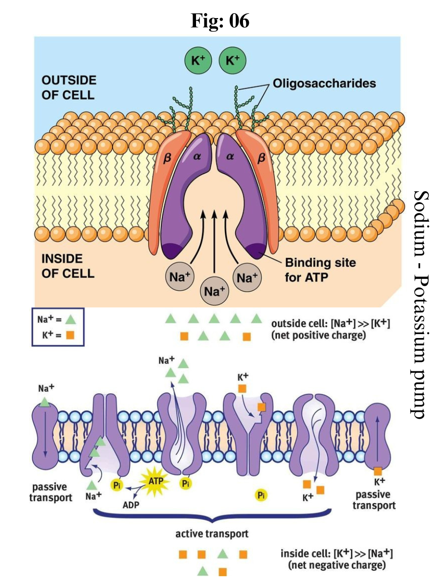
Active transport is the energy-requiring process of pumping molecules and ions across membranes "uphill" - against a concentration gradient. To move these molecules against their concentration gradient, a carrier protein is needed. Carrier proteins can work with a concentration gradient (during passive transport), but some carrier proteins can move solutes against the concentration gradient (from low concentration to high concentration), with an input of energy. 

In active transport, as carrier proteins are used to move materials against their concentration gradient, these proteins are known as pumps. As in other types of cellular activities, ATP supplies the energy for most active transport. One way ATP powers active transport is by transferring a phosphate group directly to a carrier protein. 

This may cause the carrier protein to change its shape, which moves the molecule or ion to the other side of the membrane. An example of this type of active transport system, as shown in Figure below, is the sodium-potassium pump, which exchanges sodium ions for potassium ions across the plasma membrane of animal cells.

The sodium-potassium pump system moves sodium and potassium ions against large concentration gradients. It moves two potassium ions into the cell where potassium levels are high, and pumps three sodium ions out of the cell and into the extracellular fluid.

The three sodium ions bind with the protein pump inside the cell. The carrier protein then gets energy from ATP and changes shape. In doing so, it pumps the three sodium ions out of the cell. 

At that point, two potassium ions from outside the cell bind to the protein pump. The potassium ions are then transported into the cell, and the process repeats. 

The sodium-potassium pump is found in the plasma membrane of almost every human cell and is common to all cellular life. It helps maintain cell potential and regulates cellular volume.

Death of Neuron

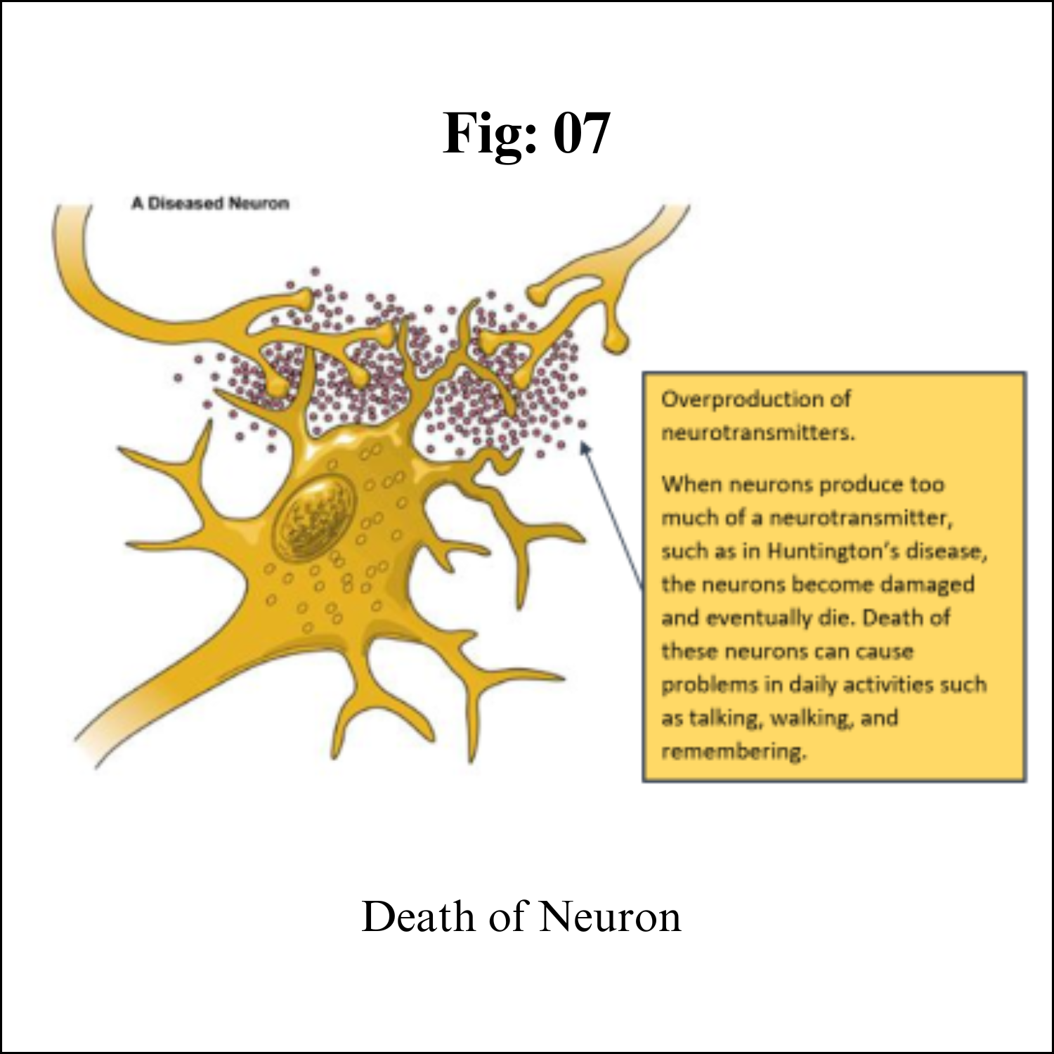
Although neurons are the longest living cells in the body, large numbers of them die during migration and differentiation. The lives of some neurons can take strange turns. Some diseases of the brain are the result of the unnatural deaths of neurons.

In Parkinson’s disease, neurons that produce the neurotransmitter dopamine die off in the basal ganglia, an area of the brain that controls body movements. This causes people with this disease to experience shaking, to move more slowly, and to have problems with balance.

In Huntington’s disease, a genetic mutation causes neurons to create too much of a neurotransmitter called glutamate, which kills neurons in the basal ganglia. As a result, people twist and move uncontrollably, and over time, they lose the ability to do many everyday tasks like walking and eating. People with this disease typically have shorter lives than those without this disease.

In Alzheimer’s disease, unusual proteins build up in and around neurons in the neocortex and hippocampus, the parts of the brain that control memory. When these neurons die, people lose their abilities to remember and do everyday tasks.

Physical damage to the brain and the spinal cord can also kill or disable neurons. Damage to the brain caused by shaking or hitting the head, or because of a stroke, can kill neurons immediately or slowly, starving them of the oxygen and nutrients they need to survive.

A spinal cord injury can cut off communication between the brain and the muscles. When neurons lose their connection to the axons (the parts of neurons that send messages to other neurons) located below the site of injury, the neurons may still live, but they lose their ability to communicate.

Functions 

Your nervous system controls such functions as your:

Heartbeat and blood pressure.
Breathing.
Muscle movements.
Thoughts, memory, learning and feelings.
Sleep, healing and aging.
Stress response.
Hormone regulation.
Digestion, sense of hunger and thirst.
Senses (response to what you see, hear, feel, touch and taste).

###

Friday, July 21, 2023

Dental Formula


Dental formula

Introduction 

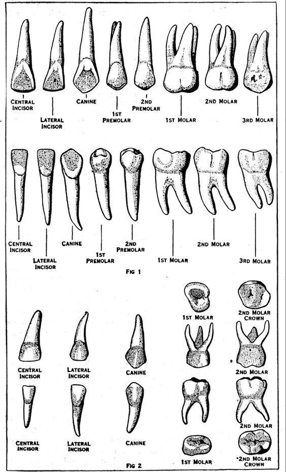
Dental formula is used to understand the number and arrangements of teeth in humans and animals. Dental formula describes the relationship of teeth within the jaws of animal teeth and human teeth. It provides information about the number and size of teeth as well as the type of teeth. Teeth are the most important element in our mouths. They help us to eat, churn and chew the food. There are two sets of teeth in mammals – deciduous and permanent. Deciduous teeth are also known as milk teeth or baby teeth. Incisors, Canines , Premolars and Molar are four types of teeth present in our mouth. 

Dentition

Dentition is used to describe the development of teeth and their arrangement in the mouth. The development of teeth depends on the characteristics of teeth as their kind, arrangement, and number of teeth in a species. The dentition of animals whose teeth succession is known as diphyodont, whereas in animals with only one set of teeth throughout life is known as monophyodont.

Each tooth in our mouth consists of a crown, a root and a cervical part. Enamel covers the crown. Apical foramen and the root canal help the root of the tooth, blood vessels and nerves to access the pulp cavity. Parodontium refers to the collection of cementum , desmodium, alveolar bone and gingival together.

Different Types of Teeth

There are four types of teeth namely Incisors, Canines, Premolars and Molars. These four types of teeth are present on the jawbones in the mouth, extending into the buccal cavity. The four types of teeth have different functions like cutting, crushing, shredding and tearing the food. 

Incisors (I) 

These are in the front row of the mouth. They help in cutting and slicing the food into the pieces to fit in the mouth. They are present in both of the jaws in mammals. 

Canines (C) 

These are most commonly known as dog teeth. In the case of Carnivores, these teeth help in holding and tearing apart food as they have pointed structures and in the case of herbivores, these teeth help in the breakdown and split hard surfaces of certain fruits such as nuts. In human beings, these provide a benefit of aiding in articulation.

Premolars (P) 

These are the transition teeth between canines and molars. They provide smooth surfaces that help in occlusion (the process of cutting, crushing, grinding the food into the rapid digestible form).

Molars (M)

These are large teeth present in mammals. They help in grinding the food. In mammals, there are 12 molars present in a set of three in both the lower and upper jaw.


Dental Formula

The dental formula describes and expresses the total number of teeth in man and animal as per the arrangements. The dental formula is expressed with letters and figures. The letters used in this formula are on the basis of the types of teeth Incisors, Canines, Premolars and Molars.

Dental Formula

(number of each type of teeth upper jaw ) / (number of teeth on one side of the lower jaw)

Dental formula = (2 Incisors 1 Canines 2 Premolars 3 Molars) /(2 Incisors 1 Canines 2 Premolars 3 Molars)

Human teeth have two dental formulas: 

The dental formula represents the arrangement of teeth in each half of the upper and the lower jaw. The entire formula is multiplied by two to represent the total number of teeth.

The dental formula for milk teeth in humans is:

The primary dentition (20 teeth)

I2/2 C1/1 M2/2 = 10 × 2 = 20 teeth

Each half of the upper jaw and the lower jaw has 2 incisors, 1 canine, and 2 molars. Premolars are absent in milk teeth hence the zero.

The permanent dentition (32 teeth):

I2/2C1/1 P2/2 M 3/3 = 16 × 2 = 32 teeth 

Each half of the upper jaw and the lower jaw has 2 incisors, 1 canine, 2 premolars, and 3 molars. An adult human has 32 permanent teeth.

Here, I- Incisors , C- Canines , P- Premolars , M- Molar.


Key Difference – Herbivores vs Carnivores Teeth
 
The key difference between herbivores and carnivores teeth is that herbivores teeth are used for cutting, gnawing, and biting while carnivores teeth are sharper and more suited to catching, killing, and tearing the prey. Based on the food habits there are three types of animals; carnivores, herbivores and omnivores. Animals that rely entirely on the flesh of other animals are called carnivores and animals that feed entirely on vegetation/ plant matters are called herbivores. Omnivores are the animals that feed on both meat and vegetation. Because of the various dietary patterns and the nutrient amount in food, the structure, the number and the location of teeth among these three groups vary widely. In this article, the difference between herbivores and carnivores teeth will be highlighted.

Herbivores Teeth

The incisors of herbivores are sharp and are used mainly to cut, gnaw and bite. Gnawing herbivores have long chisel-like incisors located in front of the skull and used for gnawing and scraping. They do not have canines. A horny pad in the upper jaw completely replaces the canines and incisors in ruminants. Moreover, their incisors and canines are similar and act as blades to cut and gather grass. Molars and premolars of herbivores have flat grinding surfaces, and they grow continuously throughout their lifetime.

Carnivores Teeth

Carnivore teeth are extremely adapted to the dietary habits of carnivores. Their upper premolar 4 and lower molar 1 are carnassial teeth and used to cut the meat away from the bone. The long, pointed canines are used to catch, kill their prey, and tear the flesh of prey. Their premolars and molars are flattened with uneven edges and are used to shear the flesh of prey into smaller pieces. Their incisors are pointed teeth and are used to catch prey.


Thursday, July 20, 2023

Human Digestive System

Human Digestive system 

Introduction:

The digestive system is made up of the gastrointestinal tract—also called the GI tract or digestive tract—and the liver, pancreas, and gallbladder. The GI tract is a series of hollow organs joined in a long, twisting tube from the mouth to the anus. The hollow organs that make up the GI tract are the mouth, esophagus, stomach, small intestine, large intestine, and anus. The liver, pancreas, and gallbladder are the solid organs of the digestive system.

The small intestine has three parts. The first part is called the duodenum. The jejunum is in the middle and the ileum is at the end. The large intestine includes the appendix, cecum, colon, and rectum. The appendix is a finger-shaped pouch attached to the cecum. The cecum is the first part of the large intestine. The colon is next. The rectum is the end of the large intestine.

Bacteria in your GI tract, also called gut flora or microbiome, help with digestion. Parts of your nervous and circulatory systems also help. Working together, nerves, hormones, bacteria, blood, and the organs of your digestive system digest the foods and liquids you eat or drink each day.

Importance of Digestive system:

Digestion is important because your body needs nutrients from food and drink to work properly and stay healthy. Proteins, fats, carbohydrates, vitamins, minerals, and water are nutrients. Your digestive system breaks nutrients into parts small enough for your body to absorb and use for energy, growth, and cell repair.

Proteins break into amino acids
Fats break into fatty acids and glycerol
Carbohydrates break into simple sugars

Digestive system work:

Each part of your digestive system helps to move food and liquid through your GI tract, break food and liquid into smaller parts, or both. Once foods are broken into small enough parts, your body can absorb and move the nutrients to where they are needed. Your large intestine absorbs water, and the waste products of digestion become stool. Nerves and hormones help control the digestive process.

Food pathway in GI tract:

Food moves through your GI tract by a process called peristalsis. The large, hollow organs of your GI tract contain a layer of muscle that enables their walls to move. The movement pushes food and liquid through your GI tract and mixes the contents within each organ. The muscle behind the food contracts and squeezes the food forward, while the muscle in front of the food relaxes to allow the food to move.


Mouth. Food starts to move through your GI tract when you eat. When you swallow, your tongue pushes the food into your throat. A small flap of tissue, called the epiglottis, folds over your windpipe to prevent choking and the food passes into your esophagus.

Esophagus. Once you begin swallowing, the process becomes automatic. Your brain signals the muscles of the esophagus and peristalsis begins.

Lower esophageal sphincter. When food reaches the end of your esophagus, a ringlike muscle—called the lower esophageal sphincter —relaxes and lets food pass into your stomach. This sphincter usually stays closed to keep what’s in your stomach from flowing back into your esophagus.

Stomach. After food enters your stomach, the stomach muscles mix the food and liquid with digestive juices. The stomach slowly empties its contents, called chyme, into your small intestine.

Small intestine. The muscles of the small intestine mix food with digestive juices from the pancreas, liver, and intestine, and push the mixture forward for further digestion. The walls of the small intestine absorb water and the digested nutrients into your bloodstream. As peristalsis continues, the waste products of the digestive process move into the large intestine.

Large intestine. Waste products from the digestive process include undigested parts of food, fluid, and older cells from the lining of your GI tract. The large intestine absorbs water and changes the waste from liquid into stool. Peristalsis helps move the stool into your rectum.

Rectum. The lower end of your large intestine, the rectum, stores stool until it pushes stool out of your anus during a bowel movement. 

How Digestive system Break food: 

As food moves through your GI tract, your digestive organs break the food into smaller parts using:

motion, such as chewing, squeezing, and mixing
digestive juices, such as stomach acid, bile, and enzymes

Mouth. The digestive process starts in your mouth when you chew. Your salivary glands make saliva, a digestive juice, which moistens food so it moves more easily through your esophagus into your stomach. Saliva also has an enzyme that begins to break down starches in your food.

Esophagus. After you swallow, peristalsis pushes the food down your esophagus into your stomach.

Stomach. Glands in your stomach lining make stomach acid and enzymes that break down food. Muscles of your stomach mix the food with these digestive juices.

Pancreas. Your pancreas makes a digestive juice that has enzymes that break down carbohydrates, fats, and proteins. The pancreas delivers the digestive juice to the small intestine through small tubes called ducts.

Liver. Your liver makes a digestive juice called bile that helps digest fats and some vitamins. Bile ducts carry bile from your liver to your gallbladder for storage, or to the small intestine for use.

Gallbladder. Your gallbladder stores bile between meals. When you eat, your gallbladder squeezes bile through the bile ducts into your small intestine.

Small intestine. Your small intestine makes digestive juice, which mixes with bile and pancreatic juice to complete the breakdown of proteins, carbohydrates, and fats. Bacteria in your small intestine make some of the enzymes you need to digest carbohydrates. Your small intestine moves water from your bloodstream into your GI tract to help break down food. Your small intestine also absorbs water with other nutrients.

Large intestine. In your large intestine, more water moves from your GI tract into your bloodstream. Bacteria in your large intestine help break down remaining nutrients and make vitamin K. Waste products of digestion, including parts of food that are still too large, become stool.

Product of Digested food: 

The small intestine absorbs most of the nutrients in your food, and your circulatory system passes them on to other parts of your body to store or use. Special cells help absorbed nutrients cross the intestinal lining into your bloodstream. Your blood carries simple sugars, amino acids, glycerol, and some vitamins and salts to the liver. Your liver stores, processes, and delivers nutrients to the rest of your body when needed.

The lymph system, a network of vessels that carry white blood cells and a fluid called lymph throughout your body to fight infection, absorbs fatty acids and vitamins.

Your body uses sugars, amino acids, fatty acids, and glycerol to build substances you need for energy, growth, and cell repair.

Digestion control: 

Your hormones and nerves work together to help control the digestive process. Signals flow within your GI tract and back and forth from your GI tract to your brain.

Hormones

Cells lining your stomach and small intestine make and release hormones that control how your digestive system works. These hormones tell your body when to make digestive juices and send signals to your brain that you are hungry or full. Your pancreas also makes hormones that are important to digestion.

Nerves

You have nerves that connect your central nervous system—your brain and spinal cord—to your digestive system and control some digestive functions. For example, when you see or smell food, your brain sends a signal that causes your salivary glands to "make your mouth water" to prepare you to eat.

You also have an enteric nervous system (ENS)—nerves within the walls of your GI tract. When food stretches the walls of your GI tract, the nerves of your ENS release many different substances that speed up or delay the movement of food and the production of digestive juices. The nerves send signals to control the actions of your gut muscles to contract and relax to push food through your intestines.

#####

Wednesday, July 19, 2023

B Cell Receptor (Immunoglobulin) Class Switching


B Cell Receptor (Immunoglobulin) Class Switching 

Immunoglobulin

Immunoglobulins, also known as antibodies, are glycoprotein molecules produced by plasma cells or white blood cells. They specifically recognize and bind to particular antigens.

Structure of immunoglobulins


Antibody (or immunoglobulin) molecules are glycoproteins composed of one or more units, each containing four polypeptide chains: two identical heavy chains (H) and two identical light chains (L). 

The amino terminal ends of the polypeptide chains show considerable variation in amino acid composition and are referred to as the variable (V) regions to distinguish them from the relatively constant (C) regions. 

Each L chain consists of one variable domain, VL, and one constant domain, CL. The H chains consist of a variable domain, VH, and three constant domains CH1, CH2 and CH3. 

Each heavy chain has about twice the number of amino acids and molecular weight (~50,000) as each light chain (~25,000), resulting in a total immunoglobulin monomer molecular weight of approximately 150,000.

Heavy and light chains are held together by a combination of non-covalent interactions and covalent interchain disulfide bonds, forming a bilaterally symmetric structure. The V regions of H and L chains comprise the antigen-binding sites of the immunoglobulin (Ig) molecules. Each Ig monomer contains two antigen-binding sites and is said to be bivalent.

The hinge region is the area of the H chains between the first and second C region domains and is held together by disulfide bonds. This flexible hinge (found in IgG, IgA, and IgD, but not IgM or IgE) region allows the distance between the two antigen-binding sites to vary.


Classes of immunoglobulins

The five primary classes of immunoglobulins are IgG, IgM, IgA, IgD, and IgE. These are distinguished by the type of heavy chain found in the molecule. IgG molecules have heavy chains known as gamma-chains; IgMs have mu-chains; IgAs have alpha-chains; IgEs have epsilon-chains; and IgDs have delta-chains.

Differences in heavy chain polypeptides allow these immunoglobulins to function in different types of immune responses and at particular stages of the immune response. 

The polypeptide protein sequences responsible for these differences are found primarily in the Fc fragment. While there are five different types of heavy chains, there are only two main types of light chains: kappa (κ) and lambda (λ).

Antibody classes differ in valency as a result of different numbers of Y-like units (monomers) that join to form the complete protein. 

For example, in humans, functioning IgM antibodies have five Y-shaped units (pentamer) containing a total of ten light chains, ten heavy chains, and ten antigen-binding.


IgG

Properties of IgG:

1. Molecular weight: 150,000 Da
2. H-chain type (MW): gamma (53,000 Da)
3. Serum concentration: 10 to 16 mg/mL
4. Percent of total immunoglobulin: 75%
5. Glycosylation (by weight): 3%
6. Distribution: intra- and extravascular
7. Function: secondary response

IgM

Properties of IgM:

1. Molecular weight: 900,000 Da
2. H-chain type (MW): mu (65,000 Da)
3. Serum concentration: 0.5 to 2 mg/mL
4. Percent of total immunoglobulin: 10%
5. Glycosylation (by weight): 12%
6. Distribution: mostly intravascular
7. Function: primary response

IgA

Properties of IgA:

1. Molecular weight: 320,000 Da (secretory)
2. H-chain type (MW): alpha (55,000 Da)
3. Serum concentration: 1 to 4 mg/mL
4. Percent of total immunoglobulin: 15%
5. Glycosylation (by weight): 10%
6. Distribution: intravascular and secretions
7. Function: protect mucus membranes

IgD

Properties of IgD:

1. Molecular weight: 180,000 Da
2. H-chain type (MW): delta (70,000 Da)
3. Serum concentration: 0 to 0.4 mg/mL
4. Percent of total immunoglobulin: 0.2%
5. Glycosylation (by weight): 13%
6. Distribution: lymphocyte surface
7. Function: unknown

IgE

Properties of IgE:

1. Molecular weight: 200,000 Da
2. H-chain type (MW): epsilon (73,000 Da)
3. Serum concentration: 10 to 400 ng/mL
4. Percent of total immunoglobulin: 0.002%
5. Glycosylation (by weight): 12%
6. Distribution: basophils and mast cells in saliva and nasal secretions
7. Function: protect against parasites

Subclasses of immunoglobulins

In addition to the major immunoglobulin classes, several Ig subclasses exist in all members of a particular animal species. 

Antibodies are classified into subclasses based on minor differences in the heavy chain type of each Ig class. In humans there are four subclasses of IgG: IgG1, IgG2, IgG3, and IgG4 (numbered in order of decreasing concentration in serum).

Variance among different subclasses is less than the variance among different classes. For example, IgG1 is more closely related to IgG2, IgG3 and IgG4 than to IgA, IgM, IgD, or IgE. 

Consequently, antibody-binding proteins (e.g., Protein A or Protein G) and most secondary antibodies used in immunodetection methods cross-react with multiple subclasses but usually not multiple classes of Ig.


Isotype Class Switching 

Key Terms

Isotype: Antibodies can come in different varieties known as isotypes, which refer to the genetic variations or differences in the constant regions of the heavy and light chains of the antibody.

Class Switch Recombination: A biological mechanism that changes a B cell’s production of antibody from one class to another; for example, from an isotype called IgM to an isotype called IgG.

Immunoglobulin (Ig) [Ab] Class Switching 

Antibodies can come in different varieties, known as isotypes or classes. In placental mammals there are five antibody isotypes: IgA, IgD, IgE, IgG and IgM. They are each named with an “Ig” prefix that stands for immunoglobulin (another name for antibody) and differ in their biological properties, functional locations, and ability to deal with different antigens.

The antibody isotype of a B cell changes during cell development and activation. Immature B cells, which have never been exposed to an antigen, are known as naïve B cells and express only the IgM isotype in a cell surface bound form. 

B cells begin to express both IgM and IgD when they reach maturity; the co-expression of both of these immunoglobulin isotypes renders the B cell ‘mature’ and ready to respond to an antigen. 

B cell activation follows engagement of the cell-bound antibody molecule with an antigen, causing the cell to divide and differentiate into an antibody-producing cell, called a plasma cell. 

In this activated form, the B cell starts to produce antibodies in a secreted form rather than a membrane-bound form. If these activated B cells encounter specific signaling molecules via their CD40 and cytokine receptors (both modulated by T helper cells), they undergo antibody class switching to produce IgG, IgA or IgE antibodies (from IgM or IgD) that have defined roles in the immune system.

Immunoglobulin class switching (or isotype switching, or isotypic commutation, or class switch recombination (CSR)) is a biological mechanism that changes a B cell’s production of antibody from one class to another; for example, from an isotype called IgM to an isotype called IgG. 

During this process, the constant region portion of the antibody-heavy chain is changed, but the variable region of the heavy chain stays the same (the terms “constant” and “variable” refer to changes or lack thereof between antibodies that target different epitopes). 

Since the variable region does not change, class switching does not affect antigen specificity. Instead, the antibody retains affinity for the same antigens, but can interact with different effector molecules. This allows different daughter cells from the same activated B cell to produce antibodies of different isotypes or subtypes (e.g. IgG1, IgG2 etc.).

Class switching occurs by a mechanism called class switch recombination (CSR) binding. Class switch recombination is a biological mechanism that allows the class of antibodies produced by an activated B cell to change during a process known as isotype or class switching. 

During CSR, portions of the antibody-heavy chain locus are removed from the chromosome, and the gene segments surrounding the deleted portion are rejoined to retain a functional antibody gene that produces antibodies of a different isotype. 

Double-stranded breaks are generated in DNA at conserved nucleotide motifs, called switch (S) regions, which are upstream from gene segments that encode the constant regions of antibody-heavy chains; these occur adjacent to all heavy chain constant region genes with the exception of the δ-chain. 

DNA is nicked and broken at two selected S-regions by the activity of a series of enzymes, including Activation-Induced (Cytidine) Deaminase (AID), uracil DNA glycosylase and apyrimidinic/apurinic (AP)-endonucleases. The intervening DNA between the S-regions is subsequently deleted from the chromosome, removing unwanted μ or δ heavy chain constant region exons and allowing substitution of a γ, α or ε constant region gene segment. 

The free ends of the DNA are rejoined by a process called non-homologous end joining (NHEJ) to link the variable domain exon to the desired downstream constant domain exon of the antibody-heavy chain. 

In the absence of non-homologous end joining, free ends of DNA may be rejoined by an alternative pathway biased toward microhomology joins. With the exception of the μ and δ genes, only one antibody class is expressed by a B cell at any point in time.


Key Points

The antibody isotype of a B cell changes during cell development and activation. Immature B cells have never been exposed to an antigen and are known as naïve B cells. B cells begin to express both IgM and IgD when they reach maturity and renders the B cell ‘mature’ and ready to respond to antigen.

If activated B cells encounter specific signaling molecules via their CD40 and cytokine receptors (both modulated by T helper cells), they undergo antibody class switching to produce IgG, IgA or IgE antibodies that have defined roles in the immune system.

During class switch recombination the constant region portion of the antibody-heavy chain is changed, but the variable region of the heavy chain stays the same; thus, class switching does not affect antigen specificity.

The antibody retains affinity for the same antigens, but can interact with different effector molecules. This allows different daughter cells from the same activated B cell to produce antibodies of different isotypes or subtypes (e.g. IgG1, IgG2 etc. ).

Making Memory B Cells

Memory B cells are a B cell subtype that are formed following a primary infection. In the wake of the first (primary response) infection involving a particular antigen, the responding naïve cells (ones which have never been exposed to the antigen) proliferate to produce a colony of cells. 

Most of them differentiate into the plasma cells, also called effector B cells (which produce the antibodies) and clear away with the resolution of infection. The rest persist as the memory cells that can survive for years, or even a lifetime.

To understand the events taking place, it is important to appreciate that the antibody molecules present on a clone (a group of genetically identical cells) of B cells have a unique paratope (the sequence of amino acids that binds to the epitope on an antigen).

Each time these cells are induced to proliferate due to an infection, the genetic region coding for the paratope undergoes spontaneous mutations with a frequency of about 1 in every 1600 cell divisions. This may not seem high, but because the cells divide so often, it ends up resulting in many mutations. The frequency of mutations in other cells is around 1 in 106, which is much lower.

All these events occur in the highly “eventful” germinal centers of lymphoid follicles, within the lymph nodes.

Some of the resulting paratopes (and the cells elaborating them) have a better affinity for the antigen (actually, the epitope) and are more likely to proliferate than the others.

Moreover, the number of different clones responding to the same antigen increases (polyclonal response) with each such exposure to the antigen and a greater number of memory cells persist. Thus, a stronger (basically, larger number of antibody molecules) and more specific antibody production is the hallmark of secondary antibody response.

The fact that all the cells of a single clone elaborate one (and only one) paratope, and that the memory cells survive for long periods, is what imparts a memory to the immune response. This is the principle behind vaccination and administration of boosters.

The paratope is the part of an antibody which recognizes an antigen, the antigen-binding site of an antibody. It is a small region (15–22 amino acids) of the antibody’s Fc region and contains parts of the antibody’s heavy and light chains. The part of the antigen to which the paratope binds is called an epitope.


Primary and Secondary Antibody Responses

The immune system is a system of biological structures and processes within an organism that protects against disease. To function properly, an immune system must detect a wide variety of agents, from viruses to parasitic worms, and distinguish them from the organism’s own healthy tissue. 

Pathogens can rapidly evolve and adapt to avoid detection and neutralization by the immune system. As a result, multiple defense mechanisms have also evolved to recognize and neutralize pathogens. Even simple unicellular organisms such as bacteria possess a rudimentary immune system, in the form of enzymes that protect against bacteriophage infections. 

Other basic immune mechanisms evolved in ancient eukaryotes and remain in their modern descendants, such as plants and insects. These mechanisms include phagocytosis, antimicrobial peptides called defensins, and the complement system. 

Jawed vertebrates, including humans, have even more sophisticated defense mechanisms, including the ability to adapt over time to recognize specific pathogens more efficiently. 

Adaptive (or acquired) immunity creates immunological memory after an initial response to a specific pathogen, leading to an enhanced response to subsequent encounters with that same pathogen. This process of acquired immunity is the basis of vaccination.

Disorders of the immune system can result in autoimmune diseases, inflammatory diseases and cancer. Immunodeficiency occurs when the immune system is less active than normal, resulting in recurring and life-threatening infections. 

In humans, immunodeficiency can either be the result of a genetic disease such as severe combined immunodeficiency, acquired conditions such as HIV/AIDS, or the use of immunosuppressive medication. 

In contrast, autoimmunity results from a hyperactive immune system attacking normal tissues as if they were foreign organisms. Common autoimmune diseases include Hashimoto’s thyroiditis, rheumatoid arthritis, diabetes mellitus type 1, and systemic lupus erythematosus. Immunology covers the study of all aspects of the immune system.

The immune system protects organisms from infection with layered defenses of increasing specificity. In simple terms, physical barriers prevent pathogens such as bacteria and viruses from entering the organism. 

If a pathogen breaches these barriers, the innate immune system provides an immediate, but non-specific response. Innate immune systems are found in all plants and animals. If pathogens successfully evade the innate response, vertebrates possess a second layer of protection, the adaptive immune system, which is activated by the innate response. 

Here, the immune system adapts its response during an infection to improve its recognition of the pathogen. This improved response is then retained after the pathogen has been eliminated, in the form of an immunological memory, and allows the adaptive immune system to mount faster and stronger attacks each time this pathogen is encountered. 

Both innate and adaptive immunity depend on the ability of the immune system to distinguish between self and non- self molecules. In immunology, self molecules are those components of an organism’s body that can be distinguished from foreign substances by the immune system. 

Conversely, non-self molecules are those recognized as foreign molecules. One class of non-self molecules are called antigens (short for antibody generators) and are defined as substances that bind to specific immune receptors and elicit an immune response.

When B cells and T cells are first activated by a pathogen, memory B-cells and T- cells develop. Throughout the lifetime of an animal these memory cells will “remember” each specific pathogen encountered, and are able to mount a strong response if the pathogen is detected again. 

This type of immunity is both active and adaptive because the body’s immune system prepares itself for future challenges. Active immunity often involves both the cell-mediated and humoral aspects of immunity as well as input from the innate immune system. 

The innate system is present from birth and protects an individual from pathogens regardless of experiences, whereas adaptive immunity arises only after an infection or immunization and hence is “acquired” during life.

Monday, July 17, 2023

Monoclonal Antibodies (mAb) Production


Monoclonal Antibodies (mAb) Production

Introduction 

Monoclonal antibody, antibody produced artificially through genetic engineering and related techniques. Production of monoclonal antibodies was one of the most important techniques of biotechnology to emerge during the last quarter of the 20th century. When activated by an antigen, a circulating B cell multiplies to form a clone of plasma cells, each secreting identical immunoglobulin molecules. It is such immunoglobulins—derived from the descendants of a single B cell—that are called monoclonal antibodies. 

The antibody response to a natural infection or an active immunization, however, is polyclonal. In other words, it involves many B cells, each of which recognizes a different antigenic determinant (epitope) of the immunizing antigen and secretes a different immunoglobulin. 

Thus the blood serum of an immunized person or animal normally contains a mixture of antibodies, all capable of combining with the same antigen but with different epitopes that appear on the surface of the antigen. 

Furthermore, even antibodies that bind to the same epitope often have different abilities to bind to that epitope. This makes isolating an appreciable quantity of a particular monoclonal antibody from the polyclonal mixture extremely difficult.

Hybridoma

An astonishingly high serum concentration of a single type of immunoglobulin is associated with multiple myeloma, a type of cancer in which a single B cell proliferates to form a tumorous clone of antibody-secreting cells that can multiply indefinitely, like all cancer cells (see immune system disorder: Cancers of the lymphocytes). 

Thus the immunoglobulins made by myelomas are monoclonal, and myeloma cells have been propagated to produce large quantities of monoclonal antibodies, which have been used to study the basic nature of immunoglobulins. Unfortunately, however, the antigen to which the myeloma antibodies bind is unknown. 

If an immunologist wanted to obtain large amounts of a particular antibody—say, the anti-Rh antibody—the induction of myelomas is useless, for it has proved impossible to specify beforehand what antibody will be secreted by any given myeloma.

However, it is possible to produce large amounts of a chosen, identifiable monoclonal antibody (see illustration). Occasionally a cultured myeloma cell line continues to grow well but loses its ability to secrete immunoglobulin. 

In 1975 the immunologists Georges Köhler and César Milstein fused non-antibody-secreting cultured myeloma cells with normal B cells from the spleen of an immunized mouse. 

The fusion of a myeloma cell from a line that has lost the ability to secrete immunoglobulin with a B cell known to secrete a particular antibody results in a remarkable hybrid cell that produces the antibody made by its B-cell component but retains the capacity of its myeloma component to multiply indefinitely. Such a hybrid cell is called a hybridoma.

Because of hybridomas, researchers can obtain monoclonal antibodies that recognize individual antigenic sites on almost any molecule, from drugs and hormones to microbial antigens and cell receptors. 

The exquisite specificity of monoclonal antibodies and their availability in quantity have made it possible to devise sensitive assays for an enormous range of biologically important substances and to distinguish cells from one another by identifying previously unknown marker molecules on their surfaces. 

For example, monoclonal antibodies that react with cancer antigens can be used to identify cancer cells in tissue samples. Moreover, if short-lived radioactive atoms are added to these antibodies and they are then administered in tiny quantities to a patient, they become attached exclusively to the cancer tissue. 

By means of instruments that detect the radioactivity, physicians can locate the cancerous sites without surgical intervention. Monoclonal antibodies also have been used experimentally to deliver cytotoxic drugs or radiation to cancer cells.

Human monoclonal antibodies

Although the preparation of monoclonal antibodies from rat or mouse cells has become routine practice, the construction of human hybridomas has not been as easy. This is partly because most human myeloma cells do not grow well in culture, and those that do have not produced stable hybridomas. 

If, however, human B cells isolated from blood are infected by the Epstein-Barr virus (the agent that causes infectious mononucleosis), they can be propagated in culture, where they continue to secrete immunoglobulin. Very few of them are likely to produce an antibody with a desired specificity, even from a subject who has been immunized; but in some instances immunologists have succeeded in identifying and selecting cells that secrete the wanted immunoglobulin. 

These cells can be grown in culture as single clones that secrete a monoclonal antibody. Researchers have used this process to obtain human monoclonal antibodies against the Rh antigen.

Producing Monoclonal Antibodies

This method for production of monoclonal antibodies is called hybridoma technology. Since then, monoclonal antibodies have been widely used as an essential tool of biomedical research and therapeutic applications.

Another method of manufacturing monoclonal antibodies is by using phage display which was discovered by G. Smith in 1985. And it has become one of the most effective techniques for producing large amounts of peptides, proteins and antibodies.

Based on hybridoma and phage display technology, Sino Biological has the capability to offer custom monoclonal antibody services as well as large scale antibody production and purification. Whatever your specifications are, Sino Biological can propose tailored solutions and its expertise to successfully complete each project.
 
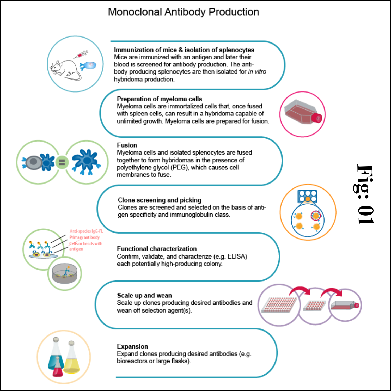
Steps to Produce Monoclonal Antibodies in Mouse 

Some assays require better antibody specificity and affinity than can be obtained using a polyclonal antiserum. To achieve this high specificity, all of the antibodies must bind with high affinity to a single epitope. This high specificity can be provided by monoclonal antibodies (mAbs). Production of monoclonal antibodies involves several critical procedures as follows.

1) Animal immunization

The very first step in hybridoma technology is to immunize an animal (usually a mouse), with appropriate antigen. The antigen, along with an adjuvant like Freund's complete or incomplete adjuvant is injected subcutaneously (adjuvants are non-specific potentiators of specific immune responses). The injections at multiple sites are repeated several times.

This enables increased stimulation of B-lymphocytes which are responding to the antigen. Three days prior to killing of the animal, a final dose of antigen is intravenously administered. The immune-stimulated cells for synthesis of antibodies have grown maximally by this approach. The concentration of the desired antibodies is assayed in the serum of the animal at frequent intervals during the course of immunization.

When the serum concentration of the antibodies is optimal, the animal is sacrificed. The spleen is aseptically removed and disrupted by mechanical or enzymatic methods to release the cells. The lymphocytes of the spleen are separated from the rest of the cells by density gradient centrifugation.

 2) Fusion & Selection 

The thoroughly washed lymphocytes are mixed with HGPRT defective myeloma cells. The mixture of cells is exposed to polyethylene glycol (PEG) for a short period (a few minutes), since it is toxic. PEG is removed by washing and the cells are kept in a fresh medium. These cells are composed of a mixture of hybridomas (fused cells), free myeloma cells and free lymphocytes.

When the cells are cultured in HAT medium, only the hybridoma cells grow, while the rest will slowly disappear. This happens in 7-10 days of culture. Selection of a single antibody producing hybrid cells is very important. This is possible if the hybridomas are isolated and grown individually. The suspension of hybridoma cells is so diluted that the individual aliquots contain on an average one cell each. These cells, when grown in a regular culture medium, produce the desired antibody.
 
 3) Antibody purification

Monoclonal antibodies may need to be purified before they are used for a variety of purposes. Protein A affinity chromatography is used to purify monoclonal antibodies. It's the golden standard for monoclonal antibody (mAb) purification, and a technology that has gained high interest because of its great performance and capabilities. If customers need higher purity, further purification methods can be performed.
 
 4) Quality control

After purification, a series of quality control tests are performed to ensure the quality of monoclonal antibodies. Antibody concentration is evaluated by absorption at 280 nm (A280). The purity of monoclonal antibodies is checked using SDS-PAGE. To estimate the monoclonal antibody titer, an ELISA test was the most suitable method.


Application of Monoclonal Antibodies

Applications of monoclonal antibodies (mAbs) have become even broader in the past few years, as availability of highest-quality immunoglobulins and IgG-derivatives increased while cost-efficiency in manufacturing processes has progressed as well.

Diagnostic applications of monoclonal antibodies

Monoclonal antibodies are widely used in medical diagnostic tests. Due to their high affinity and high selectivity, they are applied as reagents and receptors in assays to detect their respective target antigen in the most demanding matrix: tissue and bodily fluids. Good examples of diagnostic application of monoclonal antibodies that everyone of us knows are the test for the detection of COVID-19 or the pregnancy test.

Examples of monoclonal antibodies in diagnostic applications:

1. Enzyme-linked immunosorbent assay (ELISA): color change in the presence of the antigen.

2. Western immunoblotting: molecular biology technique to detect and quantify proteins in complex mixtures.

3. Immunohistochemistry: fluorescently staining tissue sections to detect and map the distribution of cancer cells, e.g. characterization of colorectal cancer, breast cancer, etc.

4. Test kits: presence of a specific antigen (e.g. a biomarker for a disease, a determinant for disease severity, or fragments of pathogens in infectious diseases) leads to a visually recognizable feature, e.g. a coloured line on the inspection field of the test.

Therapeutic uses of monoclonal antibodies

Antibody therapy generally relies on the highest-quality monoclonal antibodies produced by biotechnology methods. Polyclonal antibodies are less expensive, but they are not specific to single epitopes and would lead to side effects. Moreover, they are produced in non-human animals and would lead to severe immunogenicity.

After the development of monoclonal antibody production by hybridoma technology by Köhler and Milstein in the 1970s, enhanced murine, chimeric, humanized and human antibodies were accessible for broader use. Subsequently, the first therapeutic monoclonal antibodies gained FDA approval after large clinical trials.

Monoclonal antibodies are particularly used for therapies in autoimmune diseases or cancer. 

mAbs against autoimmune diseases

A class of diseases stems from a malfunction of the immune system, which then attacks the body’s own cells and its tissues. Several lines of treatments are routinely used to regulate immune responses, such as nonsteroidal antiinflammatory drugs, immunosuppressants and monoclonal antibodies.

Such mAbs’ pharmacology may be optimized at the Fab region to bind and inactivate certain pro-inflammatory peptides like cytokines or even specifically target the dysregulated immune cells.

Anti inflammatory mAbs that act as inhibitors of the physiologic TNF-response are routinely used in conditions such as Crohn’s disease, ulcerative colitis and rheumatoid arthritis. Monoclonal antibodies may be used as preventive treatments after organ transplants to avoid the development of the graft-versus-host-disease (GVHD), in which T lymphocytes (T cells) of the donated tissue recognize the host’s cells as foreign and begin immune responses.

Application of mAbs in cancer therapy

Monoclonal antibodies (mAbs) have revolutionized cancer therapy and are widely used in the treatment of various types of cancer. Here are some applications of mAbs in cancer therapy:

Targeted Therapy: Monoclonal antibodies are a type of targeted drug therapy that specifically recognize and bind to proteins on cancer cells. By targeting these specific proteins, mAbs can interfere with the growth and survival of cancer cells.

Immunotherapy: Some mAbs work by triggering the immune system to attack and kill cancer cells. They can attach themselves to cancer cells, making it easier for the immune system to identify and destroy them. This process is known as antibody-dependent cell-mediated cytotoxicity (ADCC). 

Delivering Therapeutic Agents: Monoclonal antibodies can be conjugated with therapeutic agents such as chemotherapy drugs or radioactive particles. These conjugated mAbs act as homing devices, delivering the therapeutic agents directly to the cancer cells, thereby enhancing their effectiveness.

Blocking Cancer Cell Signals: Certain cancer cells have receptors that signal them to divide and multiply. Monoclonal antibodies can block these signals, preventing cancer cells from proliferating.

Inhibiting Immune Checkpoints: Immune checkpoints are molecules that regulate the immune response. Some monoclonal antibodies can inhibit these checkpoints, allowing the immune system to recognize and eliminate cancer cells more effectively.

The field of mAb research and development in cancer therapy is constantly evolving, with new mAbs being developed and tested for different types of cancer.

Further areas of application of monoclonal antibodies

Monoclonal antibodies are successfully used in therapies for various diseases, such as AIDS, asthma or COVID-19. Here’s a summary of their use in each of these areas:

AIDS: mAbs have been explored as a potential treatment for HIV/AIDS. They can target specific viral proteins and inhibit viral replication. However, the development of effective mAbs for HIV has been challenging due to the high mutation rate of the virus.

COVID-19: Monoclonal antibodies have emerged as a promising therapeutic approach for COVID-19. Several mAbs have been authorized for emergency use or received full approval for the treatment of COVID-19. They can neutralize the SARS-CoV-2 virus, reduce viral load, and potentially prevent severe disease progression.

Asthma: mAbs have shown promise in the treatment of asthma. They can target specific molecules involved in the inflammatory response, such as immunoglobulin E (IgE) or interleukins, to reduce airway inflammation and improve asthma control.

Viral Infections: Monoclonal antibodies have been investigated for their potential in combating various viral infections. They can directly neutralize viruses, enhance immune responses, and provide prophylactic benefits. mAbs have been studied for viruses such as Ebola, respiratory syncytial virus (RSV), cytomegalovirus (CMV), and others.

Advantages of monoclonal antibodies

• Same quality of the antibody is maintained amongst the different production batches.
• Highly reproducible and scalable, unlimited production source.
• Speed and sensitivity and specificity of assays.
• Can produce antibodies when needed.
• No need to worry about maintaining the animals.
• Antigen or immunogen need not be pure.
• Selection helps to identify the right clones against the specific antigen.

Reverse Transcription

Reverse Transcription Introduction Reverse transcription is the synthesis of DNA from an RNA template. This process is driven by RNA-depende...