Neuron - An Overview
Introduction
Neurons are nerve cells that send messages all over your body to allow you to do everything from breathing to talking, eating, walking, and thinking. Until recently, most neuroscientists (scientists who study the brain) thought we were born with all the neurons we were ever going to have. As children, we might grow some new neurons to help build the pathways—called neural circuits—that act as information highways between different areas of the brain. However, scientists believed that once a neural circuit was in place, adding any new neurons would change the flow of information and break the brain’s communication system
Discovery of Neurogenesis
In 1962, scientist Joseph Altman challenged this belief when he saw evidence of neurogenesis (the birth of neurons) in a region of the adult rat brain called the hippocampus. He later reported that newborn neurons traveled from their birthplace in the hippocampus to other parts of the brain. In 1979, another scientist, Michael Kaplan, confirmed Altman’s findings in the rat brain; and in 1983, he found special kinds of cells—called neural precursor cells—with the ability to become brain cells like neurons, in adult monkeys.
These discoveries about neurogenesis in the adult brain were surprising to other researchers who thought they were not true in humans. Fortunately, in the early 1980s, a scientist trying to understand how birds learn to sing began to see how neurogenesis in the adult brain might make sense. In a series of experiments, Fernando Nottebohm and his research team showed that the numbers of neurons in the forebrains (areas controlling complex behaviors) of male canaries dramatically increased during the mating season, when the birds learn new songs to attract females.
Why did these bird brains add neurons at such an important time in learning? Nottebohm believed it was because newborn neurons helped store new song patterns within the pathways of the forebrain; these new neurons made learning new songs possible! If birds made new neurons to help them remember and learn, Nottebohm thought the brains of mammals—like humans—might too.
Other scientists, like Elizabeth Gould, later found evidence of newborn neurons in a distinct area of the brain in monkeys, and Fred Gage and Peter Eriksson showed that the adult human brain produce new neurons in a similar area.
Neurogenesis in the adult human brain is still tricky for neuroscientists to show, let alone learn about, how it impacts the brain and its functions. Still, scientists are intrigued by current research on neurogenesis and the possible role of new neurons in the adult brain for learning and memory.
Architecture of Neuron
Neurons, also known as nerve cells, send and receive signals from your brain. While neurons have a lot in common with other types of cells, they’re structurally and functionally unique.
Specialized projections called axons allow neurons to transmit electrical and chemical signals to other cells. Neurons can also receive these signals via rootlike extensions known as dendrites.
A 2009 study estimated that the human brain houses about 86 billion neurons trusted source. The creation of new nerve cells is called neurogenesis. While this process isn’t well understood, we know that it’s much more active when you’re an embryo. However, 2013 evidenceTrusted Source suggests that some neurogenesis occurs in adult brains throughout our lives.
As researchers gain insight into both neurons and neurogenesis, many are also working to uncover links to neurodegenerative diseases such as Alzheimer’s and Parkinson’s.
Parts of a Neuron
Neurons vary in size, shape, and structure depending on their role and location. However, nearly all neurons have three essential parts: a cell body, an axon, and dendrites.
Cell body
Also known as a soma, the cell body is the core section of the neuron. The cell body contains genetic information, maintains the neuron’s structure, and provides energy to drive activities.
Like other cell bodies, a neuron’s soma contains a nucleus and specialized organelles. It’s enclosed by a membrane that both protects it and allows it to interact with its immediate surroundings.
Axon
An axon is a long, tail-like structure. It joins the cell body at a specialized junction called the axon hillock. Many axons are insulated with a fatty substance called myelin. Myelin helps axons to conduct an electrical signal.
Neurons usually have one main axon.
Dendrites
Dendrites are fibrous roots that branch out from the cell body. Like antennae, dendrites receive and process signals from the axons of other neurons. Neurons can have more than one set of dendrites, known as dendritic trees.
How many they have generally depends on their role. For instance, Purkinje cells are a special type of neuron found in a part of the brain called the cerebellum. These cells have highly developed dendritic trees which allow them to receive thousands of signals.
Types of neurons
Neurons vary in structure, function, and genetic makeup. Given the sheer number of neurons, there are thousands of different types, much like there are thousands of species of living organisms on Earth.
However, there are five major neuron forms. Each combines several elements of the basic neuron shape.
Multipolar neurons. These neurons have a single axon and symmetrical dendrites that extend from it. This is the most common form of neuron in the central nervous system.
Unipolar neurons. Usually only found in invertebrate species, these neurons have a single axon.
Bipolar neurons. Bipolar neurons have two extensions extending from the cell body. At the end of one side is the axon, and the dendrites are on the other side. These types of neurons are mostly found in the retina of the eye. But they can also be found in parts of the nervous system that help the nose and ear function.
Pyramidal neurons. These neurons have one axon but several dendrites to form a pyramid type shape. These are the largest neuron cells and are mostly found in the cortex. The cortex is the part of the brain responsible for conscious thoughts.
Purkinje neurons. Purkinje neurons have multiple dendrites that fan out from the cell body. These neurons are inhibitory neurons, meaning they release neurotransmitters that keep other neurons from firing.
In terms of function, scientists classify neurons into three broad types: sensory, motor, and interneurons.
Sensory neurons
Sensory neurons help you:
. taste
. smell
. hear
. see
. feel things around you
Sensory neurons are triggered by physical and chemical inputs from your environment. Sound, touch, heat, and light are physical inputs. Smell and taste are chemical inputs.
For example, stepping on hot sand activates sensory neurons in the soles of your feet. Those neurons send a message to your brain, which makes you aware of the heat.
Motor neurons
Motor neurons play a role in movement, including voluntary and involuntary movements. These neurons allow the brain and spinal cord to communicate with muscles, organs, and glands all over the body.
There are two types of motor neurons: lower and upper. Lower motor neurons carry signals from the spinal cord to the smooth muscles and skeletal muscles. Upper motor neurons carry signals between your brain and spinal cord.
When you eat, for instance, lower motor neurons in your spinal cord send signals to the smooth muscles in your esophagus, stomach, and intestines. These muscles contract, which allows food to move through your digestive tract.
Interneurons
Interneurons are neural intermediaries found in your brain and spinal cord. They’re the most common type of neuron. They pass signals from sensory neurons and other interneurons to motor neurons and other interneurons. Often, they form complex circuits that help you to react to external stimuli.
For instance, when you touch something sharp like a cactus, sensory neurons in your fingertips send a signal to interneurons in your spinal cord. Some interneurons pass the signal on to motor neurons in your hand, which allows you to move your hand away. Other interneurons send a signal to the pain center in your brain, and you experience pain.
Neuron Birth
Many neuroscientists disagree about how many and how often new neurons are created in the brain. Most of the brain’s neurons are already created by the time we’re born, but there is evidence to support the theory that neurogenesis is a lifelong process.
Neurons are born in areas of the brain that are full of neural stem cells, or precursor cells. Stem cells have the potential to make most, if not all, of the different types of neurons and glia found in the brain.
Neuroscientists have observed how neural stem cells behave during experiments in the laboratory. Although this may not be exactly how these cells act when they’re in the brain, it gives us information about how they might function when they’re in the brains of humans or other animals.
The science of stem cells is still very new and could change with additional discoveries, but researchers have learned enough to be able to describe how neural stem cells create the other cells of the brain. The way that stem cells can become other types of brain cells is similar to the idea of a family tree.
Neural stem cells increase by dividing in two. Then they can become either two new stem cells, or two early progenitor cells (parent cells to new neurons or glia), or one of each.
When a stem cell divides to produce another stem cell, it is said to self-renew. This new cell has the potential to make more stem cells.
When a stem cell divides to produce an early progenitor cell, it is said to differentiate. Differentiation means that the new cell is more specialized in how it’s formed and what it can do.
Early progenitor cells can make other progenitor cells, self-renew like stem cells, or can change in either of two ways. One way will make new astrocytes. The other way will make neurons or oligodendrocytes.
Neuron - A Road trip
Once a neuron is born, it must travel to the place in the brain where it will do its work.
How does a neuron know where to go? What helps it get there?
Scientists have seen that neurons use at least two different methods to travel:
Some neurons travel, or migrate, by following the long fibers of cells called radial glia. These fibers stretch from the inner layers to the outer layers of the brain. Neurons glide along the fibers until they reach their destination.
Neurons also travel by using chemical signals. Scientists have found special molecules on the surface of neurons—adhesion molecules—that attach to similar molecules on nearby glial cells or nerve axons. These chemical signals guide the neurons to their final location.
Not all neurons are successful in their journey. Scientists think that only a third reach their destination. Some cells die in development or while traveling.
Other neurons survive the trip but end up where they should not be. Accidental changes (called mutations) in the genes that control migration create brain areas of misplaced or oddly formed neurons that can cause disorders like childhood epilepsy. Some researchers think that certain disorders, such as schizophrenia and dyslexia, are partly the result of misguided neurons.
Differentiation - Neuron gain its Structure
Once a neuron reaches its destination, it must settle into work. There is still a lot that scientists don’t understand about the part of neurogenesis called differentiation.
Neurons are responsible for sending and receiving neurotransmitters—chemicals that carry information between brain cells.
Depending on its location, a neuron can perform the job of a sensory neuron, a motor neuron, or an interneuron, sending and receiving specific neurotransmitters.
In the developing brain, a neuron depends on molecular signals from other cells, such as astrocytes, to determine its shape and location, the kind of transmitter it produces, and the other neurons it will connect to. These freshly born cells create neural circuits—or information pathways connecting neurons to neurons—that will be in place throughout adulthood.
But in the adult brain, neural circuits are already developed, and neurons must find a way to fit in. As new neurons settle in, they start to look like the surrounding cells. They develop axons and dendrites and begin to communicate with their neighbors through synapses.
Synapse
Synapse, also called Neuronal Junction, the site of transmission of electric nerve impulses between two nerve cells (neurons) or between a neuron and a gland or muscle cell (effector). A synaptic connection between a neuron and a muscle cell is called a neuromuscular junction.
At a chemical synapse each ending, or terminal, of a nerve fiber (presynaptic fiber) swells to form a knoblike structure that is separated from the fiber of an adjacent neuron, called a postsynaptic fiber, by a microscopic space called the synaptic cleft. The typical synaptic cleft is about 0.02 micron wide.
The arrival of a nerve impulse at the presynaptic terminals causes the movement toward the presynaptic membrane of membrane-bound sacs, or synaptic vesicles, which fuse with the membrane and release a chemical substance called a neurotransmitter.
This substance transmits the nerve impulse to the postsynaptic fiber by diffusing across the synaptic cleft and binding to receptor molecules on the postsynaptic membrane. The chemical binding action alters the shape of the receptors, initiating a series of reactions that open channel-shaped protein molecules.
Electrically charged ions then flow through the channels into or out of the neuron. This sudden shift of electric charge across the postsynaptic membrane changes the electric polarization of the membrane, producing the postsynaptic potential, or PSP.
If the net flow of positively charged ions into the cell is large enough, then the PSP is excitatory; that is, it can lead to the generation of a new nerve impulse, called an action potential.
Once they have been released and have bound to postsynaptic receptors, neurotransmitter molecules are immediately deactivated by enzymes in the synaptic cleft; they are also taken up by receptors in the presynaptic membrane and recycled. This process causes a series of brief transmission events, each one taking place in only 0.5 to 4.0 milliseconds.
A single neurotransmitter may elicit different responses from different receptors. For example, norepinephrine, a common neurotransmitter in the autonomic nervous system, binds to some receptors that excite nervous transmission and to others that inhibit it.
The membrane of a postsynaptic fiber has many different kinds of receptors, and some presynaptic terminals release more than one type of neurotransmitter. Also, each postsynaptic fiber may form hundreds of competing synapses with many neurons.
These variables account for the complex responses of the nervous system to any given stimulus. The synapse, with its neurotransmitter, acts as a physiological valve, directing the conduction of nerve impulses in regular circuits and preventing random or chaotic stimulation of nerves.
Electric synapses allow direct communications between neurons whose membranes are fused by permitting ions to flow between the cells through channels called gap junctions. Found in invertebrates and lower vertebrates, gap junctions allow faster synaptic transmission as well as the synchronization of entire groups of neurons.
Gap junctions are also found in the human body, most often between cells in most organs and between glial cells of the nervous system. Chemical transmission seems to have evolved in large and complex vertebrate nervous systems, where transmission of multiple messages over longer distances is required.
Neurotransmitters
Neurotransmitters are chemical messengers that your body can’t function without. Their job is to carry chemical signals (“messages”) from one neuron (nerve cell) to the next target cell. The next target cell can be another nerve cell, a muscle cell or a gland.
Your body has a vast network of nerves (your nervous system) that send and receive electrical signals from nerve cells and their target cells all over your body. Your nervous system controls everything from your mind to your muscles, as well as organ functions.
In other words, nerves are involved in everything you do, think and feel. Your nerve cells send and receive information from all body sources. This constant feedback is essential to your body’s optimal function.
Neurotransmitters are located in a part of the neuron called the axon terminal. They’re stored within thin-walled sacs called synaptic vesicles. Each vesicle can contain thousands of neurotransmitter molecules.
As a message or signal travels along a nerve cell, the electrical charge of the signal causes the vesicles of neurotransmitters to fuse with the nerve cell membrane at the very edge of the cell.
The neurotransmitters, which now carry the message, are then released from the axon terminal into a fluid-filled space that’s between one nerve cell and the next target cell (another nerve cell, muscle cell or gland).
In this space, called the synaptic junction, the neurotransmitters carry the message across less than 40 nanometers (nm) wide (by comparison, the width of a human hair is about 75,000 nm).
Each type of neurotransmitter lands on and binds to a specific receptor on the target cell (like a key that can only fit and work in its partner lock).
After binding, the neurotransmitter that triggers a change or action in the target cell, like an electrical signal in another nerve cell, a muscle contraction or the release of hormones from a cell in a gland.
Types of Neurotransmitters
Scientists know of at least 100 neurotransmitters and suspect there are many others that have yet to be discovered. They can be grouped into types based on their chemical nature. Some of the better-known categories and neurotransmitter examples and their functions include the following:
Amino acids neurotransmitters
These neurotransmitters are involved in most functions of your nervous system.
1. Glutamate. This is the most common excitatory neurotransmitter of your nervous system. It’s the most abundant neurotransmitter in your brain. It plays a key role in cognitive functions like thinking, learning and memory. Imbalances in glutamate levels are associated with Alzheimer’s disease, dementia, Parkinson’s disease and seizures.
2. Gamma-aminobutryic acid (GABA). GABA is the most common inhibitory neurotransmitter of your nervous system, particularly in your brain. It regulates brain activity to prevent problems in the areas of anxiety, irritability, concentration, sleep, seizures and depression.
3. Glycine. Glycine is the most common inhibitory neurotransmitter in your spinal cord. Glycine is involved in controlling hearing processing, pain transmission and metabolism.
Monoamines neurotransmitters
These neurotransmitters play a lot of different roles in your nervous system and especially in your brain. Monoamines neurotransmitters regulate consciousness, cognition, attention and emotion. Many disorders of your nervous system involve abnormalities of monoamine neurotransmitters, and many drugs that people commonly take affect these neurotransmitters.
1. Serotonin. Serotonin is an inhibitory neurotransmitter. Serotonin helps regulate mood, sleep patterns, sexuality, anxiety, appetite and pain. Diseases associated with serotonin imbalance include seasonal affective disorder, anxiety, depression, fibromyalgia and chronic pain. Medications that regulate serotonin and treat these disorders include selective serotonin reuptake inhibitors (SSRIs) and serotonin-norepinephrine reuptake inhibitors (SNRIs).
2. Histamine. Histamine regulates body functions including wakefulness, feeding behavior and motivation. Histamine plays a role in asthma, bronchospasm, mucosal edema and multiple sclerosis.
3. Dopamine. Dopamine plays a role in your body’s reward system, which includes feeling pleasure, achieving heightened arousal and learning. Dopamine also helps with focus, concentration, memory, sleep, mood and motivation. Diseases associated with dysfunctions of the dopamine system include Parkinson’s disease, schizophrenia, bipolar disease, restless legs syndrome and attention deficit hyperactivity disorder (ADHD). Many highly addictive drugs (cocaine, methamphetamines, amphetamines) act directly on the dopamine system.
4. Epinephrine. Epinephrine (also called adrenaline) and norepinephrine (see below) are responsible for your body’s so-called “fight-or-flight response” to fear and stress. These neurotransmitters stimulate your body’s response by increasing your heart rate, breathing, blood pressure, blood sugar and blood flow to your muscles, as well as heighten attention and focus to allow you to act or react to different stressors. Too much epinephrine can lead to high blood pressure, diabetes, heart disease and other health problems. As a drug, epinephrine is used to treat anaphylaxis, asthma attacks, cardiac arrest and severe infections.
5. Norepinephrine. Norepinephrine (also called noradrenaline) increases blood pressure and heart rate. It’s most widely known for its effects on alertness, arousal, decision-making, attention and focus. Many medications (stimulants and depression medications) aim to increase norepinephrine levels to improve focus or concentration to treat ADHD or to modulate norepinephrine to improve depression symptoms.
Peptide neurotransmitters
Peptides are polymers or chains of amino acids.
Endorphins. Endorphins are your body’s natural pain reliever. They play a role in our perception of pain. Release of endorphins reduces pain, as well as causes “feel good” feelings. Low levels of endorphins may play a role in fibromyalgia and some types of headaches.
Acetylcholine
This excitatory neurotransmitter does a number of functions in your central nervous system (CNS [brain and spinal cord]) and in your peripheral nervous system (nerves that branch from the CNS). Acetylcholine is released by most neurons in your autonomic nervous system regulating heart rate, blood pressure and gut motility. Acetylcholine plays a role in muscle contractions, memory, motivation, sexual desire, sleep and learning. Imbalances in acetylcholine levels are linked with health issues, including Alzheimer’s disease, seizures and muscle spasms.
Table
Change do neurotransmitters transmit
Neurotransmitters transmit one of three possible actions in their messages, depending on the specific neurotransmitter.
1. Excitatory. Excitatory neurotransmitters “excite” the neuron and cause it to “fire off the message,” meaning, the message continues to be passed along to the next cell. Examples of excitatory neurotransmitters include glutamate, epinephrine and norepinephrine.
2. Inhibitory. Inhibitory neurotransmitters block or prevent the chemical message from being passed along any farther. Gamma-aminobutyric acid (GABA), glycine and serotonin are examples of inhibitory neurotransmitters.
3. Modulatory. Modulatory neurotransmitters influence the effects of other chemical messengers. They “tweak” or adjust how cells communicate at the synapse. They also affect a larger number of neurons at the same time.
The Sodium-Potassium Pump
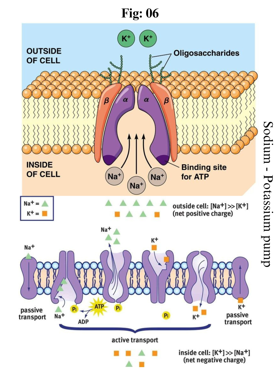
Active transport is the energy-requiring process of pumping molecules and ions across membranes "uphill" - against a concentration gradient. To move these molecules against their concentration gradient, a carrier protein is needed. Carrier proteins can work with a concentration gradient (during passive transport), but some carrier proteins can move solutes against the concentration gradient (from low concentration to high concentration), with an input of energy.
In active transport, as carrier proteins are used to move materials against their concentration gradient, these proteins are known as pumps. As in other types of cellular activities, ATP supplies the energy for most active transport. One way ATP powers active transport is by transferring a phosphate group directly to a carrier protein.
This may cause the carrier protein to change its shape, which moves the molecule or ion to the other side of the membrane. An example of this type of active transport system, as shown in Figure below, is the sodium-potassium pump, which exchanges sodium ions for potassium ions across the plasma membrane of animal cells.
The sodium-potassium pump system moves sodium and potassium ions against large concentration gradients. It moves two potassium ions into the cell where potassium levels are high, and pumps three sodium ions out of the cell and into the extracellular fluid.
The three sodium ions bind with the protein pump inside the cell. The carrier protein then gets energy from ATP and changes shape. In doing so, it pumps the three sodium ions out of the cell.
At that point, two potassium ions from outside the cell bind to the protein pump. The potassium ions are then transported into the cell, and the process repeats.
The sodium-potassium pump is found in the plasma membrane of almost every human cell and is common to all cellular life. It helps maintain cell potential and regulates cellular volume.
Death of Neuron
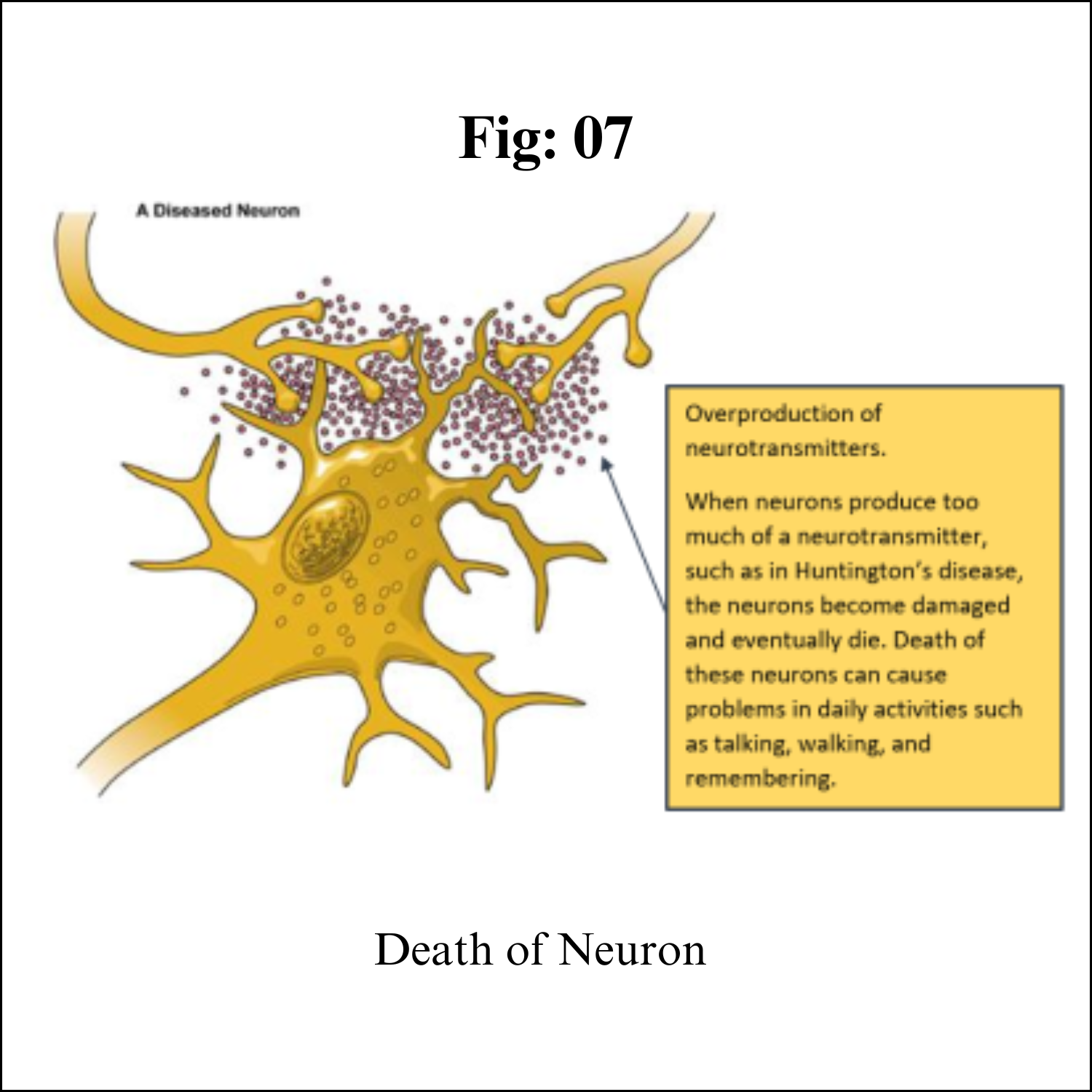
Although neurons are the longest living cells in the body, large numbers of them die during migration and differentiation. The lives of some neurons can take strange turns. Some diseases of the brain are the result of the unnatural deaths of neurons.
In Parkinson’s disease, neurons that produce the neurotransmitter dopamine die off in the basal ganglia, an area of the brain that controls body movements. This causes people with this disease to experience shaking, to move more slowly, and to have problems with balance.
In Huntington’s disease, a genetic mutation causes neurons to create too much of a neurotransmitter called glutamate, which kills neurons in the basal ganglia. As a result, people twist and move uncontrollably, and over time, they lose the ability to do many everyday tasks like walking and eating. People with this disease typically have shorter lives than those without this disease.
In Alzheimer’s disease, unusual proteins build up in and around neurons in the neocortex and hippocampus, the parts of the brain that control memory. When these neurons die, people lose their abilities to remember and do everyday tasks.
Physical damage to the brain and the spinal cord can also kill or disable neurons. Damage to the brain caused by shaking or hitting the head, or because of a stroke, can kill neurons immediately or slowly, starving them of the oxygen and nutrients they need to survive.
A spinal cord injury can cut off communication between the brain and the muscles. When neurons lose their connection to the axons (the parts of neurons that send messages to other neurons) located below the site of injury, the neurons may still live, but they lose their ability to communicate.
Functions
Your nervous system controls such functions as your:
Heartbeat and blood pressure.
Breathing.
Muscle movements.
Thoughts, memory, learning and feelings.
Sleep, healing and aging.
Stress response.
Hormone regulation.
Digestion, sense of hunger and thirst.
Senses (response to what you see, hear, feel, touch and taste).
###


