Wednesday, July 26, 2023

Physiology of Learning and Memory


Physiology of Learning and Memory  

Learning and the development of memory are processes that cannot be separated from psychology and sociology. This blog deals with the physiology of learning and memory. Topics include understanding the relationship between experience and the storage of acquired knowledge, how the brain deals with “useless” knowledge, and how infants start to understand their surroundings.

Learning

Learning is the process of acquiring new knowledge or skills by study, instruction, or experience. Learning is the precondition for the brain to store experiences and use those experiences in our actions to gain benefits and prevent damage.

Humans adapt to their environment by learning. When an individual learns to expect and prepare for a significant event such as the arrival of food or pain, it is called learning by classical conditioning. When a person learns to repeat a behavior that brings a reward and understands how to avoid doing something that brings unwanted results, the process is termed learning by operant conditioning. When an individual learns about things they have neither experienced nor observed merely based on observation and through language, the process is termed cognitive learning. This chapter will discuss how a person learns.


Formation of synapses

Learning processes during the first 6 months of an infant’s life hold great significance in the development of the nervous system. Environmental stimuli and experiences also play a role as they lead to the formation of new synapses and the improvement of already existing synaptic connections. The ability of the brain to form and improve these connections are referred to as neuroplasticity, which is based on physiological or neuroanatomical conditions.

Infants learning

Most brain cells are already formed in utero. The 1st neurons and synapses begin to develop in the spinal cord approximately 7 weeks after conception. However, most nerve cells of infants are incapable of communication at the time of birth as they are not yet connected. These connections develop during the first 3 years of life by the formation of dendrites, which enable cells to absorb information. Moreover, synaptic connections are formed, which are responsible for relaying information. The extent of these connections is far greater than what is required, making it easy to adjust later if needed.

Learning capacity of infants

Babies react strongly to stimuli, which is an important indication that the learning process is being carried out. Besides, these stimuli are necessary for the brain to develop. Thus, an environment with few stimuli hinders the development of infants.

Infants can recognize faces and vocal sounds during their developmental stage, enabling them to differentiate between familiar faces and strangers. After imprinting their personal surroundings, infants lose a certain amount of flexibility related to their mental abilities. However, learning processes become more specific. This development occurs during the first 6 months of life.

Brain Work

The brain uses neurons to communicate and, to a large extent, manage itself. However, the development of neurons and the resulting brain capacities depend on environmental and sensory stimuli. The brain works in conjunction with the spinal cord to send and receive information via neurons. The brain processes perceptions, which are also connected; accordingly, it uses experiences that are already stored. However, most perceptions are suppressed. The brain differentiates perceptions that must be processed during the learning process based on the following aspects:

1. Relevance
2. Value of new knowledge
3. Significance
4. Meaningfulness

Importance of Emotions During the Learning Process

Cognition and emotions play a major role in the learning process. Sensations are used as somatic markers, which influence processing, storage, and memory. Learning also includes the strengthening of the most used neuronal pathways such that they can be used longer and, above all, faster.

Learning as Formation and Deformation in the Brain

The learning process starts with the processing of external influences. Learning leads to changes in the brain, which can be classified under 4 categories, namely, expanding, tuning, reconstructing, and pruning.

Expanding refers to improving the number and strength of neuronal connections by developing a network of already existing information. Tuning describes the process of creating new connections. Relearning occurs during the process of reconstructing. In this time-consuming and exhaustive process, pre-existing learning achievements (motor patterns and routine processes) are replaced by new ones that are better suited for the respective tasks. Pruning refers to regression of the neuronal pathways that are seldom or never used. During this process, connections can be changed such that they can no longer be activated.

Learning can be categorized as follows:

a. Intentional learning
b. Individual learning
c. Collective learning
d. Physical learning
e. Social learning

Significance of social interaction and physical activity

Humans need social interactions, and this also applies to the brain. Mirror neurons in the brain are responsible for development of the required cognitive orientation patterns. A mirror neuron is a type of sensory-motor cell located in the brain that is activated when an individual performs an action or observes another individual performing the same action. Thus, the neuron “mirrors” others’ actions. Physical activity is also important for brain performance, and this particularly applies in the 1st few years of life.

Localization of Learning Processes and Memory

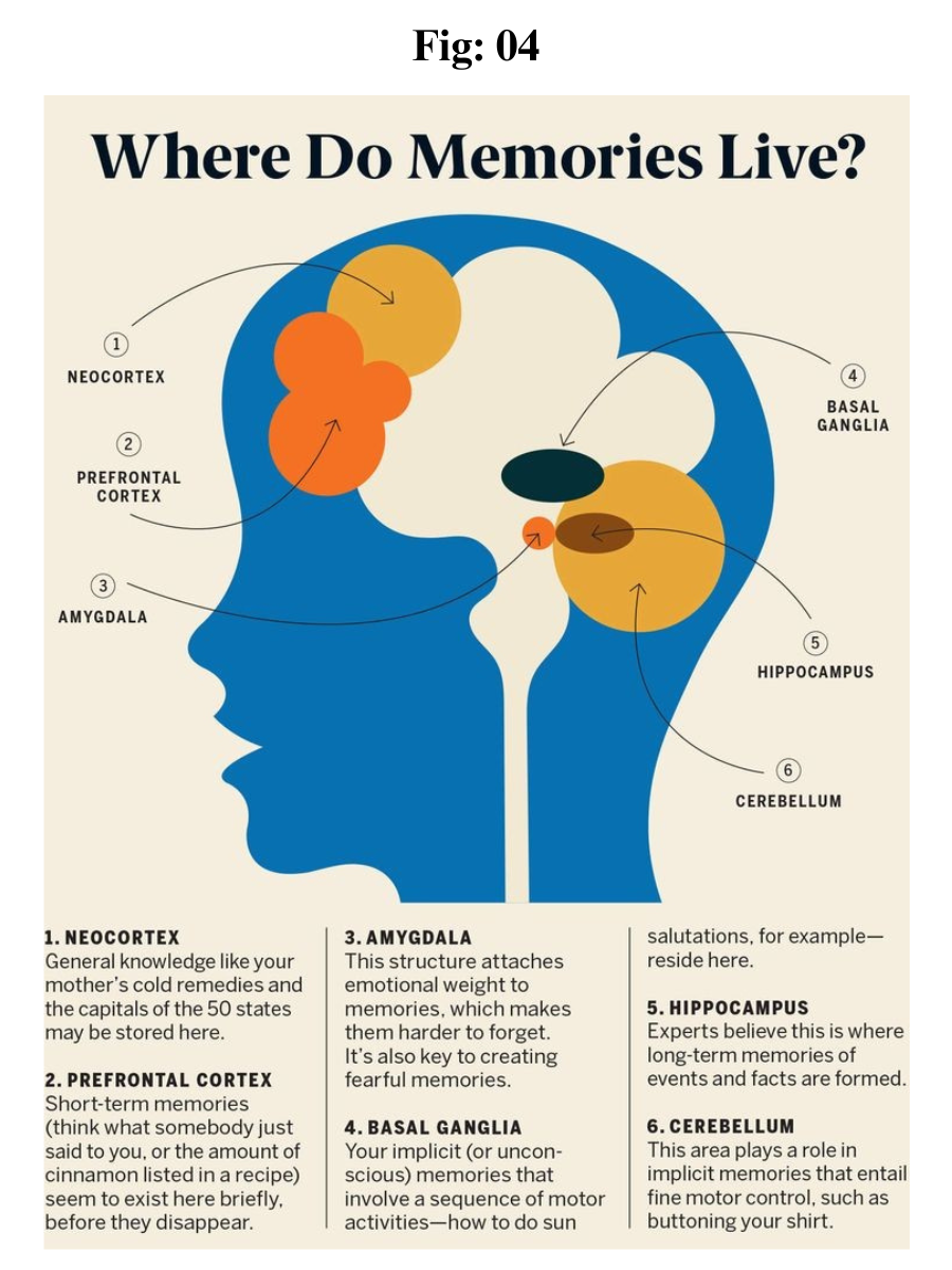
Neurons of the cerebellum and basal ganglia are responsible for motor learning. Declarative memory is located in the medial temporal lobe. Hippocampal lesions lead to anterograde amnesia, a condition in which new information cannot be stored.

Process of Learning in the Brain

The hippocampus plays an important role in learning. The physiologic substrate of learning consists of continuous electrophysiologic, morphologic, and molecular changes in nerve cells. Long-term potentiation is necessary for the long-term availability of information. It facilitates the stimulation of afferent axons over a period of weeks and also leads to greater calcium influx.

Thoughts stored in the long-term memory are available during the entire lifespan of that individual. The process of creating long-term memories is mediated by neurotransmitters such as glutamate (glutamic acid).

Types of Learning

1. Motor Learning: Our day to day activities like walking, running, driving, etc, must be learnt for ensuring a good life. These activities to a great extent involve muscular coordination.

2. Verbal Learning: It is related with the language which we use to communicate and various other forms of verbal communication such as symbols, words, languages, sounds, figures and signs.

3. Concept Learning: This form of learning is associated with higher order cognitive processes like intelligence, thinking, reasoning, etc, which we learn right from our childhood. Concept learning involves the processes of abstraction and generalization, which is very useful for identifying or recognizing things.

4. Discrimination Learning: Learning which distinguishes between various stimuli with its appropriate and different responses is regarded as discrimination stimuli.

5. Learning of Principles: Learning which is based on principles helps in managing the work most effectively. Principles based learning explains the relationship between various concepts.

6. Attitude Learning: Attitude shapes our behavior to a very great extent, as our positive or negative behavior is based on our attitudinal predisposition.


Memory Systems

The striatum is associated with procedural memory and uses the pathway of the neocortex. Associative learning for emotional and motor processes occurs in the amygdala and cerebellum, respectively. Nonassociative learning occurs in the form of habituation and sensitization (both via reflex circuits).

Hebbian theory

The Hebbian theory explains how connections between certain neurons can be strengthened. This theory asserts that “neurons that fire together, wire together,” which means that when activity in a cell repeatedly elicits action potentials in a 2nd cell, the synaptic strength is potentiated.

If an axon of neuron A is located close enough to neuron B such that neuron B can be stimulated by neuron A repeatedly or continuously, the efficiency of neuron A for the stimulation by neuron B is increased by growth processes or changes in metabolism in 1 or both neurons. This suggests that experience-related changes in the nervous systems depend on certain conditions.

Development of Memory and the Papez Circuit

The Papez circuit exists in all mammals and is important for memory development. It is located in the center of the 
limbic system, which is present above the brain stem. The Papez circuit plays a vital role in social behavior, solicitude, love, fear, and learning by imitation.

Papez circuit

The Papez circuit is a chain of neurons named after its discoverer James Papez. Research on the tasks of the Papez circuit for memory performance is ongoing. However, the assumption that the circuit controls anger and rage is already outdated, as it has been discovered that the circuit is even more complex than Papez had thought.

It is currently believed that the Papez circuit plays a role in memory storage by transferring information from the primary memory (short-term memory) to the secondary (long-term memory) or tertiary memory (an independent component of long-term memory).

The Papez circuit proceeds as follows:

hippocampus → fornix → mammillary body in the hypothalamus (corpora mammillaria) → cingulate cortex → hippocampus. 

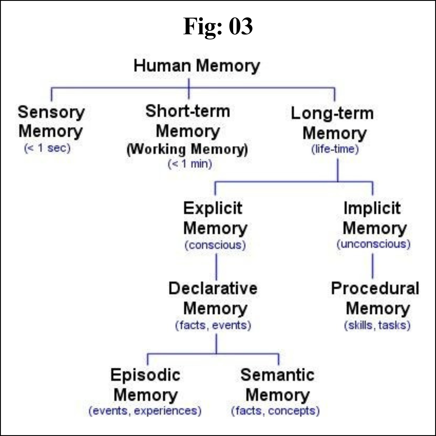
Different Types of Memory

The specialist term for mind and memory is called the mnestic function. Some things are easier to remember than others. For example, important events are easier to remember than those that hold no meaning, and positive experiences are easier to remember than neutral experiences. Moreover, the process of remembering is easier in a prevailing positive mood, which also means that remembering things is more difficult in a state of 
fatigue or grief.

Process of encoding information

Encoding is the process of transferring sensory information into a construct, which is then stored in our memory system. Working memory stores information for immediate use as part of mental activity (i.e., learning or problem solving).

It is believed to include a phonological loop (i.e., a component of the working memory model that deals with spoken and written material), a visuospatial sketchpad, a central executive, and an episodic buffer.

It allows for the manipulation and organization of information as opposed to short-term memory.

The primary effect is a cognitive bias that results in a subject recalling the 1st items on a list.

Items that have been encoded for the longest duration are transferred to the long-term memory.

The recency effect is a cognitive bias that results in a subject recalling the last items on a list. 

Items in the phonological loop are highly accessible.

Humans have declarative memory (explicit memory) and 
procedural memory (implicit memory). 

Declarative memory stores information that can be reproduced because humans are conscious of the experience. In contrast, procedural memory stores experiences for which one has no direct memory of the learning process. Nevertheless, this type of memory influences our behavior. A classic example of procedural memory is the learning of a new language.


Sensory (ultra-short-term) memory

The ultra-short-term memory receives stimuli from sensory organs in the form of neuronal excitation. This process has a duration of < 1 second, and perception is via the eyes or ears. Ultra-short-term memory via the eye is referred to as iconic memory, whereas that via the ears is known as echoic memory (it perishes just as fast). Only the stimuli that reach the short-term memory remain, as ultra-short-term memory has no storage capabilities.

Short-term/working memory (primary memory)

Memories in the primary (short-term/working) memory are available for as long as we occupy ourselves with them. If that process is interrupted, the memory is lost too. Memories that begin in the primary memory can be available permanently, but only if they are transferred to the long-term memory. Short-term memory is the usual transitory path for experiences to pass into long-term memory; however, this is occasionally short-circuited so that information can pass directly from the sensory memory to the long-term memory.

The hippocampus, located in the cerebral cortex, is involved in transmitting information from the primary memory to the long-term memory. The hippocampus is thought to be involved in this process because when lesions appear in the hippocampus, only the short-term memory remains intact. Another term for primary memory is labile memory, as it is very unstable. A single distraction is enough to forget the information that has been perceived or heard. Calcium plays a major role in these processes.

Long-term memory

Repetition is particularly important for storing memories in long-term memory. This concept is easily understandable when the high amount of repetition required to learn new movement patterns is considered, e.g., when learning a new sport.

Semantic networks and spreading networks

Information is stored in our long-term memory as an organized network.

Individual ideas or hubs are called nodes (e.g., cities on a map).

Nodes are connected by links or associations (e.g., roads between cities).

The strength of the association depends on how frequently and deeply the connection is made.

Processing material in different ways leads to the establishment of multiple connections.

Nodes are activated only when they reach a response threshold.

The response threshold is reached by the summation of input signals from multiple nodes.

Activation of a node leads to stimulation of neighboring connecting nodes.

Activation of a few nodes can lead to a pattern of activation within the network that spreads inward (spreading activation).

It explains contextual cues, priming, and associations.


Pathology of Memory Performance

The highly complex system of learning and memory is susceptible to malfunctions. If anomalies occur, the differential diagnosis must be made with the greatest care, because even changes in the mineral balance of the body can lead to disorders that give the impression of a disease (e.g., calcium deficiency). Furthermore, cases of 
dementia are increasing, which is partly due to the increase in life expectancy.

Amnesia

Amnesia is a memory disorder in which an individual loses access to stored information. “Amnesia” is derived from the Greek words a (without) and mnémē (memory). Amnesia is not an independent disease, but the symptom of a disease or the consequence of an influence on the brain. The influence can be internal or external. Individuals with amnesia cannot recall prior experiences or knowledge; this can affect either all or only certain parts and types of information. For example, patients can lose access to memories from certain stages of their lives. In most cases, they can remember events that occurred long ago rather than events that occurred recently. Several forms of amnesia cannot be differentiated from each other; however, the following variants are of importance:

Retrograde amnesia: a type of amnesia in which a person cannot recall memories that were formed before the event that caused the amnesia. Only recently stored memories are affected and not memories from years before. 

Transient global amnesia: a self-limiting clinical syndrome lasting < 24 hours and characterized by the acute onset of anterograde amnesia. Affected individuals are disoriented and ask repetitive questions. There may be an inability to recall general or personal information (retrograde amnesia) while the episode lasts.

Anterograde amnesia: an inability to form new memories/acquire new knowledgePsychogenic amnesia: refers to cases of memory loss presumed to have a psychological rather than a neurological cause Alzheimer disease (AD) 

Alzheimer disease is characterized by variable degrees of cortical atrophy, seen as gyral narrowing and sulcal widening, mostly in the frontal, temporal, and parietal lobes. If there is marked atrophy, there will be compensatory ventricular enlargement (hydrocephalus ex vacuo) secondary to the reduced brain volume. The outcome is the impaired relay of information. Furthermore, AD may make information processing or learning nearly impossible. The basic abnormality in AD is the accumulation of Aβ and tau proteins in specific regions of the brain due to excessive production and defective removal. Amyloid plaques and neurofibrillary tangles are the 2 pathologic hallmarks of AD.

The regions of the brain involved in processing information and memory performance are particularly affected by AD.

Monday, July 24, 2023

Nervous System


Nervous system
 

Introduction 

Although terminology seems to indicate otherwise, there is really only one nervous system in the body. Although each subdivision of the system is also called a "nervous system," all of these smaller systems belong to the single, highly integrated nervous system. Each subdivision has structural and functional characteristics that distinguish it from the others. The nervous system as a whole is divided into two subdivisions: the central nervous system (CNS) and the peripheral nervous system (PNS).

The Central Nervous System (CNS)

The brain and spinal cord are the organs of the central nervous system. Because they are so vitally important, the brain and spinal cord, located in the dorsal body cavity, are encased in bone for protection. The brain is in the cranial vault, and the spinal cord is in the vertebral canal of the vertebral column. Although considered to be two separate organs, the brain and spinal cord are continuous at the foramen magnum.

The Peripheral Nervous System (PNS)

The organs of the peripheral nervous system are the nerves and ganglia. Nerves are bundles of nerve fibers, much like muscles are bundles of muscle fibers. Cranial nerves and spinal nerves extend from the CNS to peripheral organs such as muscles and glands. Ganglia are collections, or small knots, of nerve cell bodies outside the CNS.

The peripheral nervous system is further subdivided into an afferent (sensory) division and an efferent (motor) division. The afferent or sensory division transmits impulses from peripheral organs to the CNS. The efferent or motor division transmits impulses from the CNS out to the peripheral organs to cause an effect or action.

Finally, the efferent or motor division is again subdivided into the somatic nervous system and the autonomic nervous system. The somatic nervous system, also called the somatomotor or somatic efferent nervous system, supplies motor impulses to the skeletal muscles. 

Because these nerves permit conscious control of the skeletal muscles, it is sometimes called the voluntary nervous system. The autonomic nervous system, also called the visceral efferent nervous system, supplies motor impulses to cardiac muscle, to smooth muscle, and to glandular epithelium. 

It is further subdivided into sympathetic and parasympathetic divisions. Because the autonomic nervous system regulates involuntary or automatic functions, it is called the involuntary nervous system.


The Central Nervous System

The CNS consists of the brain and spinal cord, which are located in the dorsal body cavity. The brain is surrounded by the cranium, and the spinal cord is protected by the vertebrae. The brain is continuous with the spinal cord at the foramen magnum. In addition to bone, the CNS is surrounded by connective tissue membranes, called meninges, and by cerebrospinal fluid.

Meninges

There are three layers of meninges around the brain and spinal cord. The outer layer, the dura mater, is tough white fibrous connective tissue. The middle layer of meninges is arachnoid, which resembles a cobweb in appearance, is a thin layer with numerous threadlike strands that attach it to the innermost layer. 

The space under the arachnoid, the subarachnoid space, is filled with cerebrospinal fluid and contains blood vessels. The pia mater is the innermost layer of meninges. This thin, delicate membrane is tightly bound to the surface of the brain and spinal cord and cannot be dissected away without damaging the surface.

Meningiomas are tumors of the nerve tissue covering the brain and spinal cord. Although meningiomas are usually not likely to spread, physicians often treat them as though they were malignant to treat symptoms that may develop when a tumor applies pressure to the brain.


Brain

The brain is divided into the cerebrum, diencephalons, brain stem, and cerebellum.


Cerebrum

The largest and most obvious portion of the brain is the cerebrum, which is divided by a deep longitudinal fissure into two cerebral hemispheres. The two hemispheres are two separate entities but are connected by an arching band of white fibers, called the corpus callosum that provides a communication pathway between the two halves.

Each cerebral hemisphere is divided into five lobes, four of which have the same name as the bone over them: the fontal lobe, the parietal lobe, the occipital lobe, and the temporal lobe. A fifth lobe, the insula or Island of Reil, lies deep within the lateral sulcus.

Diencephalon

The diencephalons is centrally located and is nearly surrounded by the cerebral hemispheres. It includes the thalamus, hypothalamus, and epithalamus. The thalamus, about 80 percent of the diencephalons, consists of two oval masses of gray matter that serve as relay stations for sensory impulses, except for the sense of smell, going to the cerebral cortex. 

The hypothalamus is a small region below the thalamus, which plays a key role in maintaining homeostasis because it regulates many visceral activities. The epithalamus is the most dorsal portion of the diencephalons. This small gland is involved with the onset of puberty and rhythmic cycles in the body. It is like a biological clock.

Brain Stem

The brainstem is the region between the diencephalons and the spinal cord. It consists of three parts: midbrain, pons, and medulla oblongata. The midbrain is the most superior portion of the brain stem. The pons is the bulging middle portion of the brain stem. 

This region primarily consists of nerve fibers that form conduction tracts between the higher brain centers and spinal cord. The medulla oblongata, or simply medulla, extends inferiorly from the pons. It is continuous with the spinal cord at the foramen magnum. 

All the ascending (sensory) and descending (motor) nerve fibers connecting the brain and spinal cord pass through the medulla.

Cerebellum

The cerebellum, the second largest portion of the brain, is located below the occipital lobes of the cerebrum. Three paired bundles of myelinated nerve fibers, called cerebellar peduncles, form communication pathways between the cerebellum and other parts of the central nervous system.

Ventricles and Cerebrospinal Fluid

A series of interconnected, fluid-filled cavities are found within the brain. These cavities are the ventricles of the brain, and the fluid is cerebrospinal fluid (CSF).

Spinal Cord

The spinal cord extends from the foramen magnum at the base of the skull to the level of the first lumbar vertebra. The cord is continuous with the medulla oblongata at the foramen magnum. Like the brain, the spinal cord is surrounded by bone, meninges, and cerebrospinal fluid.

The spinal cord is divided into 31 segments with each segment giving rise to a pair of spinal nerves. At the distal end of the cord, many spinal nerves extend beyond the conus medullaris to form a collection that resembles a horse's tail. This is the cauda equina. In the cross section, the spinal cord appears oval in shape.


The spinal cord has two main functions:

. Serving as a conduction pathway for impulses going to and from the brain. Sensory impulses travel to the brain on ascending tracts in the cord. Motor impulses travel on descending tracts.

. Serving as a reflex center. The reflex arc is the functional unit of the nervous system. Reflexes are responses to stimuli that do not require conscious thought and consequently, they occur more quickly than reactions that require thought processes. For example, with the withdrawal reflex, the reflex action withdraws the affected part before you are aware of the pain. Many reflexes are mediated in the spinal cord without going to the higher brain centers.


The Peripheral Nervous System


The peripheral nervous system consists of the nerves that branch out from the brain and spinal cord. These nerves form the communication network between the CNS and the body parts. The peripheral nervous system is further subdivided into the somatic nervous system and the autonomic nervous system. The somatic nervous system consists of nerves that go to the skin and muscles and is involved in conscious activities. The autonomic nervous system consists of nerves that connect the CNS to the visceral organs such as the heart, stomach, and intestines. It mediates unconscious activities.

Structure of a Nerve

A nerve contains bundles of nerve fibers, either axons or dendrites, surrounded by connective tissue. Sensory nerves contain only afferent fibers, long dendrites of sensory neurons. Motor nerves have only efferent fibers, long axons of motor neurons. Mixed nerves contain both types of fibers.

A connective tissue sheath called the epineurium surrounds each nerve. Each bundle of nerve fibers is called a fasciculus and is surrounded by a layer of connective tissue called the perineurium. Within the fasciculus, each individual nerve fiber, with its myelin and neurilemma, is surrounded by connective tissue called the endoneurium. A nerve may also have blood vessels enclosed in its connective tissue wrappings.

Cranial Nerves

Twelve pairs of cranial nerves emerge from the inferior surface of the brain. All of these nerves, except the vagus nerve, pass through foramina of the skull to innervate structures in the head, neck, and facial region.

The cranial nerves are designated both by name and by Roman numerals, according to the order in which they appear on the inferior surface of the brain. Most of the nerves have both sensory and motor components. Three of the nerves are associated with the special senses of smell, vision, hearing, and equilibrium and have only sensory fibers. Five other nerves are primarily motor in function but do have some sensory fibers for proprioception. The remaining four nerves consist of significant amounts of both sensory and motor fibers.

Acoustic neuromas are benign fibrous growths that arise from the balance nerve, also called the eighth cranial nerve or vestibulocochlear nerve. These tumors are non-malignant, meaning that they do not spread or metastasize to other parts of the body. The location of these tumors is deep inside the skull, adjacent to vital brain centers in the brain stem. As the tumors enlarge, they involve surrounding structures which have to do with vital functions. In the majority of cases, these tumors grow slowly over a period of years. In other cases, the growth rate is more rapid and patients develop symptoms at a faster pace. Usually, the symptoms are mild and many patients are not diagnosed until some time after their tumor has developed. Many patients also exhibit no tumor growth over a number of years when followed by yearly MRI scans.

Spinal Nerves

Thirty-one pairs of spinal nerves emerge laterally from the spinal cord. Each pair of nerves corresponds to a segment of the cord and they are named accordingly. This means there are 8 cervical nerves, 12 thoracic nerves, 5 lumbar nerves, 5 sacral nerves, and 1 coccygeal nerve.

Each spinal nerve is connected to the spinal cord by a dorsal root and a ventral root. The cell bodies of the sensory neurons are in the dorsal root ganglion, but the motor neuron cell bodies are in the gray matter. The two roots join to form the spinal nerve just before the nerve leaves the vertebral column. Because all spinal nerves have both sensory and motor components, they are all mixed nerves.

Autonomic Nervous System

The autonomic nervous system is a visceral efferent system, which means it sends motor impulses to the visceral organs. It functions automatically and continuously, without conscious effort, to innervate smooth muscle, cardiac muscle, and glands. It is concerned with heart rate, breathing rate, blood pressure, body temperature, and other visceral activities that work together to maintain homeostasis.

The autonomic nervous system has two parts, the sympathetic division and the parasympathetic division. Many visceral organs are supplied with fibers from both divisions. In this case, one stimulates and the other inhibits. This antagonistic functional relationship serves as a balance to help maintain homeostasis.

Conclusion

Key notes 

The nervous system is the major controlling, regulatory, and communicating system in the body. It is the center of all mental activity including thought, learning, and memory.

The various activities of the nervous system can be grouped together as three general, overlapping functions: sensory, integrative, and motor.

Neurons are the nerve cells that transmit impulses. Supporting cells are neuroglia.

The three components of a neuron are a cell body or soma, one or more afferent processes called dendrites, and a single efferent process called an axon.

The central nervous system consists of the brain and spinal cord. Cranial nerves, spinal nerves, and ganglia make up the peripheral nervous system.

The afferent division of the peripheral nervous system carries impulses to the CNS; the efferent division carries impulses away from the CNS.

There are three layers of meninges around the brain and spinal cord. The outer layer is dura mater, the middle layer is arachnoid, and the innermost layer is pia mater.

The spinal cord functions as a conduction pathway and as a reflex center. Sensory impulses travel to the brain on ascending tracts in the cord. Motor impulses travel on descending tracts.


# # # # # 

Saturday, July 22, 2023

Neurons - An Overview


Neuron - An Overview 

Introduction

Neurons are nerve cells that send messages all over your body to allow you to do everything from breathing to talking, eating, walking, and thinking. Until recently, most neuroscientists (scientists who study the brain) thought we were born with all the neurons we were ever going to have. As children, we might grow some new neurons to help build the pathways—called neural circuits—that act as information highways between different areas of the brain. However, scientists believed that once a neural circuit was in place, adding any new neurons would change the flow of information and break the brain’s communication system

Discovery of Neurogenesis

In 1962, scientist Joseph Altman challenged this belief when he saw evidence of neurogenesis (the birth of neurons) in a region of the adult rat brain called the hippocampus. He later reported that newborn neurons traveled from their birthplace in the hippocampus to other parts of the brain. In 1979, another scientist, Michael Kaplan, confirmed Altman’s findings in the rat brain; and in 1983, he found special kinds of cells—called neural precursor cells—with the ability to become brain cells like neurons, in adult monkeys.

These discoveries about neurogenesis in the adult brain were surprising to other researchers who thought they were not true in humans. Fortunately, in the early 1980s, a scientist trying to understand how birds learn to sing began to see how neurogenesis in the adult brain might make sense. In a series of experiments, Fernando Nottebohm and his research team showed that the numbers of neurons in the forebrains (areas controlling complex behaviors) of male canaries dramatically increased during the mating season, when the birds learn new songs to attract females.

Why did these bird brains add neurons at such an important time in learning? Nottebohm believed it was because newborn neurons helped store new song patterns within the pathways of the forebrain; these new neurons made learning new songs possible! If birds made new neurons to help them remember and learn, Nottebohm thought the brains of mammals—like humans—might too.

Other scientists, like Elizabeth Gould, later found evidence of newborn neurons in a distinct area of the brain in monkeys, and Fred Gage and Peter Eriksson showed that the adult human brain produce new neurons in a similar area.

Neurogenesis in the adult human brain is still tricky for neuroscientists to show, let alone learn about, how it impacts the brain and its functions. Still, scientists are intrigued by current research on neurogenesis and the possible role of new neurons in the adult brain for learning and memory.

Architecture of Neuron 

Neurons, also known as nerve cells, send and receive signals from your brain. While neurons have a lot in common with other types of cells, they’re structurally and functionally unique.

Specialized projections called axons allow neurons to transmit electrical and chemical signals to other cells. Neurons can also receive these signals via rootlike extensions known as dendrites.

A 2009 study estimated that the human brain houses about 86 billion neurons trusted source. The creation of new nerve cells is called neurogenesis. While this process isn’t well understood, we know that it’s much more active when you’re an embryo. However, 2013 evidenceTrusted Source suggests that some neurogenesis occurs in adult brains throughout our lives.

As researchers gain insight into both neurons and neurogenesis, many are also working to uncover links to neurodegenerative diseases such as Alzheimer’s and Parkinson’s.


Parts of a Neuron 

Neurons vary in size, shape, and structure depending on their role and location. However, nearly all neurons have three essential parts: a cell body, an axon, and dendrites.

Cell body

Also known as a soma, the cell body is the core section of the neuron. The cell body contains genetic information, maintains the neuron’s structure, and provides energy to drive activities.

Like other cell bodies, a neuron’s soma contains a nucleus and specialized organelles. It’s enclosed by a membrane that both protects it and allows it to interact with its immediate surroundings.

Axon

An axon is a long, tail-like structure. It joins the cell body at a specialized junction called the axon hillock. Many axons are insulated with a fatty substance called myelin. Myelin helps axons to conduct an electrical signal.

Neurons usually have one main axon.

Dendrites

Dendrites are fibrous roots that branch out from the cell body. Like antennae, dendrites receive and process signals from the axons of other neurons. Neurons can have more than one set of dendrites, known as dendritic trees.

How many they have generally depends on their role. For instance, Purkinje cells are a special type of neuron found in a part of the brain called the cerebellum. These cells have highly developed dendritic trees which allow them to receive thousands of signals.

Types of neurons

Neurons vary in structure, function, and genetic makeup. Given the sheer number of neurons, there are thousands of different types, much like there are thousands of species of living organisms on Earth.

However, there are five major neuron forms. Each combines several elements of the basic neuron shape.

Multipolar neurons. These neurons have a single axon and symmetrical dendrites that extend from it. This is the most common form of neuron in the central nervous system.

Unipolar neurons. Usually only found in invertebrate species, these neurons have a single axon.

Bipolar neurons. Bipolar neurons have two extensions extending from the cell body. At the end of one side is the axon, and the dendrites are on the other side. These types of neurons are mostly found in the retina of the eye. But they can also be found in parts of the nervous system that help the nose and ear function.

Pyramidal neurons. These neurons have one axon but several dendrites to form a pyramid type shape. These are the largest neuron cells and are mostly found in the cortex. The cortex is the part of the brain responsible for conscious thoughts.

Purkinje neurons. Purkinje neurons have multiple dendrites that fan out from the cell body. These neurons are inhibitory neurons, meaning they release neurotransmitters that keep other neurons from firing.

In terms of function, scientists classify neurons into three broad types: sensory, motor, and interneurons.

Sensory neurons

Sensory neurons help you:

. taste
. smell
. hear
. see
. feel things around you

Sensory neurons are triggered by physical and chemical inputs from your environment. Sound, touch, heat, and light are physical inputs. Smell and taste are chemical inputs.

For example, stepping on hot sand activates sensory neurons in the soles of your feet. Those neurons send a message to your brain, which makes you aware of the heat.

Motor neurons

Motor neurons play a role in movement, including voluntary and involuntary movements. These neurons allow the brain and spinal cord to communicate with muscles, organs, and glands all over the body.

There are two types of motor neurons: lower and upper. Lower motor neurons carry signals from the spinal cord to the smooth muscles and skeletal muscles. Upper motor neurons carry signals between your brain and spinal cord.

When you eat, for instance, lower motor neurons in your spinal cord send signals to the smooth muscles in your esophagus, stomach, and intestines. These muscles contract, which allows food to move through your digestive tract.

Interneurons

Interneurons are neural intermediaries found in your brain and spinal cord. They’re the most common type of neuron. They pass signals from sensory neurons and other interneurons to motor neurons and other interneurons. Often, they form complex circuits that help you to react to external stimuli.

For instance, when you touch something sharp like a cactus, sensory neurons in your fingertips send a signal to interneurons in your spinal cord. Some interneurons pass the signal on to motor neurons in your hand, which allows you to move your hand away. Other interneurons send a signal to the pain center in your brain, and you experience pain.


Neuron Birth 

Many neuroscientists disagree about how many and how often new neurons are created in the brain. Most of the brain’s neurons are already created by the time we’re born, but there is evidence to support the theory that neurogenesis is a lifelong process.

Neurons are born in areas of the brain that are full of neural stem cells, or precursor cells. Stem cells have the potential to make most, if not all, of the different types of neurons and glia found in the brain.

Neuroscientists have observed how neural stem cells behave during experiments in the laboratory. Although this may not be exactly how these cells act when they’re in the brain, it gives us information about how they might function when they’re in the brains of humans or other animals.

The science of stem cells is still very new and could change with additional discoveries, but researchers have learned enough to be able to describe how neural stem cells create the other cells of the brain. The way that stem cells can become other types of brain cells is similar to the idea of a family tree.

Neural stem cells increase by dividing in two. Then they can become either two new stem cells, or two early progenitor cells (parent cells to new neurons or glia), or one of each.

When a stem cell divides to produce another stem cell, it is said to self-renew. This new cell has the potential to make more stem cells.

When a stem cell divides to produce an early progenitor cell, it is said to differentiate. Differentiation means that the new cell is more specialized in how it’s formed and what it can do.

Early progenitor cells can make other progenitor cells, self-renew like stem cells, or can change in either of two ways. One way will make new astrocytes. The other way will make neurons or oligodendrocytes.


Neuron - A Road trip 

Once a neuron is born, it must travel to the place in the brain where it will do its work.

How does a neuron know where to go? What helps it get there?

Scientists have seen that neurons use at least two different methods to travel:

Some neurons travel, or migrate, by following the long fibers of cells called radial glia. These fibers stretch from the inner layers to the outer layers of the brain. Neurons glide along the fibers until they reach their destination.

Neurons also travel by using chemical signals. Scientists have found special molecules on the surface of neurons—adhesion molecules—that attach to similar molecules on nearby glial cells or nerve axons. These chemical signals guide the neurons to their final location.

Not all neurons are successful in their journey. Scientists think that only a third reach their destination. Some cells die in development or while traveling.

Other neurons survive the trip but end up where they should not be. Accidental changes (called mutations) in the genes that control migration create brain areas of misplaced or oddly formed neurons that can cause disorders like childhood epilepsy. Some researchers think that certain disorders, such as schizophrenia and dyslexia, are partly the result of misguided neurons. 


Differentiation - Neuron gain its Structure 

Once a neuron reaches its destination, it must settle into work. There is still a lot that scientists don’t understand about the part of neurogenesis called differentiation.

Neurons are responsible for sending and receiving neurotransmitters—chemicals that carry information between brain cells.

Depending on its location, a neuron can perform the job of a sensory neuron, a motor neuron, or an interneuron, sending and receiving specific neurotransmitters.

In the developing brain, a neuron depends on molecular signals from other cells, such as astrocytes, to determine its shape and location, the kind of transmitter it produces, and the other neurons it will connect to. These freshly born cells create neural circuits—or information pathways connecting neurons to neurons—that will be in place throughout adulthood.

But in the adult brain, neural circuits are already developed, and neurons must find a way to fit in. As new neurons settle in, they start to look like the surrounding cells. They develop axons and dendrites and begin to communicate with their neighbors through synapses.

Synapse 

Synapse, also called Neuronal Junction, the site of transmission of electric nerve impulses between two nerve cells (neurons) or between a neuron and a gland or muscle cell (effector). A synaptic connection between a neuron and a muscle cell is called a neuromuscular junction.

At a chemical synapse each ending, or terminal, of a nerve fiber (presynaptic fiber) swells to form a knoblike structure that is separated from the fiber of an adjacent neuron, called a postsynaptic fiber, by a microscopic space called the synaptic cleft. The typical synaptic cleft is about 0.02 micron wide. 

The arrival of a nerve impulse at the presynaptic terminals causes the movement toward the presynaptic membrane of membrane-bound sacs, or synaptic vesicles, which fuse with the membrane and release a chemical substance called a neurotransmitter. 

This substance transmits the nerve impulse to the postsynaptic fiber by diffusing across the synaptic cleft and binding to receptor molecules on the postsynaptic membrane. The chemical binding action alters the shape of the receptors, initiating a series of reactions that open channel-shaped protein molecules. 

Electrically charged ions then flow through the channels into or out of the neuron. This sudden shift of electric charge across the postsynaptic membrane changes the electric polarization of the membrane, producing the postsynaptic potential, or PSP. 

If the net flow of positively charged ions into the cell is large enough, then the PSP is excitatory; that is, it can lead to the generation of a new nerve impulse, called an action potential.

Once they have been released and have bound to postsynaptic receptors, neurotransmitter molecules are immediately deactivated by enzymes in the synaptic cleft; they are also taken up by receptors in the presynaptic membrane and recycled. This process causes a series of brief transmission events, each one taking place in only 0.5 to 4.0 milliseconds.

A single neurotransmitter may elicit different responses from different receptors. For example, norepinephrine, a common neurotransmitter in the autonomic nervous system, binds to some receptors that excite nervous transmission and to others that inhibit it. 

The membrane of a postsynaptic fiber has many different kinds of receptors, and some presynaptic terminals release more than one type of neurotransmitter. Also, each postsynaptic fiber may form hundreds of competing synapses with many neurons. 

These variables account for the complex responses of the nervous system to any given stimulus. The synapse, with its neurotransmitter, acts as a physiological valve, directing the conduction of nerve impulses in regular circuits and preventing random or chaotic stimulation of nerves.

Electric synapses allow direct communications between neurons whose membranes are fused by permitting ions to flow between the cells through channels called gap junctions. Found in invertebrates and lower vertebrates, gap junctions allow faster synaptic transmission as well as the synchronization of entire groups of neurons. 

Gap junctions are also found in the human body, most often between cells in most organs and between glial cells of the nervous system. Chemical transmission seems to have evolved in large and complex vertebrate nervous systems, where transmission of multiple messages over longer distances is required.


Neurotransmitters

Neurotransmitters are chemical messengers that your body can’t function without. Their job is to carry chemical signals (“messages”) from one neuron (nerve cell) to the next target cell. The next target cell can be another nerve cell, a muscle cell or a gland.

Your body has a vast network of nerves (your nervous system) that send and receive electrical signals from nerve cells and their target cells all over your body. Your nervous system controls everything from your mind to your muscles, as well as organ functions. 

In other words, nerves are involved in everything you do, think and feel. Your nerve cells send and receive information from all body sources. This constant feedback is essential to your body’s optimal function.

Neurotransmitters are located in a part of the neuron called the axon terminal. They’re stored within thin-walled sacs called synaptic vesicles. Each vesicle can contain thousands of neurotransmitter molecules.

As a message or signal travels along a nerve cell, the electrical charge of the signal causes the vesicles of neurotransmitters to fuse with the nerve cell membrane at the very edge of the cell. 

The neurotransmitters, which now carry the message, are then released from the axon terminal into a fluid-filled space that’s between one nerve cell and the next target cell (another nerve cell, muscle cell or gland).

In this space, called the synaptic junction, the neurotransmitters carry the message across less than 40 nanometers (nm) wide (by comparison, the width of a human hair is about 75,000 nm). 

Each type of neurotransmitter lands on and binds to a specific receptor on the target cell (like a key that can only fit and work in its partner lock). 

After binding, the neurotransmitter that triggers a change or action in the target cell, like an electrical signal in another nerve cell, a muscle contraction or the release of hormones from a cell in a gland.

Types of Neurotransmitters 

Scientists know of at least 100 neurotransmitters and suspect there are many others that have yet to be discovered. They can be grouped into types based on their chemical nature. Some of the better-known categories and neurotransmitter examples and their functions include the following:

Amino acids neurotransmitters

These neurotransmitters are involved in most functions of your nervous system.

1. Glutamate. This is the most common excitatory neurotransmitter of your nervous system. It’s the most abundant neurotransmitter in your brain. It plays a key role in cognitive functions like thinking, learning and memory. Imbalances in glutamate levels are associated with Alzheimer’s disease, dementia, Parkinson’s disease and seizures.

2. Gamma-aminobutryic acid (GABA). GABA is the most common inhibitory neurotransmitter of your nervous system, particularly in your brain. It regulates brain activity to prevent problems in the areas of anxiety, irritability, concentration, sleep, seizures and depression.

3. Glycine. Glycine is the most common inhibitory neurotransmitter in your spinal cord. Glycine is involved in controlling hearing processing, pain transmission and metabolism.

Monoamines neurotransmitters

These neurotransmitters play a lot of different roles in your nervous system and especially in your brain. Monoamines neurotransmitters regulate consciousness, cognition, attention and emotion. Many disorders of your nervous system involve abnormalities of monoamine neurotransmitters, and many drugs that people commonly take affect these neurotransmitters.

1. Serotonin. Serotonin is an inhibitory neurotransmitter. Serotonin helps regulate mood, sleep patterns, sexuality, anxiety, appetite and pain. Diseases associated with serotonin imbalance include seasonal affective disorder, anxiety, depression, fibromyalgia and chronic pain. Medications that regulate serotonin and treat these disorders include selective serotonin reuptake inhibitors (SSRIs) and serotonin-norepinephrine reuptake inhibitors (SNRIs).

2. Histamine. Histamine regulates body functions including wakefulness, feeding behavior and motivation. Histamine plays a role in asthma, bronchospasm, mucosal edema and multiple sclerosis.

3. Dopamine. Dopamine plays a role in your body’s reward system, which includes feeling pleasure, achieving heightened arousal and learning. Dopamine also helps with focus, concentration, memory, sleep, mood and motivation. Diseases associated with dysfunctions of the dopamine system include Parkinson’s disease, schizophrenia, bipolar disease, restless legs syndrome and attention deficit hyperactivity disorder (ADHD). Many highly addictive drugs (cocaine, methamphetamines, amphetamines) act directly on the dopamine system.

4. Epinephrine. Epinephrine (also called adrenaline) and norepinephrine (see below) are responsible for your body’s so-called “fight-or-flight response” to fear and stress. These neurotransmitters stimulate your body’s response by increasing your heart rate, breathing, blood pressure, blood sugar and blood flow to your muscles, as well as heighten attention and focus to allow you to act or react to different stressors. Too much epinephrine can lead to high blood pressure, diabetes, heart disease and other health problems. As a drug, epinephrine is used to treat anaphylaxis, asthma attacks, cardiac arrest and severe infections.

5. Norepinephrine. Norepinephrine (also called noradrenaline) increases blood pressure and heart rate. It’s most widely known for its effects on alertness, arousal, decision-making, attention and focus. Many medications (stimulants and depression medications) aim to increase norepinephrine levels to improve focus or concentration to treat ADHD or to modulate norepinephrine to improve depression symptoms.

Peptide neurotransmitters

Peptides are polymers or chains of amino acids.

Endorphins. Endorphins are your body’s natural pain reliever. They play a role in our perception of pain. Release of endorphins reduces pain, as well as causes “feel good” feelings. Low levels of endorphins may play a role in fibromyalgia and some types of headaches.

Acetylcholine

This excitatory neurotransmitter does a number of functions in your central nervous system (CNS [brain and spinal cord]) and in your peripheral nervous system (nerves that branch from the CNS). Acetylcholine is released by most neurons in your autonomic nervous system regulating heart rate, blood pressure and gut motility. Acetylcholine plays a role in muscle contractions, memory, motivation, sexual desire, sleep and learning. Imbalances in acetylcholine levels are linked with health issues, including Alzheimer’s disease, seizures and muscle spasms.

Table


Change do neurotransmitters transmit

Neurotransmitters transmit one of three possible actions in their messages, depending on the specific neurotransmitter.

1. Excitatory. Excitatory neurotransmitters “excite” the neuron and cause it to “fire off the message,” meaning, the message continues to be passed along to the next cell. Examples of excitatory neurotransmitters include glutamate, epinephrine and norepinephrine.

2. Inhibitory. Inhibitory neurotransmitters block or prevent the chemical message from being passed along any farther. Gamma-aminobutyric acid (GABA), glycine and serotonin are examples of inhibitory neurotransmitters.

3. Modulatory. Modulatory neurotransmitters influence the effects of other chemical messengers. They “tweak” or adjust how cells communicate at the synapse. They also affect a larger number of neurons at the same time.

The Sodium-Potassium Pump

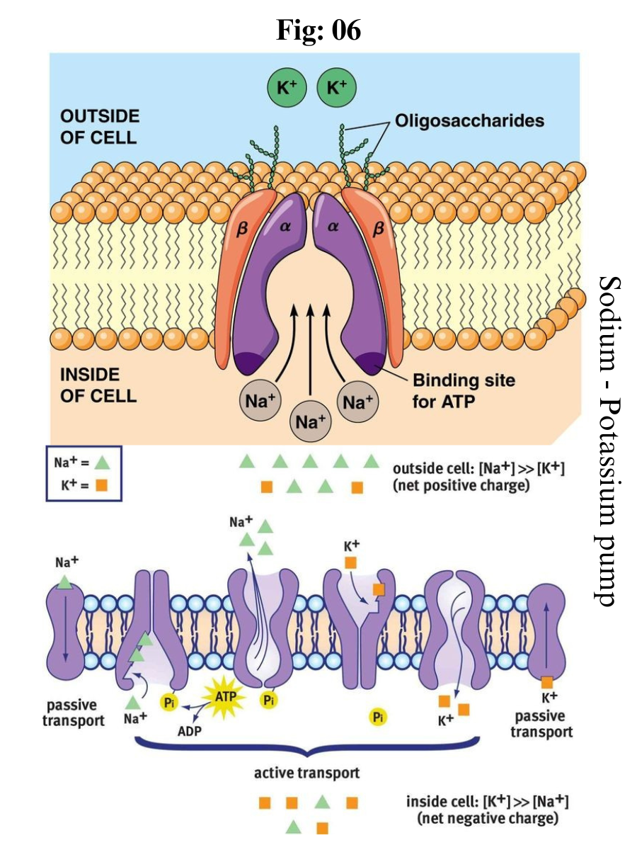
Active transport is the energy-requiring process of pumping molecules and ions across membranes "uphill" - against a concentration gradient. To move these molecules against their concentration gradient, a carrier protein is needed. Carrier proteins can work with a concentration gradient (during passive transport), but some carrier proteins can move solutes against the concentration gradient (from low concentration to high concentration), with an input of energy. 

In active transport, as carrier proteins are used to move materials against their concentration gradient, these proteins are known as pumps. As in other types of cellular activities, ATP supplies the energy for most active transport. One way ATP powers active transport is by transferring a phosphate group directly to a carrier protein. 

This may cause the carrier protein to change its shape, which moves the molecule or ion to the other side of the membrane. An example of this type of active transport system, as shown in Figure below, is the sodium-potassium pump, which exchanges sodium ions for potassium ions across the plasma membrane of animal cells.

The sodium-potassium pump system moves sodium and potassium ions against large concentration gradients. It moves two potassium ions into the cell where potassium levels are high, and pumps three sodium ions out of the cell and into the extracellular fluid.

The three sodium ions bind with the protein pump inside the cell. The carrier protein then gets energy from ATP and changes shape. In doing so, it pumps the three sodium ions out of the cell. 

At that point, two potassium ions from outside the cell bind to the protein pump. The potassium ions are then transported into the cell, and the process repeats. 

The sodium-potassium pump is found in the plasma membrane of almost every human cell and is common to all cellular life. It helps maintain cell potential and regulates cellular volume.


Death of Neuron

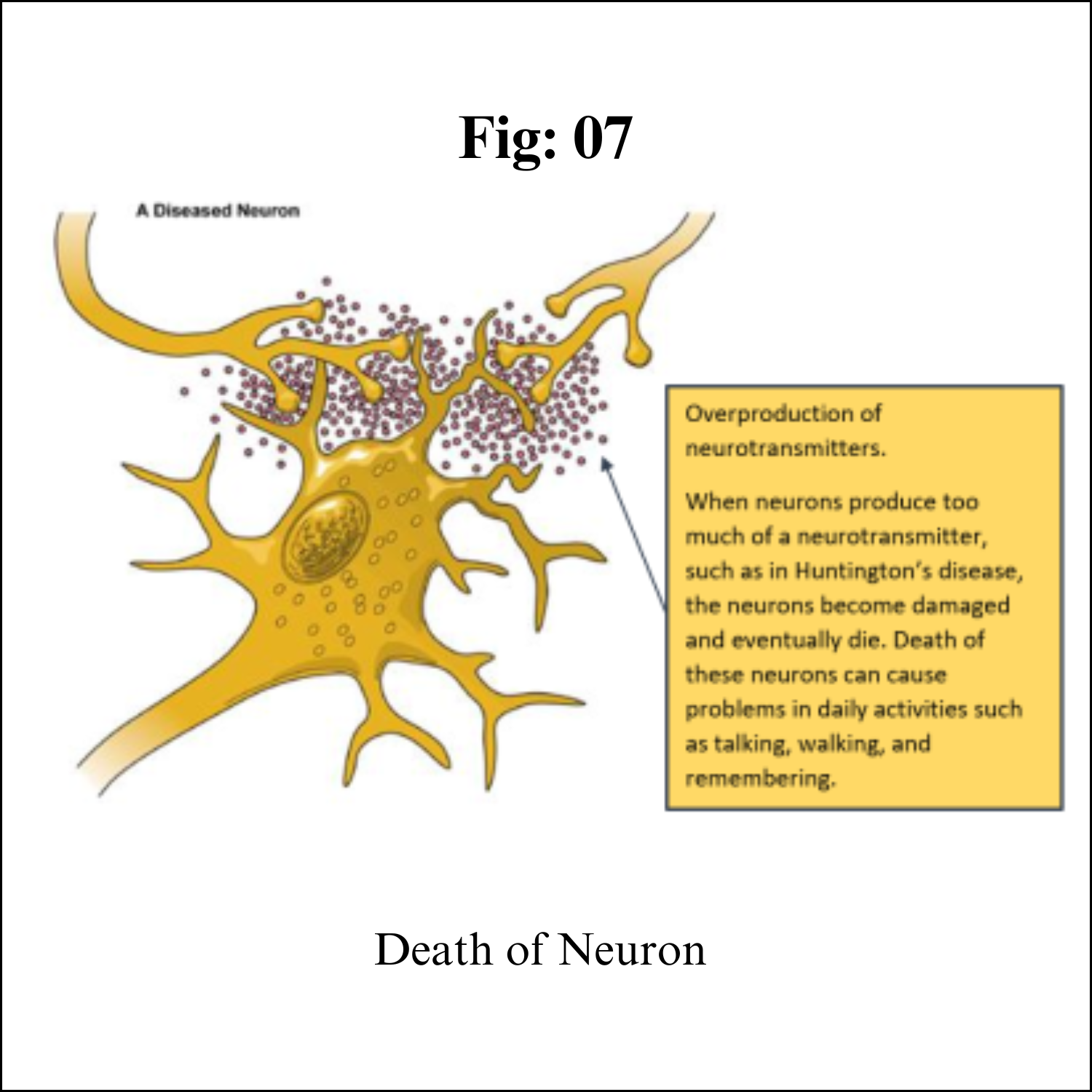
Although neurons are the longest living cells in the body, large numbers of them die during migration and differentiation. The lives of some neurons can take strange turns. Some diseases of the brain are the result of the unnatural deaths of neurons.

In Parkinson’s disease, neurons that produce the neurotransmitter dopamine die off in the basal ganglia, an area of the brain that controls body movements. This causes people with this disease to experience shaking, to move more slowly, and to have problems with balance.

In Huntington’s disease, a genetic mutation causes neurons to create too much of a neurotransmitter called glutamate, which kills neurons in the basal ganglia. As a result, people twist and move uncontrollably, and over time, they lose the ability to do many everyday tasks like walking and eating. People with this disease typically have shorter lives than those without this disease.

In Alzheimer’s disease, unusual proteins build up in and around neurons in the neocortex and hippocampus, the parts of the brain that control memory. When these neurons die, people lose their abilities to remember and do everyday tasks.

Physical damage to the brain and the spinal cord can also kill or disable neurons. Damage to the brain caused by shaking or hitting the head, or because of a stroke, can kill neurons immediately or slowly, starving them of the oxygen and nutrients they need to survive.

A spinal cord injury can cut off communication between the brain and the muscles. When neurons lose their connection to the axons (the parts of neurons that send messages to other neurons) located below the site of injury, the neurons may still live, but they lose their ability to communicate.


Functions 

Your nervous system controls such functions as your:

Heartbeat and blood pressure.
Breathing.
Muscle movements.
Thoughts, memory, learning and feelings.
Sleep, healing and aging.
Stress response.
Hormone regulation.
Digestion, sense of hunger and thirst.
Senses (response to what you see, hear, feel, touch and taste).

###

Friday, July 21, 2023

Dental Formula


Dental formula

Introduction 

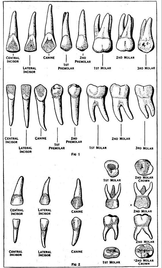
Dental formula is used to understand the number and arrangements of teeth in humans and animals. Dental formula describes the relationship of teeth within the jaws of animal teeth and human teeth. It provides information about the number and size of teeth as well as the type of teeth. Teeth are the most important element in our mouths. They help us to eat, churn and chew the food. There are two sets of teeth in mammals – deciduous and permanent. Deciduous teeth are also known as milk teeth or baby teeth. Incisors, Canines , Premolars and Molar are four types of teeth present in our mouth. 

Dentition

Dentition is used to describe the development of teeth and their arrangement in the mouth. The development of teeth depends on the characteristics of teeth as their kind, arrangement, and number of teeth in a species. The dentition of animals whose teeth succession is known as diphyodont, whereas in animals with only one set of teeth throughout life is known as monophyodont.

Each tooth in our mouth consists of a crown, a root and a cervical part. Enamel covers the crown. Apical foramen and the root canal help the root of the tooth, blood vessels and nerves to access the pulp cavity. Parodontium refers to the collection of cementum , desmodium, alveolar bone and gingival together.

Different Types of Teeth

There are four types of teeth namely Incisors, Canines, Premolars and Molars. These four types of teeth are present on the jawbones in the mouth, extending into the buccal cavity. The four types of teeth have different functions like cutting, crushing, shredding and tearing the food. 

Incisors (I) 

These are in the front row of the mouth. They help in cutting and slicing the food into the pieces to fit in the mouth. They are present in both of the jaws in mammals. 

Canines (C) 

These are most commonly known as dog teeth. In the case of Carnivores, these teeth help in holding and tearing apart food as they have pointed structures and in the case of herbivores, these teeth help in the breakdown and split hard surfaces of certain fruits such as nuts. In human beings, these provide a benefit of aiding in articulation.

Premolars (P) 

These are the transition teeth between canines and molars. They provide smooth surfaces that help in occlusion (the process of cutting, crushing, grinding the food into the rapid digestible form).

Molars (M)

These are large teeth present in mammals. They help in grinding the food. In mammals, there are 12 molars present in a set of three in both the lower and upper jaw.


Dental Formula

The dental formula describes and expresses the total number of teeth in man and animal as per the arrangements. The dental formula is expressed with letters and figures. The letters used in this formula are on the basis of the types of teeth Incisors, Canines, Premolars and Molars.

Dental Formula

(number of each type of teeth upper jaw ) / (number of teeth on one side of the lower jaw)

Dental formula = (2 Incisors 1 Canines 2 Premolars 3 Molars) /(2 Incisors 1 Canines 2 Premolars 3 Molars)

Human teeth have two dental formulas: 

The dental formula represents the arrangement of teeth in each half of the upper and the lower jaw. The entire formula is multiplied by two to represent the total number of teeth.

The dental formula for milk teeth in humans is:

The primary dentition (20 teeth)

I2/2 C1/1 M2/2 = 10 × 2 = 20 teeth

Each half of the upper jaw and the lower jaw has 2 incisors, 1 canine, and 2 molars. Premolars are absent in milk teeth hence the zero.

The permanent dentition (32 teeth):

I2/2C1/1 P2/2 M 3/3 = 16 × 2 = 32 teeth 

Each half of the upper jaw and the lower jaw has 2 incisors, 1 canine, 2 premolars, and 3 molars. An adult human has 32 permanent teeth.

Here, I- Incisors , C- Canines , P- Premolars , M- Molar.


Key Difference – Herbivores vs Carnivores Teeth
 
The key difference between herbivores and carnivores teeth is that herbivores teeth are used for cutting, gnawing, and biting while carnivores teeth are sharper and more suited to catching, killing, and tearing the prey. Based on the food habits there are three types of animals; carnivores, herbivores and omnivores. Animals that rely entirely on the flesh of other animals are called carnivores and animals that feed entirely on vegetation/ plant matters are called herbivores. Omnivores are the animals that feed on both meat and vegetation. Because of the various dietary patterns and the nutrient amount in food, the structure, the number and the location of teeth among these three groups vary widely. In this article, the difference between herbivores and carnivores teeth will be highlighted.

Herbivores Teeth

The incisors of herbivores are sharp and are used mainly to cut, gnaw and bite. Gnawing herbivores have long chisel-like incisors located in front of the skull and used for gnawing and scraping. They do not have canines. A horny pad in the upper jaw completely replaces the canines and incisors in ruminants. Moreover, their incisors and canines are similar and act as blades to cut and gather grass. Molars and premolars of herbivores have flat grinding surfaces, and they grow continuously throughout their lifetime.

Carnivores Teeth

Carnivore teeth are extremely adapted to the dietary habits of carnivores. Their upper premolar 4 and lower molar 1 are carnassial teeth and used to cut the meat away from the bone. The long, pointed canines are used to catch, kill their prey, and tear the flesh of prey. Their premolars and molars are flattened with uneven edges and are used to shear the flesh of prey into smaller pieces. Their incisors are pointed teeth and are used to catch prey.


Thursday, July 20, 2023

Human Digestive System

Human Digestive system 

Introduction:

The digestive system is made up of the gastrointestinal tract—also called the GI tract or digestive tract—and the liver, pancreas, and gallbladder. The GI tract is a series of hollow organs joined in a long, twisting tube from the mouth to the anus. The hollow organs that make up the GI tract are the mouth, esophagus, stomach, small intestine, large intestine, and anus. The liver, pancreas, and gallbladder are the solid organs of the digestive system.

The small intestine has three parts. The first part is called the duodenum. The jejunum is in the middle and the ileum is at the end. The large intestine includes the appendix, cecum, colon, and rectum. The appendix is a finger-shaped pouch attached to the cecum. The cecum is the first part of the large intestine. The colon is next. The rectum is the end of the large intestine.

Bacteria in your GI tract, also called gut flora or microbiome, help with digestion. Parts of your nervous and circulatory systems also help. Working together, nerves, hormones, bacteria, blood, and the organs of your digestive system digest the foods and liquids you eat or drink each day.

Importance of Digestive system:

Digestion is important because your body needs nutrients from food and drink to work properly and stay healthy. Proteins, fats, carbohydrates, vitamins, minerals, and water are nutrients. Your digestive system breaks nutrients into parts small enough for your body to absorb and use for energy, growth, and cell repair.

Proteins break into amino acids
Fats break into fatty acids and glycerol
Carbohydrates break into simple sugars

Digestive system work:

Each part of your digestive system helps to move food and liquid through your GI tract, break food and liquid into smaller parts, or both. Once foods are broken into small enough parts, your body can absorb and move the nutrients to where they are needed. Your large intestine absorbs water, and the waste products of digestion become stool. Nerves and hormones help control the digestive process.

Food pathway in GI tract:

Food moves through your GI tract by a process called peristalsis. The large, hollow organs of your GI tract contain a layer of muscle that enables their walls to move. The movement pushes food and liquid through your GI tract and mixes the contents within each organ. The muscle behind the food contracts and squeezes the food forward, while the muscle in front of the food relaxes to allow the food to move.


Mouth. Food starts to move through your GI tract when you eat. When you swallow, your tongue pushes the food into your throat. A small flap of tissue, called the epiglottis, folds over your windpipe to prevent choking and the food passes into your esophagus.

Esophagus. Once you begin swallowing, the process becomes automatic. Your brain signals the muscles of the esophagus and peristalsis begins.

Lower esophageal sphincter. When food reaches the end of your esophagus, a ringlike muscle—called the lower esophageal sphincter —relaxes and lets food pass into your stomach. This sphincter usually stays closed to keep what’s in your stomach from flowing back into your esophagus.

Stomach. After food enters your stomach, the stomach muscles mix the food and liquid with digestive juices. The stomach slowly empties its contents, called chyme, into your small intestine.

Small intestine. The muscles of the small intestine mix food with digestive juices from the pancreas, liver, and intestine, and push the mixture forward for further digestion. The walls of the small intestine absorb water and the digested nutrients into your bloodstream. As peristalsis continues, the waste products of the digestive process move into the large intestine.

Large intestine. Waste products from the digestive process include undigested parts of food, fluid, and older cells from the lining of your GI tract. The large intestine absorbs water and changes the waste from liquid into stool. Peristalsis helps move the stool into your rectum.

Rectum. The lower end of your large intestine, the rectum, stores stool until it pushes stool out of your anus during a bowel movement. 

How Digestive system Break food

As food moves through your GI tract, your digestive organs break the food into smaller parts using:

motion, such as chewing, squeezing, and mixing
digestive juices, such as stomach acid, bile, and enzymes

Mouth. The digestive process starts in your mouth when you chew. Your salivary glands make saliva, a digestive juice, which moistens food so it moves more easily through your esophagus into your stomach. Saliva also has an enzyme that begins to break down starches in your food.

Esophagus. After you swallow, peristalsis pushes the food down your esophagus into your stomach.

Stomach. Glands in your stomach lining make stomach acid and enzymes that break down food. Muscles of your stomach mix the food with these digestive juices.

Pancreas. Your pancreas makes a digestive juice that has enzymes that break down carbohydrates, fats, and proteins. The pancreas delivers the digestive juice to the small intestine through small tubes called ducts.

Liver. Your liver makes a digestive juice called bile that helps digest fats and some vitamins. Bile ducts carry bile from your liver to your gallbladder for storage, or to the small intestine for use.

Gallbladder. Your gallbladder stores bile between meals. When you eat, your gallbladder squeezes bile through the bile ducts into your small intestine.

Small intestine. Your small intestine makes digestive juice, which mixes with bile and pancreatic juice to complete the breakdown of proteins, carbohydrates, and fats. Bacteria in your small intestine make some of the enzymes you need to digest carbohydrates. Your small intestine moves water from your bloodstream into your GI tract to help break down food. Your small intestine also absorbs water with other nutrients.

Large intestine. In your large intestine, more water moves from your GI tract into your bloodstream. Bacteria in your large intestine help break down remaining nutrients and make vitamin K. Waste products of digestion, including parts of food that are still too large, become stool.

Product of Digested food

The small intestine absorbs most of the nutrients in your food, and your circulatory system passes them on to other parts of your body to store or use. Special cells help absorbed nutrients cross the intestinal lining into your bloodstream. Your blood carries simple sugars, amino acids, glycerol, and some vitamins and salts to the liver. Your liver stores, processes, and delivers nutrients to the rest of your body when needed.

The lymph system, a network of vessels that carry white blood cells and a fluid called lymph throughout your body to fight infection, absorbs fatty acids and vitamins.

Your body uses sugars, amino acids, fatty acids, and glycerol to build substances you need for energy, growth, and cell repair.

Digestion control

Your hormones and nerves work together to help control the digestive process. Signals flow within your GI tract and back and forth from your GI tract to your brain.

Hormones

Cells lining your stomach and small intestine make and release hormones that control how your digestive system works. These hormones tell your body when to make digestive juices and send signals to your brain that you are hungry or full. Your pancreas also makes hormones that are important to digestion.

Nerves

You have nerves that connect your central nervous system—your brain and spinal cord—to your digestive system and control some digestive functions. For example, when you see or smell food, your brain sends a signal that causes your salivary glands to "make your mouth water" to prepare you to eat.

You also have an enteric nervous system (ENS)—nerves within the walls of your GI tract. When food stretches the walls of your GI tract, the nerves of your ENS release many different substances that speed up or delay the movement of food and the production of digestive juices. The nerves send signals to control the actions of your gut muscles to contract and relax to push food through your intestines.

#####

Female orgasms

Female orgasms A female orgasm can be a highly pleasurable experience during masturbation or sexual activity. While orgasms may ...