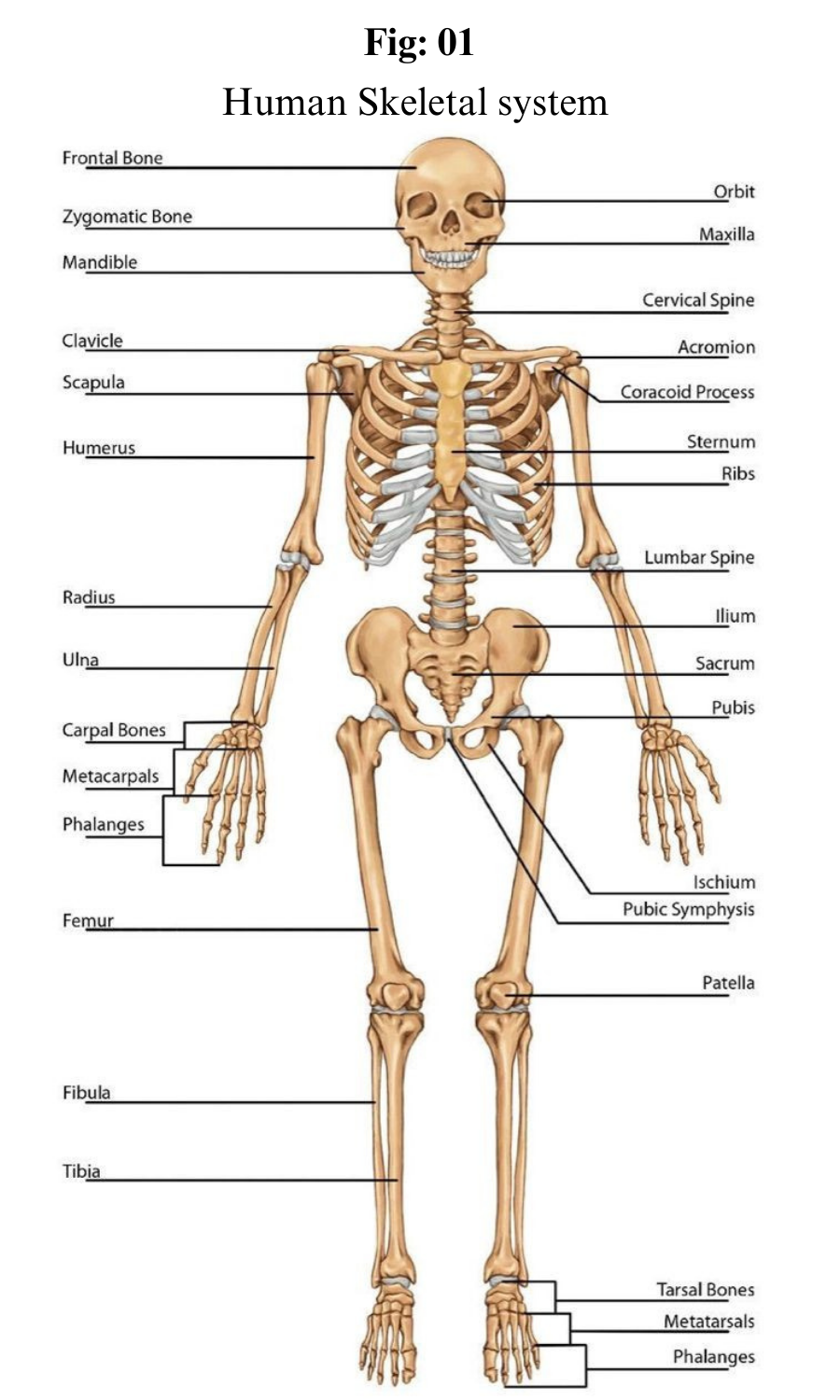
The skeletal system is the organ system that provides an internal framework for the human body.
Why do you need a skeletal system? Try to imagine what you would look like without it. You would be a soft, wobbly pile of skin containing muscles and internal organs but no bones. You might look something like a very large slug. Not that you would be able to see yourself — folds of skin would droop down over your eyes and block your vision because of your lack of skull bones. You could push the skin out of the way if you could only move your arms, but you need bones for that as well.
Components of Skeletal system:
The skeletal system includes 206 bones. Bones are organs made of dense connective tissues, mainly the tough protein collagen. Bones contain blood vessels, nerves, and other tissues.
Bones are hard and rigid due to deposits of calcium and other mineral salts within their living tissues. Locations, where two or more bones meet, are called joints.
Many joints allow bones to move like levers. For example, your elbow is a joint that allows you to bend and straighten your arm.
Our bones are made up of a combination of hard minerals, such as calcium that make up about 70 per cent of the body.
It's hard, smooth, and solid. Inside the cortical bone is a previous, spongy bone material called the trabecular or cancellous bone.
This way our bones will not break so fluently. At the center of bones is a softer substance called gist.
Bone Attribute:
Bones are calcified connective tissue present in our body. It forms the maximum portion of the skeleton. Bones are dense, semirigid and porus. It consists of an organic matrix and various mineral components inside them. Bones are tough structures.
Parts of the Skeletal System:
Aside from bones, it also consists of cartilage, tendons, ligaments, and joints. Let us discuss these components -
Bones: They are the most basic part of the skeletal system. Bones are made up of a special kind of tissue known as connective tissue which is made stronger with the help of a mineral called calcium. Inside the hard covering of the bone there is a soft sponge part. This part of the bone is lighter and allows for blood vessels to pass through. At the center of the bone lies the Bone Marrow.
Bone marrow is a soft spongy substance mainly responsible for the production of blood cells.
Ligaments: These are tough elastic bands of tissues that are responsible for attaching one bone to another and keeping them stable
Cartilage: Cartilage refers to a smooth protective covering found over joints. It and synovial fluid allows movement in the joints while protecting the bones from rubbing against one another and wearing down. It is also found between the ribs.
Tendons: Tendons are rope-like structures that join bone to muscle. They also facilitate movement while holding the skeletal system together. They also prevent injury to the muscles by absorbing impact when performing strenuous activities such as jumping
Joints
Joints refer to the areas within the body where two bones meet or connect. They are essential for movement. Some joints allow for a wide range of motion while others allow more restricted or no movement at all.
Based on the kind of movement they allow, there are three types of joints-
Fibrous Joints: These allow no movement and are also called immovable joints. For example the ones present in the skull.
Cartilaginous Joints- These allow for some movement, and thus are called Partially moveable joints. For example the spine
Synovial Joints- these joints have a wide range of motion and can be subdivided into subcategories based on the kind of movement they allow. These are also called movable joints.
For example the shoulder (Ball and socket joints), the knee (Hinge joint), the neck (pivot joint), etc. They are also filled with a liquid known as the synovial fluid which lubricates the joints and makes movement easier and smoother.
Axial and Appendicular Skeletons:
The skeleton is traditionally divided into two major parts: the axial skeleton and the appendicular skeleton.
The axial skeleton forms the axis of the body. It includes the skull, vertebral column (spine), and rib cage. The bones of the axial skeleton, along with ligaments and muscles, allow the human body to maintain its upright posture. The axial skeleton also transmits weight from the head, trunk, and upper extremities down the back to the lower extremities. In addition, the bones protect the brain and organs in the chest.
The appendicular skeleton forms the appendages and their attachments to the axial skeleton. It includes the bones of the arms and legs, hands and feet, and shoulder and pelvic girdles. The bones of the appendicular skeleton make possible locomotion and other movements of the appendages. They also protect the major organs of digestion, excretion, and reproduction.
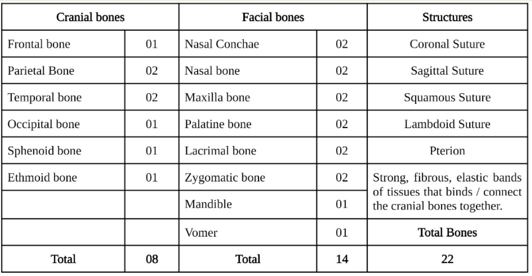
The skull (also known as cranium) consists of 22 bones which can be subdivided into 8 cranial bones and 14 facial bones.
The main function of the bones of the skull along with the surrounded meninges, is to provide protection and structure.
Protection to the brain (cerebellum, cerebrum, brainstem) and orbits of the eyes. Structurally it provides an anchor for tendinous and muscular attachments of the muscles of the scalp and face. The skull also protects various nerves and vessels that feed and innervate the brain, facial muscles, and skin.
Ribs
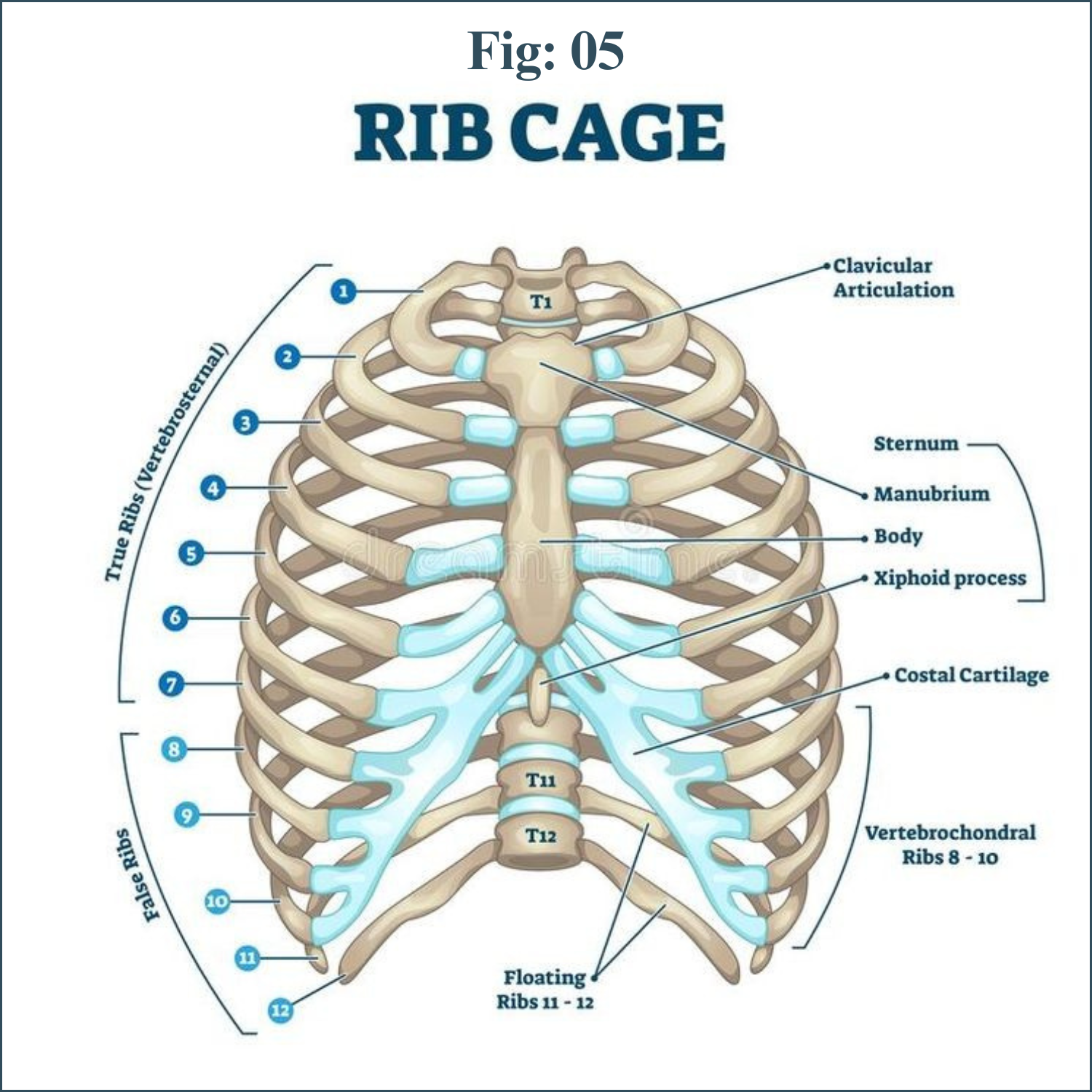
There are two classifications of ribs – atypical and typical. The typical ribs have a generalized structure, while the atypical ribs have variations on this structure.
Typical Ribs
The typical rib consists of a head, neck and body:
The head is wedge shaped, and has two articular facets separated by a wedge of bone. One facet articulates with the numerically corresponding vertebra, and the other articulates with the vertebra above.
The neck contains no bony prominences, but simply connects the head with the body. Where the neck meets the body there is a roughed tubercle, with a facet for articulation with the transverse process of the corresponding vertebra.
The body, or shaft of the rib is flat and curved. The internal surface of the shaft has a groove for the neurovascular supply of the thorax, protecting the vessels and nerves from damage.
Atypical Ribs
Ribs 1, 2, 10 11 and 12 can be described as ‘atypical’ – they have features that are not common to all the ribs.
Rib 1 is shorter and wider than the other ribs. It only has one facet on its head for articulation with its corresponding vertebra (there isn’t a thoracic vertebra above it). The superior surface is marked by two grooves, which make way for the subclavian vessels.
Rib 2 is thinner and longer than rib 1, and has two articular facets on the head as normal. It has a roughened area on its upper surface, from which the serratus anterior muscle originates.
Rib 10 only has one facet – for articulation with its numerically corresponding vertebra.
Ribs 11 and 12 have no neck, and only contain one facet, which is for articulation with their corresponding vertebra.
Articulations
The majority of the ribs have an anterior and posterior articulation.
Posterior
All the twelve ribs articulate posteriorly with the vertebra of the spine. Each rib forms two joints:
Costotransverse joint – Between the tubercle of the rib, and the transverse costal facet of the corresponding vertebra.
Costovertebral joint – Between the head of the rib, superior costal facet of the corresponding vertebra, and the inferior costal facet of the vertebra above.
Anterior
The anterior attachment of the ribs vary:
Ribs 1-7 attach independently to the sternum.
Ribs 8 – 10 attach to the costal cartilages superior to them.
Ribs 11 and 12 do not have an anterior attachment and end in the abdominal musculature. Because of this, they are sometimes called ‘floating ribs’.
Vertebral Column
The vertebral column is a series of approximately 33 bones called vertebrae, which are separated by intervertebral discs.
The column can be divided into five different regions, with each region characterised by a different vertebral structure.
In this article, we shall look at the anatomy of the vertebral column – its function, structure, and clinical significance.
Structure of a Vertebrae
All vertebrae share a basic common structure. They each consist of an anterior vertebral body, and a posterior vertebral arch.
Vertebral Body
The vertebral body forms the anterior part of each vertebrae.
It is the weight-bearing component, and vertebrae in the lower portion of the column have larger bodies than those in the upper portion (to better support the increased weight).
The superior and inferior aspects of the vertebral body are lined with hyaline cartilage. Adjacent vertebral bodies are separated by a fibrocartilaginous intervertebral disc.
Vertebral Arch
The vertebral arch forms the lateral and posterior aspect of each vertebrae.
In combination with the vertebral body, the vertebral arch forms an enclosed hole – the vertebral foramen. The foramina of all the vertebrae line up to form the vertebral canal, which encloses the spinal cord.
The vertebral arches have several bony prominences, which act as attachment sites for muscles and ligaments:
Spinous processes – each vertebra has a single spinous process, centered posteriorly at the point of the arch.
Transverse processes – each vertebra has two transverse processes, which extend laterally and posteriorly from the vertebral body. In the thoracic vertebrae, the transverse processes articulate with the ribs.
Pedicles – connect the vertebral body to the transverse processes.
Lamina – connect the transverse and spinous processes.
Articular processes – form joints between one vertebra and its superior and inferior counterparts. The articular processes are located at the intersection of the laminae and pedicles.
Classifications of Vertebrae
Cervical Vertebrae
There are seven cervical vertebrae in the human body. They have three main distinguishing features:
Bifid spinous process – the spinous process bifurcates at its distal end.
Exceptions to this are C1 (no spinous process) and C7 (spinous process is longer than that of C2-C6 and may not bifurcate).
Transverse foramina – an opening in each transverse process, through which the vertebral arteries travel to the brain.
Triangular vertebral foramen
Two cervical vertebrae that are unique. C1 and C2 (called the atlas and axis respectively), are specialized to allow for the movement of the head.
Thoracic Vertebrae
The twelve thoracic vertebrae are medium-sized, and increase in size from superior to inferior. Their specialized function is to articulate with ribs, producing the bony thorax.
Each thoracic vertebra has two ‘demi facets,’ superiorly and inferiorly placed on either side of its vertebral body. The demi facets articulate with the heads of two different ribs.
On the transverse processes of the thoracic vertebrae, there is a costal facet for articulation with the shaft of a single rib. For example, the head of Rib 2 articulates with the inferior demi facet of thoracic vertebra 1 (T1) and the superior demi facet of T2, while the shaft of Rib 2 articulates with the costal facets of T2.
The spinous processes of thoracic vertebrae are oriented obliquely inferiorly and posteriorly. In contrast to the cervical vertebrae, the vertebral foramen of thoracic vertebrae is circular.
Lumbar Vertebrae
There are five lumbar vertebrae in most humans, which are the largest in the vertebral column. They are structurally specialised to support the weight of the torso.
Lumbar vertebrae have very large vertebral bodies, which are kidney shaped. They lack the characteristic features of other vertebrae, with no transverse foramina, costal facets, or bifid spinous processes.
However, like the cervical vertebrae, they have a triangular-shaped vertebral foramen. Their spinous processes are shorter than those of thoracic vertebrae and do not extend inferiorly below the level of the vertebral body.
Their size and orientation permits needle access to the spinal canal and spinal cord (which would not be possible between thoracic vertebrae). Examples include epidural anesthesia administration and lumbar puncture.
Sacrum and Coccyx
The sacrum is a collection of five fused vertebrae. It is described as an inverted triangle, with the apex pointing inferiorly. On the lateral walls of the sacrum are facets for articulation with the pelvis at the sacroiliac joints.
The coccyx is a small bone which articulates with the apex of the sacrum. It is recognised by its lack of vertebral arches. Due to the lack of vertebral arches, there is no vertebral canal.
Separation of S1 from the sacrum is termed “lumbarisation”, while fusion of L5 to the sacrum is termed “sacralisation”. These conditions are congenital abnormalities.
Hip Bones
The left and right hip bones (innominate bones, pelvic bones) are two irregularly shaped bones that form part of the pelvic girdle – the bony structure that attaches the axial skeleton to the lower limbs.
The hip bones have three main articulations:
Sacroiliac joint – articulation with the sacrum.
Pubic symphysis – articulation between the left and right hip bones.
Hip joint – articulation with the head of femur.
The Ilium
The ilium is the widest and largest of the three parts of the hip bone, and is located superiorly. The body of the ilium forms the superior part of the acetabulum (acetabular roof). Immediately above the acetabulum, the ilium expands to form the wing (or ala).
The wing of the ilium has two surfaces:
Inner surface – has a concave shape, which produces the iliac fossa (site of origin of the iliacus muscle).
External surface (gluteal surface) – has a convex shape and provides attachments to the gluteal muscles.
The superior margin of the wing is thickened, forming the iliac crest. It extends from the anterior superior iliac spine (ASIS) to the posterior superior iliac spine (PSIS).
On the posterior aspect of the ilium there is an indentation known as the greater sciatic notch.
The Pubis
The pubis is the most anterior portion of the hip bone. It consists of a body, superior ramus and inferior ramus (ramus = branch).
Pubic body – located medially, it articulates with the opposite pubic body at the pubic symphysis. Its superior aspect is marked by a rounded thickening (the pubic crest), which extends laterally as the pubic tubercle.
Superior pubic ramus – extends laterally from the body to form part of the acetabulum.
Inferior pubic ramus – projects towards the ischium.
Together, the superior and inferior rami enclose part of the obturator foramen – through which the obturator nerve, artery and vein pass through to reach the lower limb.
The Ischium
The ischium forms the posteroinferior part of the hip bone. Much like the pubis, it is composed of a body, an inferior ramus and superior ramus.
The inferior ischial ramus combines with the inferior pubic ramus forming the ischiopubic ramus, which encloses part of the obturator foramen. The posterorinferior aspect of the ischium forms the ischial tuberosities and when sitting, it is these tuberosities on which our body weight falls.
Near the junction of the superior ramus and body is a posteromedial projection of bone; the ischial spine.
Two important ligaments attach to the ischium:
Sacrospinous ligament – runs from the ischial spine to the sacrum, thus creating the greater sciatic foramen through which lower limb neurovasculature (including the sciatic nerve) transcends.
Sacrotuberous ligament – runs from the sacrum to the ischial tuberosity, forming the lesser sciatic foramen.
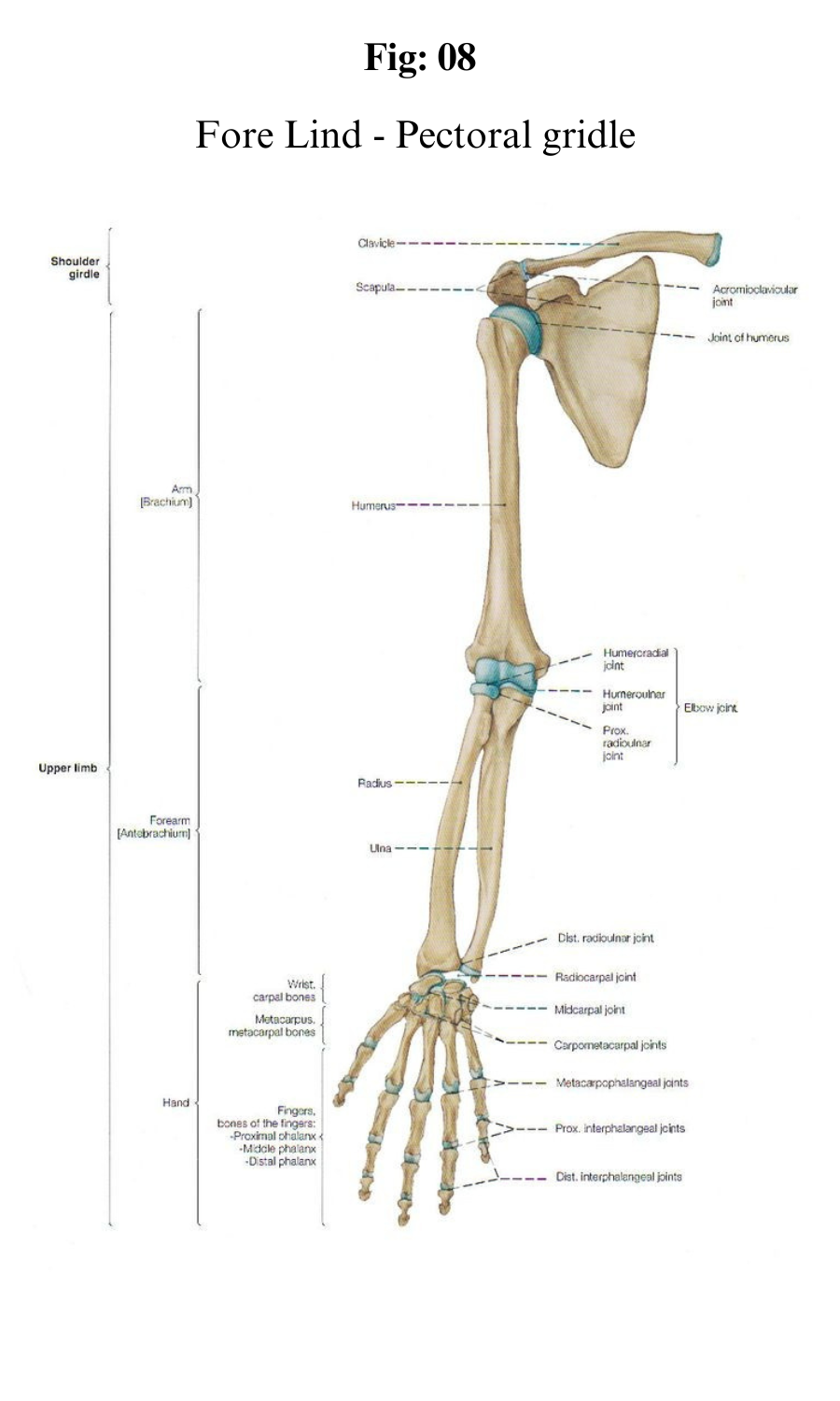
Fore Limb
Limbs are arms and legs in the human body. Forelimbs are the ones that are found in the front part of the body i.e arms. Hind limbs are those that are found in the back part of the body I.e legs.
Human arms have hands which are specialized organs allowing them to grasp and manipulate the objects. Fore limbs are shorter than the hind limbs. This is due to taller bones present in the hind limbs as compared to fore limbs.
Forelimb is located between the wrist and elbow.
The bones present in the forelimbs are — scapula, humerus, radius, ulna, carpals (8), metacarpals (5) and phalanges (14).
The humerus is a single bone present in the upper arm. Ulna and radius are two paired bones. Ulna lies medially and radius lies laterally.
Hind limb
The bones of the limb along with their respective girdles constitute the appendicular skeleton. The hind-limb contains the following bones :- Femur( the longest bone ), Tibia, Fibula, Tarsal, Metatarsal, Patella and Phalanges. Counting the number of bones, these are
Femur 1
Fibula 1
Tibia 1
Patella 1
Tarsals 7
Metatarsals 5
Phalanges 14
Thus, the Hind-limb of humans consists of a total of 30 bones.
Functions of Bones:
1. Bones support our bodies and help us to move.
2. Bones protect our internal organs like the heart, lungs, etc.
3. Some bones produce our red blood cells, white blood cells, and platelets.
4. Certain bones store fats present in our body and provide us when we need them.
5. Bones can also store important minerals for our bodies.
**********__________**********


