Saturday, September 2, 2023

Gastrulation


Gastrulation 

Introduction

Gastrulation is a critical process during week 3 of human development. Gastrulation is an early developmental process in which an embryo transforms from a one-dimensional layer of epithelial cells, a blastula, and reorganizes into a multilayered and multidimensional structure called the gastrula. In triploblastic organisms such as reptiles, avians, and mammals, gastrulation attains a three tissue-layered organism composed of endoderm, mesoderm, and ectoderm. Each germ layer corresponds to the development of specific primitive systems during organogenesis.

In addition to setting the embryo up for organ formation, gastrulation provides a mechanism to develop a multileveled body plan that demarcates anatomical axis formation. These axes are the dorsal/ventral axis, also termed the anterior/posterior or rostral/posterior axis, and the cranial/caudal or superior/inferior axis. Gastrulation also promotes the retention of global left and right symmetry and the loss of bilateral symmetry in specific organs such as the heart.

Development

After fertilization, the single-celled zygote undergoes multiple mitotic cleavages of the blastomeres to change from a two-celled to a 16-celled ball or morula. The morula begins as a solid mass of totipotent blastomeres that undergoes compaction and cavitation to transform into the blastula (non-mammalian term) or blastocyst (human development). Within the blastocyst, two tissue layers differentiate: an outer shell, known as the trophoblast, and an inner collection of cells termed the inner cell mass (ICM). Cells within the outer shell bind together via gap junctions and desmosomes to undergo compaction, ultimately forming a water-tight shell called the trophoblast.

The outer trophoblast will develop into structures that provide nutrients, help the growing embryo implant in the uterine lining, and contribute to the formation of the placenta. Additionally, the trophoblast cells are essential in the cavitation of the solid morula into a hollowed ball of cells with an internal cavity. Trophoblast cells utilize the active transport of sodium ions and osmosis of water to form a fluid-filled cavity known as a blastocoel.

The cells remaining after blastocoel formation are pluripotent ICM progenitor cells, which give rise to the distinctive formation of the fetus. Rather than being arranged as a solid sphere of cells, the ICM is pushed off to one side of the sphere formed by the trophoblast. Together the trophoblastic layer, blastocoel, and inner cell mass are the characteristic features of the human blastocyst.

From zygote to blastocyst formation, the organism is surrounded by the zona pellucida, a layer of the extracellular matrix that plays a role in the protection and prevention of implantation into the uterine tubes. During blastocyst formation, the zona pellucida begins to disappear, allowing the ball of cells to proliferate, differentiate, change shape, and eventually implant into the uterine wall. 

During implantation, the trophoblastic layer surrounding the blastocyst further differentiates into two functionally distinct layers. The outer trophoblast, known as the syncytiotrophoblast, releases proteolytic enzymes to assist with endometrial implantation. This layer also releases human chorionic gonadotropin (hCG, necessary in regulating progesterone secretion), the protein used in many pregnancy tests. The inner trophoblast layer, known as the cytotrophoblast, is a single sheet of cells surrounding the extraembryonic mesoderm. Within the cytotrophoblast is the ICM. During the second week of human development, the cells of the ICM spread into a flattened tissue layer and differentiate into a two-layered tissue containing epiblast (columnar epithelial cells) and hypoblast (cuboidal epithelial cells), which are together known as the bilaminar embryonic disc.

The formation of the bilaminar embryonic disc sets the dorsal/ventral axis as the epiblast cell layer is positioned dorsal to the hypoblast. The anatomical location of the bilaminar disc is found between the amniotic cavity and the primitive yolk sac. The cells of the epiblast stretch to form a semi-sphere known as the amniotic cavity, while the cells of the hypoblast extend to surround the yolk sac. On the hypoblast is a raised area of columnar cells known as the prechordal plate; this is the earliest delineation of cranial from caudal. Development of the bilaminar embryonic disc directly precedes gastrulation, the stage in week 3 of development that involves the transformation of the human blastocyst into a multilayered gastrula with endoderm, mesoderm, and ectoderm.


Cellular: 

The beginning of gastrulation is marked by the appearance of the primitive streak, a groove in the caudal end of the epiblast layer. Thus, the formation of the primitive streak firmly establishes the cranial/caudal axis. The primitive streak initially forms via a thickening of cells near the connecting stalk. As cells proliferate and migrate toward the midline of the embryo, the thickening elongates to become linear in shape, thus the term primitive steak.

The cranial end of the embryo seems to play an important role in initiating gastrulation. At the cranial end of the primitive streak, epiblast cells ingress at a greater rate forming a circular cavity known as the primitive pit. As the primitive streak elongates, migrating epiblast cells join the streak at the cranial end, forming a mass of cells around the primitive pit. This mass is called the primitive node, which becomes the primary tissue organizer where transcription factors and chemical signaling drive the induction of tissue formation. Known signaling factors and pathways in primitive streak formation include transforming growth factor-beta (TGFB), Wnt, Nodal, and bone morphogenetic protein (BMP), which are discussed in more detail in the molecular section. 

Epithelial cells in the lateral edge of the epiblast layer undergo an epithelial-to-mesenchymal cellular transition to delaminate (detach) and migrate down or into the primitive streak. The movement of epiblastic mesenchymal cells down the primitive streak is known as ingression. The first set of cells to move down the primitive streak integrate into the hypoblast layer and form endoderm, the first of the three germ layers. The second set of cells to detach and ingress will fill the space between the endoderm and epiblast layer to form the second germ layer, mesoderm. Multiple mesodermal structures will develop: cells that move into the body stalk will help to form extraembryonic mesoderm, and later the umbilical cord, cells passing through the primitive pit become the notochord or paraxial mesoderm, and other cells coming through the primitive streak become lateral plate or extraembryonic mesoderm. Finally, the remaining epiblast cells will transform into the final germ layer, ectoderm.

Cell proliferation and ingression continue in all directions as the embryo grows; however, the primitive streak will always expand directionally from the caudal to the cranial end and then regress in the opposite fashion. Regression occurs after the formation of the intra-embryonic mesoderm, and the primitive streak should completely disappear by the end of the fourth week — a lack of primitive streak regression results in clinical abnormalities.

After the three germ layers have formed, the newly produced structure (the trilaminar embryonic disc or gastrula) is primed for organ system formation, which relies highly on direct interaction and induction events between the endoderm, mesoderm, and ectoderm. Cells continue to invaginate through what is now called the primitive node. The cells begin to form a hollow tube extending from the cranial end to the prechordal plate, known as the notochordal process. As the embryo grows in each direction, the notochordal process grows longer until it fuses with the endoderm to form the notochordal plate. Once the fusion is complete, there is a free passageway between the amniotic cavity and the yolk sac, known as the neurenteric canal, or canal of Kovalevsky.

Cellular movement (Morphogenetic movement):

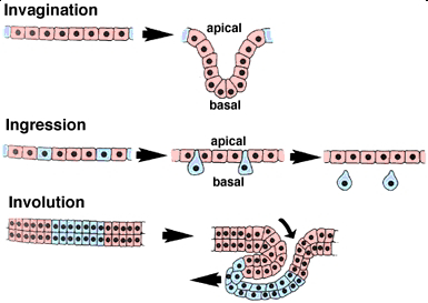
Gastrulation is complicated, Because of this, it is helpful to break the movements of gastrulation down into their component events wherever possible. In general, sheets of cells can engage in only a limited number of morphogenetic movements. This "morphogenetic repertoire" is helpful to keep in mind when we are presented with what seems to be an incomprehensible change in the shape of the embryo. Through careful observation and experimental manipulation, gastrulation can be analyzed in convenient organisms such as sea urchins. On this page, the various major morphogenetic movements that occur during gastrulation in diverse organisms are schematically represented. Some of these movements are only performed by epithelial cells, while others can be performed by both bona fide epithelial cells and by deeper, non-epithelial cells that nevertheless behave as integrated sheets of cells. The latter are poorly understood, but are common in amphibians as well as in higher vertebrates.

Invagination

During invagination, an epithelial sheet bends inward to form an inpocketing. One way to think of this in three dimensions is to imagine that you are poking a partially deflated beach ball inward with your finger. The resulting bulge or tube is an invagination. If the apical side of the epithelium forms the lumen (central empty space) of the tube, then the movement is termed invagination. If the lumen is formed by basal surfaces, then the movement is termed an evagination.

Ingression

During ingression, cells leave an epithellial sheet by transforming from well-behaved epithellial cells into freely migrating mesenchyme cells. To do so, they must presumably alter their cellular architecture, alter their program of motility, and alter their adhesive relationship(s) to the surrounding cells. Primary mesenchyme cells are an example of a mesenchymal cell type that emigrates out of an epithelium (do you know which one?).

Involution

During involution, a tissue sheet rolls inward to form an underlying layer via bulk movement of tissue. One helpful image here is of a tank tread or conveyor belt. As material moves in from the edges of the sheet, material originally at the sites of inward rolling (shown in blue here) is free to move further up underneath the exterior tissue.

Epiboly

During epiboly, a sheet of cells spreads by thinning. i.e., the sheet thins, while its overall surface area increases in the other two directions. Epiboly can involve a monolayer (i.e. a sheet of cells one cell layer thick), in which case the individual cells must undergo a change in shape. In other cases, however, a sheet that has several cell layer can thin by changes in position of its cells. In this case, epiboly occurs via intercalation, one of the other movements described on this page.

Intercalation

During intercalation, two or more rows of cells move between one another, creating an array of cells that is longer (in one or more dimensions) but thinner. The overall change in shape of the tissue results from cell rearrangement. Intercalation can be a powerful means of expanding a tissue sheet. A specialized form of intercalation is convergent extension, which is described on this page.

Convergent Extension

During convergent extension, two or more rows of cells intercalate, but the intercalation is highly directional. Cells converge by intercalating perpendicular to the axis of extension, resulting in the overall extension of the tissue in a preferred direction. If we had a way to label cells from rows on either side of the axis of extension, they would be found to mix with one another as a result of these oriented intercalation events.

Molecular level: 

Primitive Streak

The initiation of the primitive streak is based upon a system of signaling pathways working to both positively and negatively regulate downstream expression. The combination of TGFB, Wnt, Nodal, and BMPs is important in primitive streak development. The interplay between Wnt and TGFB signaling seems to be the inducer of the formation of the primitive streak. Specifically, Vg1 (a member of the TGFB family) has been shown to induce streak formation and to prevent formation with Vg1 misexpression at the posterior marginal zone. Vg1 acts on Nodal to continue the chemical cascade to streak formation. To ensure the proper location of the streak on the epiblast, the hypoblast releases antagonists of Nodal signaling.

Additionally, the induction of streak formation can be regulated by Wnt factors; not only has upregulated Wnt induced streak formation, but the use of Wnt antagonists such as Dkk-1 and Crescent prevents the formation of the streak. Finally, BMP signaling has been shown to regulate streak formation. Closer to the streak, BMP concentration is low, with the surrounding embryo exhibiting higher levels of active BMP. In addition to this, BMP inhibitors cause the formation of a streak in chick embryos. As seen in BMP and other signaling, concentration gradients are typical through most of gastrulation. Different concentrations of signaling factors allow cells to differentiate into unique tissues.

Endoderm

Endoderm is the embryonic precursor to the thyroid, lungs, pancreas, liver, and intestines, which evolve from four consecutive steps developmental steps: proliferation and induction of pluripotent stem cells, the separation of stem cell-derived endoderm versus mesoderm germ layers, anterior-posterior patterning, and bifurcation of the liver and pancreas. Cells near the anterior portion of the primitive streak will express Forkhead box A2 (Foxa2) to become definitive endoderm (DE). The DE will pattern itself into the foregut, midgut, and hindgut via mesodermal induction during embryonic folding with foregut cells expressing Hhex, Sox2, and Foxa2 and the hindgut expressing different homeobox genes Cdx1, Cdx2, and Cdx4. The upregulation of TGFB signaling promotes pancreas formation with BMP and FGF/MAPK signaling to specify the liver. The specification of the respiratory bud starts with the expression of the Nbx1-2gene. Complex signaling between the respiratory bud epithelium and mesoderm involves FGF and FGFR interactions to promote the growth of the respiratory bud. 

Mesoderm

Epiblast cells invaginating through the primitive streak that express high levels of a fibroblast growth factor (FGF2) are fated down a path towards becoming mesodermal cells. More specifically, they will end up as paraxial, intermediate, or lateral plate mesoderm, which will correlate to different tissues as the embryo develops.

Notochord

Progenitor cells from the primitive node and primitive pit migrate to initiate notochord formation. Epiblast cells from the floor plate of the amniotic cavity fill in the notochord to form a thick rod-like structure down the midline of the embryo. Providing support and serving as an induction center for surrounding cells, the notochord in vertebrates extends throughout the entire length of what will be the vertebral column and reaches as far as the midbrain. The notochord develops first, then mesodermal cells grow medially to surround it. The notochord is only present in developing organisms with the primary goal of patterning the surrounding tissues. The notochord secretes Sonic Hedgehog, Chordin, and Noggin in a morphogenic gradient pattern where the highest concentration is near the notochord with diffusion outward. These bind to receptors on target cells to induce specification and differentiation events in the neural plate, somites, and ectoderm.

Mesoderm divides into three main categories: paraxial (or axial), intermediate, and lateral (or lateral plate) mesoderm, which are the embryonic precursors to a large variety of cells and tissues, including smooth, cardiac, and skeletal muscle, kidney, reproductive organs, the muscles of the tongue and the pharyngeal arches, connective tissue, bone, cartilage, the dermis and subcutaneous layers of the skin, dura mater, vascular endothelium, blood cells, microglia, and adrenal cortex.

Cells of the paraxial mesoderm cells first organize to form somitomeres. As the somitomeres develop into somites in a cranial-to-caudal fashion, the outer cells undergo a mesenchymal to epithelial transition, which serves as a distinct boundary between individual somites. Individual somites then separate into cranial and caudal portions, followed by the cranial portion of each fusing with the caudal portion of the somite directly anterior to it. Distinct regions of each somite (sclerotome, dermatome, myotome) become specific tissue and cell types as the body matures. The skull, vertebral column, and brain meninges develop from the mesoderm surrounding the neural tube and notochord.

The intermediate mesoderm connects the paraxial mesoderm with the lateral plate and differentiates into urogenital structures.

The lateral plate mesoderm splits into a parietal (or somatic) layer to aid lateral body fold wall formation and a visceral (or splanchnic) layer involved in gut tube formation. 

Ectoderm

The interplay between BMPs and Hox genes is integral to differentiating the remaining epiblast tissue into the ectoderm. This is especially important for the surface ectoderm and what will become the neuroectoderm, setting up the brain and spinal cord.

The notochord is the main inductive tissue delineating neuroectoderm from the remaining ectoderm that will become skin. The entire presumptive ectoderm plate expresses BMP and TGFB. Noggin and Chordin secretion from the notochord diffuses into the ectoderm directly anterior to the notochord and binds to receptors in the overlying ectoderm to block BMP. The blockade of BMP specifies the tissue to neural ectoderm, while the remaining ectoderm, which still expresses BMP, will become skin.

It is theorized that the neurenteric canal forms to maintain pressure equilibrium between both chambers. Later in development, the two edges of the notochordal plate will fuse into a solid mesodermal rod known as the notochord. The notochord is a critical embryologic structure that provides structural support and marks the midline of the embryo. Chemical and physical interactions between the notochord and dorsally situated ectoderm produce neuroectoderm and, eventually, the nervous system.

Function: 

Gastrulation occurs during week 3 of human development. The process of gastrulation generates the three primary germ layers ectoderm, endoderm, and mesoderm. Gastrulation primes the system for organogenesis and is one of the most critical steps of development.

The endoderm is the innermost layer, which gives rise to the gastrointestinal tract, the lining of the gut, the liver, the pancreas, and portions of the lungs and glandular tissues. The mesoderm gives rise to the musculoskeletal system, including its connective tissue, the non-epidermal portions of the integumentary system, the circulatory system, the kidney, and the internal sex organs. The ectoderm is the outer layer of the embryo, which gives rise to the external ectoderm (epidermis, hair, nails) and the neuroectoderm (neural crest and neural tube), along with the lens of the eyes and the inner ear.

Another important function of gastrulation is to establish directionality within the developing embryo. Cranial/caudal directionality is established by the placement of the prechordal plate and the path of the primitive groove. The layering of the epiblast and hypoblast establishes the dorsal/ventral axis.

Fate Map


Fate Map 

Introduction:

A fate map is a diagram of an egg or blastula, indicating the fate of each cell or region, at a later stage of development. Fate maps are an essential tool in most embryological experiments. They provide researchers with information on which portions of the embryo will normally become which larval or adult structure. The analysis of the fate of each blasto­mere after first and second cleavage is called cytogeny or cell lineage study.

Construction of Fate Map:

Fate map of different types of animals have been constructed by the following methods:

i. Observing Living Embryos:

In some invertebrates, the embryos being transparent and having relatively few daughter cells that remain close to one another, it has been pos­sible to look through the microscope and trace the descendants of a particular cell to the organ they subsequently formed.

This type of study was performed by Edwin G. Conklin (1905) in the tunicate, Styela partita, where the different cells contain different pigments. For example, the muscle-forming cells always have a yellow colour.

ii. Vital Dye Marking:

Most embryos, however, do not have the facilities (trans­parent, few cells, different colours etc.) as described above in Styela partita. It was in 1929 that Vogt was able to trace the fate of different areas of amphibian eggs by apply­ing vital dyes. These vital dyes stain the cells without killing them.

iii. Radioactive Labelling and Fluores­cent Dyes:

A variation of the dye marking technique is to make one area of the embryo radioactive. A donor embryo is taken and grown in a solution containing radioactive thymidine. This thymidine base is subse­quently incorporated into the DNA of the dividing embryo.

A second embryo, acting as the host embryo, is grown under normal con­ditions. The region of interest is cut off from the host embryo and is replaced by a radioac­tive graft from the donor embryo. The cells that are radioactive will be the descendants of the cells of the graft, and are distinguished by autoradiography.

iv. Genetic Marking:

Radioactive and vital dye marking have their own drawbacks such as dilution over many cell divisions and the laborious preparation of slides. One per­manent way of cell marking is to create mosaic embryos having different genetic constitutions. The best example of such a marking is to graft quail cells inside a chick embryo. By doing so, fine-structure maps of the chick brain and skeletal system can be made. tool in most embryological experiments. They provide researchers with information on which portions of the embryo will normally become which larval or adult structure. The analysis of the fate of each blasto­mere after first and second cleavage is called cytogeny or cell lineage study.

Fate Map of Vertebrates:

i. Fate Map of Amphioxus:

The fate map of Amphioxus can be traced at an early stage prior to the onset of cleavage. The pre­sumptive organ forming areas in the un-cleaved egg is given in (Fig. 01). The future endodermal cells lie at the vegetal pole and would subsequently form the floor or hypoblast of the blastula. The area at the animal pole would form the presumptive ectodermal cells.


The ventral grey crescent area at the future posterior end of the blas­tula, in between the future ectoderm and endoderm, forms the future mesoderm. Another area, the dorsal crescent, in between the ectoderm and endoderm on the anterior side, gives rise to the notochord and neural cells. The presumptive ectodermal, mesoder­mal, notochordal and neural cells would sub­sequently form the epiblast of the blastula.

ii. Fate Map of Frog:

The blastula of Xenopus at the 32 cell stage gives no indica­tion as to how the different regions will develop. However, by following the fate of individual cell, or group of cells, the fate map of the blastula can be made. One way of making the fate map is by staining the vari­ous parts of the early embryo with a lipophilic dye such as dil and observe where the labelled regions end up.

Another sophis­ticated way of labelling the blastomeres is by injection of high molecular weight molecules such as rhodamine-labelled dextran, which cannot pass through cell membrane and are, therefore, restricted to the injected cell and its progeny. These cells can be easily detected later, under a UV microscope.


The fate map of the Xenopus blastula (Fig. 02) shows the presence of yolky macromeres at the vegetal pole which gives rise to the endoderm. Depending upon the position of the blastopore, the endodermal area can be divided into the sub-blastoporal and supra-blastoporal endoderm.

The cells toward the animal pole gives rise to the ecto­derm, which becomes further subdivided into epidermis and the future nervous tissue. The epidermal ectoderm forms at the ventral side of the animal hemisphere, while the neu­ral ectoderm forms at the dorsal side. The mesoderm forms a belt-like region, known as the marginal zone, around the equator of the blastula.

The mesoderm becomes subdivided along the dorsoventral axis of the blastula. The most dorsal mesoderm gives rise to the notochord. From this ventrally, the- meso­derm is differentiated by the somites (which gives rise to muscle tissue), lateral plate (which contains heart and kidney mesoderm) and blood islands.

In Xenopus, a thin outer layer of pre­sumptive endoderm overlies the presump­tive mesoderm in the marginal zone (Fig. 02).


iii. Fate Map of Chick:

Before going through the fate map of chick one should go through the formation of area pellucida and area opaca, and also through the format iod of hypoblast and epiblast. From the study of the above formations, it becomes clear that the hypoblast does not contribute any cells to the formation of the embryo proper, rather they contribute to the formation of a portion of the external membranes.

Recent studies with cell adhesion molecules (CAMs), it has become possible to construct the fate map of chick epiblast (Fig. 3) . All the three germ layers of the embryo proper is formed by the epiblastic cells. The epiblast also forms a considerable amount of extra-embryonic (mesoderm) membrane.

The fate map of chick (Fig. 3) reveals that the cells of the epiblast are organised around the notochord and nervous system. The neural ectoderm is present as a knob-like structure facing towards the anterior side. The cells at the anterior part of the epiblast form the ectoderm, while the cells at the pos­terior side gives rise to mesoderm (body proper), endoderm and extra-embryonic mesoderm.


Usefulness of Fate Map:

The fate map of organisms is helpful in tracing the morphogenetic movements of the cells and the ultimate positions they take up. However, they tell us nothing about the tissue developmental potentialities during morphogenesis.

Blastulation


Blastulation
 

Introduction:

The repeated process of cleavage results in a heap of cohering, sticky blastomeres which are loosely arranged and consist of narrow crevices in between them. This stage of development is called morula because of its superficial resemblance to a mulberry (morula is the Latin word for mulberry). In the megalecithal ova, such a ball of cells is not formed, instead, a planoconvex disc-like mass of blastomeres is formed (as in fishes, reptiles and birds) which may be called a morula.

The arrangement of blastomeres in the morula stage may vary in different groups of animals. The morula successively is followed by the next phase of development called blastula. The development of blastula is called blastulation. As the cleavage is going on, a central cavity develops among the dividing blastomeres. This cavity is called blastocoele. It begins to develop from the four-cell stage. It is filled with a gelatinous substance called blastocoele jelly. This jelly gradually absorbs water from the surrounding medium and the cavity increases in its volume. The embryo in this stage is called blastula.

Types of Blastula:


The types vary a great deal among different animals depending upon a variety of factors such as the size of the egg, the amount and distribution pattern of yolk, the rate and number of cleavage division.

1. Coeloblastula: The blastula of echinoderms and Amphioxus is called coeloblastula. It is in the form of a hollow sphere. The blastocoele is filled with mucopolysaccharides and the blastoderm is formed of a single layer of cells.


2. Stereoblastula: In many spirally cleaving eggs of annelids, mollusks, nemerteans and some of the planarians, no blastocoel cavity appears in the blastula. In them the smaller micromeres accumulate as a cluster of cells over the larger vegetally placed macromeres. At the end of cleavage, this results in a solid blastula known as a stereoblastula.

3. Periblastula or superficial blastula: The superficially cleaving eggs of insects produce a periblastula without a blastocoele cavity as nuclei collect in the peripheral layer. In a sense, a cavity is present but filled with yolk from the beginning of cleavage.

4. Discoblastula: It is found in reptiles, birds and fishes which have large yolky eggs. It is called discoblastula because it appears at the animal pole as a small multilayered flat disc separated from the yolk by a narrow segmentation or subgerminal cavity.

5. Amphiblastula: Such a blastula is formed of two types of structurally different blastomeres. Ex. in Svcon the anterior half of the blastula is formed of flagellated cells, while the posterior half is formed of large rounded granular cells. In amphibians, the blastula contains two types of cells micromeres and macromeres.

6. Blastocyst: Such blastula found in mammals. Cleavage is regular and a small cavity appears inside the dividing cells. The cavity is called blastocoele and becomes progressively larger. The cluster of cells differentiates into two distinct groups, an epithelium-like layer of trophoblast cells or nutritive cells, surrounding the expanding cavity and an inner cell mass of formative cells of embryo which are displaced to one pole of the sphere. Though mammalian egg has no yolk, they form a discoidal blastula.


The inner cell mass spreads inside the cavity as a flat disc, resembling the chick blastoderm. This stage of mammalian development is known as the blastocyst, and it is at this stage that the embryo implants into the uterine wall. Trophoblast layer surrounding the blastocyst cavity, the blastocoele, is not part of the embryo proper but eventually becomes part of the placenta or extraembryonic membranes as the embryo implants.

Blastulation in Egg:


Blastulation in Centrolecithal Egg

The centrolecithal egg has superficial cleavage. Here any cavity comparable to blastocoel does not form. However, the formation of the epithelium on the surface of the egg, after the nuclei have migrated to the exterior can be compared to the formation of the blastula. The layer of the cells thus formed on the surface of the embryo is the blastoderm. Instead of surrounding the cavity, the blastoderm envelopes the mass of uncleaved yolk. The blastoderm thus formed is known as superficial blastula or periblastula, e.g., insects.

Blastulation in a Isolecithal or Microlecithal Egg

In microlecithal egg with holoblastic cleavage, the blastomeres at the end of the cleavage are not of exactly equal size due to slight yolk inhibition of vegetal cells. These cells arrange themselves into a simple columnar epithelium enclosing the centrally located blastocoel. Since the vegetal cells are larger than the animal cells, the blastoderm is not of equal thickness throughout. At the vegetal pole, the epithelium is thicker and at the animal pole it is thinner. Thus, the polarity of the egg persists in the polarity of the blastula. This blastula is called as coeloblastula, e.g.. Amphioxus.

Blastulation in Telolecithal Egg

The amount of accumulated yolk in the vegetal hemisphere has resulted in inhibition of cleavage and displacement of all the horizontal cleavage divisions towards the animal pole. The combined result of these developmental modifications is the formation of a blastula with an eccentric blastocoel. The blastocoel is dorsally displaced. and the vegetal blastomeres, which are much larger than those of the animal hemisphere. contain within them a considerable amount of nutritive yolk.

Furthermore. the blastoderm is no longer a simple epithelium but is two or more cells thick. The cells in the interior are rather loosely connected to one another, but at the external surface of the blastula, the cells adhere to one another very firmly. because of the presence of a cementing substance joining the surface of adjoining cells. This type of blastula is known as amphiblastula and it occurs in amphibians.

Blastulation in Macrolecithal or Polylecithal Egg

This type of blastulation occurs in birds, reptiles and fishes. 

At the end of segmentation, the embryo of polylecithal egg reaches the morula stage, which consists of a disc-shaped mass of cells, several strata in thickness called the blastodisc lying applied to the yolk.

In the later stage of cleavage, the blastomeres of the central area of the blastodisc become separated from the underlying yolk and enclosed between itself and the uncleaved residue of the egg is a fluid-filled cavity, the subgerminal cavity, or the segmentation cavity.

The blastomeres of the central area are further divided by many vertical and horizontal cleavage furrows so that the blastodisc has four to five cells thick central region. The peripheral cells and the syncytial layer lying underneath the subgerminal cavity remain adhered to the yolk and is called periblast. The peripheral area of the discoblastula is opaque because the larger yolk-laden blastomeres rest directly on the underlying yolk. This outer zone is known as the area opaca. The central mass of blastomeres overlying the fluid-filled subgerminal cavity are smaller and relatively free from the yolk, appearing translucent. They constitute the area pellucida.

Soon, the larger yolk-laden blastomeres of the area pellucida segregate from the blastodisc and move inward in the subgerminal space one by one or in loose clusters and form a less organized thin flat epithelial layer called the hypoblast. The small-sized blastomeres with poor yolk left behind at the surfaces as a thin superficial layer are known as the epiblast. In a narrow horizontal cavity, the blastocoel appears between the epiblast and the hypoblast.

So far, the stages of development of the eggs are identical in fishes, reptiles, and birds. stiles and birds. However, at the time when blastoderm is being formed, a very essential difference becomes apparent. In fishes, the loosely connected blastomeres form only a single-layered epithelium above the blastocoel, but in most reptiles (except tortoises) especially in lizards and snakes the blastulation is identical to that of birds. The blastocoelic cavity corresponds to the blastocoel of amphibians and the space between the hypoblast and yolk may then be distinguished as the sub germinal cavity.

Thus, up to the blastula stage, the embryo preserves almost the same general shape as the uncleaved egg. In pigmented eggs, the pigment remains as before, more or less restricted to the upper hemisphere of the egg. During cleavage, no qualitative changes can be discovered in the chemical composition of the developing embryo. No new substances, either chemically defined or microscopically detectable have been found to appear during cleavage. However, the substances present during cleavage are essentially important for further development.

Blastulation in Mammals

With the disappearance of yolk, mammalian eggs have reverted to holoblastic cleavage. But their subsequent development bears ample evidence of the former presence of yolk.

Although the cleavage is complete, it is not as regular as in lower chordates. The blastomeres are of different sizes from the start and the numbers of blastomeres do not increase by a regular doubling sequence but tend to show arithmetic progression. Here the speed of cleavage is slower than the other animals. At about 72 to 96 hours after fertilization, the ovum reaches 16 celled stage. At this stage the 16 blastomeres are arranged in the shape of a ball called a morula. It is still surrounded by the zona pellucida, while the cells divide the morula travels towards the uterus, probably because of the peristaltic mover of the fallopian tube.

In the meantime, some fluid accumulates inside the morula to transform it into a blastocyst or blastodermic vesicle (blastula), which is a spherical structure. The blastocyst has a wall composed of blastomeres and this wall is known as trophoblast (a nourishing layer) which serves to attach the blastocyst to the uterine wall. It also has a small globular mass of blastomeres inside. These inner blastomeres stick to one side of the blastocyst wall. This inner mass of blastomeres is called the inner cell mass, which provides materials for the formation of the embryo properly. They may therefore be called the formative cells.

Sooner or later a cavity appears inside the compact mass of cells of the morula. The cavity is formed of crevices, which appear between the inner cell mass and the cells of the trophoblast. This cavity is called the blastocyst cavity and is filled with fluid. The cavity enlarges and the whole embryo becomes inflated. The trophoblast becomes lifted off the inner cell mass on most of its inner surface, but it remains attached on one side only which forms the embryonic pole and later on becomes the dorsal side of the embryo. The fully developed blastula of rabbit is disc-shaped, so is called discoblastula.

Significance of Blastulation:

Regardless of their shape, all blastulae represent an association of presumptive organ forming areas. During blastulation, these major presumptive organ forming areas of the future embryonic body are segregated into definite parts of the blastoderm. The exact pattern of arrangement of these presumptive organ forming areas varies from species to species. The blastocoel permits the migration and rearrangement of the major presumptive organ forming areas during gastrulation.


Zygote - Patterns of Cleavage


Patterns of Cleavage 

Cleavage:

After fertilization, the development of a multicellular organism proceeds by a process called cleavage, a series of mitotic divisions whereby the enormous volume of egg cytoplasm is divided into numerous smaller, nucleated cells. 

These cleavage-stage cells are called blastomeres. In most species (mammals being the chief exception), the rate of cell division and the placement of the blastomeres with respect to one another is completely under the control of the proteins and mRNAs stored in the oocyte by the mother. 

The zygotic genome, transmitted by mitosis to all the new cells, does not function in early-cleavage embryos. Few, if any, mRNAs are made until relatively late in cleavage, and the embryo can divide properly even when chemicals are used experimentally to inhibit transcription. 

During cleavage, however, cytoplasmic volume does not increase. Rather, the enormous volume of zygote cytoplasm is divided into increasingly smaller cells. 

First the egg is divided in half, then quarters, then eighths, and so forth. This division of egg cytoplasm without increasing its volume is accomplished by abolishing the growth period between cell divisions (that is, the G1 and G2 phases of the cell cycle). 

Meanwhile, the cleavage of nuclei occurs at a rapid rate never seen again (not even in tumor cells). A frog egg, for example, can divide into 37,000 cells in just 43 hours. 

Mitosis in cleavage-stage Drosophila embryos occurs every 10 minutes for over 2 hours and in just 12 hours forms some 50,000 cells. 

This dramatic increase in cell number can be appreciated by comparing cleavage with other stages of development.

Once consequence of this rapid cell division is that the ratio of cytoplasmic to nuclear volume gets increasingly smaller as cleavage progresses. In many types of embryos (such as those of Xenopus and Drosophila, but not those of C. elegans or mammals), this decrease in the cytoplasmic to nuclear volume ratio is crucial in timing the activation of certain genes. 

For example, in the frog Xenopus laevis, transcription of new messages is not activated until after 12 divisions. At that time, the rate of cleavage decreases, the blastomeres become motile, and nuclear genes begin to be transcribed. 

This stage is called the mid-blastula transition. It is thought that some factor in the egg is being titrated by the newly made chromatin, because the time of this transition can be changed by experimentally altering the ratio of chromatin to cytoplasm in the cell (Newport and Kirschner 1982a,b; Edgar et al. 1986). 

Thus, cleavage begins soon after fertilization and ends shortly after the stage when the embryo achieves a new balance between nucleus and cytoplasm.

From fertilization to cleavage:

The transition from fertilization to cleavage is caused by the activation of mitosis promoting factor (MPF). MPF was first discovered as the major factor responsible for the resumption of meiotic cell divisions in the ovulated frog egg. 

It continues to play a role after fertilization, regulating the biphasic cell cycle of early blastomeres. Blastomeres generally progress through a cell cycle consisting of just two steps: M (mitosis) and S (DNA synthesis) (Figure 8.2). Gerhart and co-workers (1984) showed that MPF undergoes cyclical changes in its level of activity in mitotic cells. 

The MPF activity of early blastomeres is highest during M and undetectable during S. Newport and Kirschner (1984) demonstrated that DNA replication (S) and mitosis (M) are driven solely by the gain and loss of MPF activity. 

Cleaving cells can be experimentally trapped in S phase by incubating them in an inhibitor of protein synthesis. When MPF is microinjected into these cells, they enter M. 

Their nuclear envelope breaks down and their chromatin condenses into chromosomes. After an hour, MPF is degraded and the chromosomes return to S phase.

Mitosis-promoting factor contains two subunits. The large subunit is called cyclin B. It is this component that shows a periodic behavior, accumulating during S and then being degraded after the cells have reached M (Evans et al. 1983; Swenson et al. 1986). 

Cyclin B is often encoded by mRNAs stored in the oocyte cytoplasm, and if the translation of this message is specifically inhibited, the cell will not enter mitosis (Minshull et al. 1989). 

The presence of cyclin B depends upon its synthesis and its degradation. Cyclin B regulates the small subunit of MPF, the cyclin-dependent kinase. 

This kinase activates mitosis by phosphorylating several target proteins, including histones, the nuclear envelope lamin proteins, and the regulatory subunit of cytoplasmic myosin. 

This brings about chromatin condensation, nuclear envelope depolymerization, and the organization of the mitotic spindle.

Without cyclin, the cyclin-dependent kinase will not function. The presence of cyclin is controlled by several proteins that ensure its periodic synthesis and degradation. 

In most species studied, the regulators of cyclin (and thus, of MPF) are stored in the egg cytoplasm. Therefore, the cell cycle is independent of the nuclear genome for numerous cell divisions. 

These early divisions tend to be rapid and synchronous. However, as the cytoplasmic components are used up, the nucleus begins to synthesize them. 

The embryo now enters the mid-blastula transition, in which several new phenomena are added to the biphasic cell divisions of the embryo. First, the growth stages (G1 and G2) are added to the cell cycle, permitting the cells to grow. 

Before this time, the egg cytoplasm was being divided into smaller and smaller cells, but the total volume of the organism remained unchanged. Xenopus embryos add those phases to the cell cycle shortly after the twelfth cleavage. Drosophila adds G2 during cycle 14 and G1 during cycle 17 (Newport and Kirschner 1982a; Edgar et al. 1986). 

Second, the synchronicity of cell division is lost, as different cells synthesize different regulators of MPF. Third, new mRNAs are transcribed. Many of these messages encode proteins that will become necessary for gastrulation. 

If transcription is blocked, cell division will occur at normal rates and at normal times in many species, but the embryo will not be able to initiate gastrulation.

Patterns of embryonic cleavage:

In 1923, embryologist E. B. Wilson reflected on how little we knew about cleavage: “To our limited intelligence, it would seem a simple task to divide a nucleus into equal parts. The cell, manifestly, entertains a very different opinion.” Indeed, different organisms undergo cleavage in distinctly different ways. 

The pattern of embryonic cleavage particular to a species is determined by two major parameters: the amount and distribution of yolk protein within the cytoplasm, and factors in the egg cytoplasm that influence the angle of the mitotic spindle and the timing of its formation.

The amount and distribution of yolk determines where cleavage can occur and the relative size of the blastomeres. When one pole of the egg is relatively yolk-free, the cellular divisions occur there at a faster rate than at the opposite pole. 

The yolk-rich pole is referred to as the vegetal pole; the yolk concentration in the animal pole is relatively low. The zygote nucleus is frequently displaced toward the animal pole. In general, yolk inhibits cleavage. 

At one extreme are the eggs of sea urchins, mammals, and snails. These eggs have sparse, equally spaced yolk and are thus isolecithal (Greek, “equal yolk”). 

In these species, cleavage is holoblastic (Greek holos, “complete”). meaning that the cleavage furrow extends through the entire egg. 

These embryos must have some other way of obtaining food. Most will generate a voracious larval form, while mammals get their nutrition from the placenta.

At the other extreme are the eggs of insects, fishes, reptiles, and birds. Most of their cell volumes are made up of yolk. 

The yolk must be sufficient to nourish these animals. Zygotes containing large accumulations of yolk undergo meroblastic cleavage, wherein only a portion of the cytoplasm is cleaved. 

The cleavage furrow does not penetrate into the yolky portion of the cytoplasm. The eggs of insects have their yolk in the center (i.e., they are centrolecithal), and the divisions of the cytoplasm occur only in the rim of cytoplasm around the periphery of the cell (i.e., superficial cleavage). 

The eggs of birds and fishes have only one small area of the egg that is free of yolk (telolecithal eggs), and therefore, the cell divisions occur only in this small disc of cytoplasm, giving rise to the discoidal pattern of cleavage. 

These are general rules, however, and closely related species can evolve different patterns of cleavage in a different environment.

However, the yolk is just one factor influencing a species' pattern of cleavage. There are also inherited patterns of cell division that are superimposed upon the constraints of the yolk. 

This can readily be seen in isolecithal eggs, in which very little yolk is present. In the absence of a large concentration of yolk, four major cleavage types can be observed: radial holoblastic, spiral holoblastic, bilateral holoblastic, and rotational holoblastic cleavage. We will see examples of these cleavage patterns below when we take a more detailed look at the early development of four different invertebrate groups.


Different organisms undergo cleavage in different ways .The pattern of embryonic cleavage is determined by two major parameters ; 

1) by the amount and distribution of yolk within the cytoplasm and 

2) by the organization of the egg. 

1. Role of yolk : The amount and distribution of yolk determines where cleavage can occur and also the relative size of the blastomeres . Yolk is a factor that controls the rate and pattern of cleavage . Yolk is concentrated toward the vegetal pole, while its concentration is relatively low towards the.animal pole .The zygote nucleus , therefore ,is frequently displaced towards the animal pole . Thus , when one pole of the egg is relatively yolk free, cleavage occurs at that pole at a faster rate than at the opposite pole . Generally , yolk inhibits cytokinesis , but it does not suppress mitotic division of the nucleus . 

Depending upon the amount and distribution of yolk the following classificatory types of cleavage are seen : 

a) Holoblastic cleavage (Greek : holos , complete) : When the amount of yolk is less , the cleavage furrow extends through the entire egg and completely divides it. This type of cleavage is called holoblastic cleavage and is generally of two types . 

i) Equal holoblastic cleavage : In microlecithal and isolecithal (homolecithal) eggs the first few
holoblastic cleavages produce blastomeres of equal size and are thus referred to as equal holoblastic cleavage . examples: Amphioxus , marsupials and placental mammals . 

ii) Unequal holoblastic cleavage : In mesolecithal and telolecithal eggs the cleavage furrows generally divide the egg into unequal sized blastomeres. The blastomeres towards the animal pole are smaller containing less amount of yolk and are called micromeres , while those towards the vegetal pole are larger, yolk laden and are called macromeres . Examples: Lower fishes and amphibians . 

b) Meroblastic cleavage : Zygotes containing large accumulation of yolk undergo meroblastic or incomplete cleavage , where only a portion of the cytoplasm is divided . The small amount of active cytoplasm near the animal pole or on the periphery of the egg divides completely , while most of the yolky portion at the vegetal pole or central area of egg remains undivided. Depending on the distribution of yolk may be mainly of the following two types of meroblastic cleavage.

i) Discoidal : In case of macrolecithal and highly telolecithal eggs , the cleavage furrow remains restricted to the disc shaped active cytoplasm of animal pole and is called discoidal meroblastic cleavage . Examples: Elasmobranchs, bony fishes,reptiles , birds and monotreme mammals. 

ii) Superficial : The centrolecithal eggs of insect have their yolk at the centre and the cleavage furrow occurs only at the rim of cytoplasm around the periphery of the cells . This type of cleavage is called superficial meroblastic cleavage . Example: Arthropods – particularly insects.

2. Organization of the egg : There are certain inherited patterns of cell division that are superimposed upon the yolk. These factors in the egg cytoplasm influences the angle of the mitotic spindle and the timing of its formation . This can be seen in isolecithal eggs, where the amount of yolk is very less. The following four major cleavage types can be observed – radial holoblastic, spiral holoblastic , bilateral holoblastic and rotational holoblastic cleavage. 

 i) Radial holoblastic cleavage : If each of the upper tier blastomeres lie over the corresponding blastomeres of the lower tier ,then the pattern of the blastomeres is radially symmetrical . This is called radial holoblastic cleavage and is seen in case of Echinoderms and Amphioxus .

ii) Spiral holoblastic cleavage : When all the blsatomeres of the upper tier are shifted in such a way , so that they come to lie not over the corresponding blastomeres at the vegetal pole , but over the junction between any two of the vegetal blastomeres . This arrangement arises not due to secondary shifting of the blastomeres , but because of oblique position of the mitotic spindles . Thus , right from the start the cleavage furrows are arranged in a sort of spiral . This type of cleavage is called spiral holoblastic cleavage and it is seen in annelids , molluscs and flat worms . 

iii) Bilateral holoblastic cleavage : In some animals such as Tunicates cleavage initially is approximately of radial type . The second cleavage plane is slightly displaced towards the posterior side of the animal vegetal axis. It results in two large anterior blastomeres and two smaller posterior blastomeres . Thus , subsequent cleavage becomes symmetrical only at the median plane and so is described as bilateral holoblastic cleavage. 

iv) Rotational holoblastic cleavage : This type of cleavage is seen in higher mammals where the first cleavage plane divides the egg into a larger and a smaller blastomere. The larger blastomere then begins to divide at a faster rate than the smaller one and this is maintained throughout resulting in rotational holoblastic cleavage . Although there is practically very less amount of yolk , the blastomere which results right from the first cleavage are unequal in size . 

Probably some conditioning effect must be present in the eggs cytoplasm which determines the size of the blastomeres and the rate of later cleavages .

Friday, September 1, 2023

Mechanism of fertilization & Myths behind fertility


Mechanism of fertilization & Myths behind fertility 

Introduction:

Fertilization, union of a sperm nucleus, of paternal origin, with an egg nucleus, of maternal origin, to form the primary nucleus of an embryo. In all organisms the essence of fertilization is, in fact, the fusion of the hereditary material of two different sex cells, or gametes, each of which carries half the number of chromosomes typical of the species. The most primitive form of fertilization, found in microorganisms and protozoans, consists of an exchange of genetic material between two cells. 

Events occur during fertilization:


The first significant event in fertilization is the fusion of the membranes of the two gametes, resulting in the formation of a channel that allows the passage of material from one cell to the other. Fertilization in advanced plants is preceded by pollination, during which pollen is transferred to, and establishes contact with, the female gamete or macrospore. Fusion in advanced animals is usually followed by penetration of the egg by a single spermatozoon. The result of fertilization is a cell (zygote) capable of undergoing cell division to form a new individual.

The fusion of two gametes initiates several reactions in the egg. One of these causes a change in the egg membrane(s), so that the attachment of and penetration by more than one spermatozoon cannot occur. In species in which more than one spermatozoon normally enters an egg (polyspermy), only one spermatozoal nucleus actually merges with the egg nucleus. 

The most important result of fertilization is egg activation, which allows the egg to undergo cell division. Activation, however, does not necessarily require the intervention of a spermatozoon; during parthenogenesis, in which fertilization does not occur, activation of an egg may be accomplished through the intervention of physical and chemical agents. Invertebrates such as aphids, bees, and rotifers normally reproduce by parthenogenesis.

In plants, certain chemicals produced by the egg may attract spermatozoa. In animals, with the possible exception of some cnidarians (coelenterates), it appears likely that contact between eggs and spermatozoa depends on random collisions. On the other hand, the gelatinous coats that surround the eggs of many animals exert a trapping action on spermatozoa, thus increasing the chances for successful sperm-egg interaction.

The eggs of marine invertebrates, especially echinoderms, are classical objects for the study of fertilization. These transparent eggs are valuable for studies observing living cells and for biochemical and molecular investigations because the time of fertilization can be accurately fixed, the development of many eggs occurs at about the same rate under suitable conditions, and large quantities of the eggs are obtainable. The eggs of some teleosts and amphibians also have been used with favorable results.

Maturation of the Egg: 

Maturation is the final step in the production of functional eggs (oogenesis) that can associate with a spermatozoon and develop a reaction that prevents the entry of more than one spermatozoon. In addition, the cytoplasm of a mature egg can support the changes that lead to fusion of spermatozoal and egg nuclei and initiate embryonic development.

Egg surface: 

Certain components of an egg’s surface, especially the cortical granules, are associated with a mature condition. Cortical granules of sea urchin eggs, aligned beneath the plasma membrane (thin, soft, pliable layer) of mature eggs, have a diameter of 0.8–1.0 micron (0.0008–0.001 millimetre) and are surrounded by a membrane similar in structure to the plasma membrane surrounding the egg. Cortical granules are formed in a cell component known as a Golgi complex, from which they migrate to the surface of the maturing egg.

The surface of a sea urchin egg has the ability to affect the passage of light unequally in different directions; this property, called birefringence, is an indication that the molecules comprising the surface layers are arranged in a definite way. Since birefringence appears as an egg matures, it is likely that the properties of a mature egg membrane are associated with specific molecular arrangements. 

A mature egg is able to support the formation of a zygote nucleus; i.e., the result of fusion of spermatozoal and egg nuclei. In most eggs the process of reduction of chromosomal number (meiosis) is not completed prior to fertilization. In such cases the fertilizing spermatozoon remains beneath the egg surface until meiosis in the egg has been completed, after which changes and movements that lead to fusion and the formation of a zygote occur. 

Egg coats: 

The surfaces of most animal eggs are surrounded by envelopes, which may be soft gelatinous coats (as in echinoderms and some amphibians) or thick membranes (as in fishes, insects, and mammals). In order to reach the egg surface, therefore, spermatozoa must penetrate these envelopes; indeed, spermatozoa contain enzymes (organic catalysts) that break them down. In some cases (e.g., fishes and insects) there is a channel, or micropyle, in the envelope, through which a spermatozoon can reach the egg.

The jelly coats of echinoderm and amphibian eggs consist of complex carbohydrates called sulfated mucopolysaccharides. The envelope of a mammalian egg is more complex. The egg is surrounded by a thick coat composed of a carbohydrate protein complex called zona pellucida. The zona is surrounded by an outer envelope, the corona radiata, which is many cell layers thick and formed by follicle cells adhering to the oocyte before it leaves the ovarian follicle.

Although it once was postulated that the jelly coat of an echinoderm egg contains a substance (fertilizin) thought to have an important role not only in the establishment of sperm-egg interaction but also in egg activation, fertilizin now has been shown identical with jelly-coat material, rather than a substance continuously secreted from it. Yet there is evidence that the egg envelopes do play a role in fertilization; i.e., contact with the egg coat elicits the acrosome reaction (described below) in spermatozoa.

Events of fertilization


Sperm-egg association

The acrosome reaction of spermatozoa is a prerequisite for the association between a spermatozoon and an egg, which occurs through fusion of their plasma membranes. After a spermatozoon comes in contact with an egg, the acrosome, which is a prominence at the anterior tip of the spermatozoa, undergoes a series of well-defined structural changes. 

A structure within the acrosome, called the acrosomal vesicle, bursts, and the plasma membrane surrounding the spermatozoon fuses at the acrosomal tip with the membrane surrounding the acrosomal vesicle to form an opening. As the opening is formed, the acrosomal granule, which is enclosed within the acrosomal vesicle, disappears. 

The dissolution of the granule releases a substance called a lysin, which breaks down the egg’s vitelline coat, allowing passage of the spermatozoon to the egg. The acrosomal membrane region opposite the opening adheres to the nuclear envelope of the spermatozoon and forms a shallow outpocketing, which rapidly elongates into a thin tube, the acrosomal tubule that extends to the egg surface and fuses with the egg plasma membrane. 

The tubule thus formed establishes continuity between the egg and the spermatozoon and provides a way for the spermatozoal nucleus to reach the interior of the egg. 

Other spermatozoal structures that may be carried within the egg include the midpiece and part of the tail; the spermatozoal plasma membrane and the acrosomal membrane, however, do not reach the interior of the egg. 

In fact, whole spermatozoa injected into unfertilized eggs cannot elicit the activation reaction or merge with the egg nucleus. As the spermatozoal nucleus is drawn within the egg, the spermatozoal plasma membrane breaks down; at the end of the process, the continuity of the egg plasma membrane is re-established. 

This description of the process of sperm-egg association, first documented for the acorn worm Saccoglossus (phylum Enteropneusta), generally applies to most eggs studied thus far.

During their passage through the female genital tract of mammals, spermatozoa undergo physiological change, called capacitation, which is a prerequisite for their participation in fertilization; they are able to undergo the acrosome reaction, traverse the egg envelopes, and reach the interior of the egg. 

Dispersal of cells in the outer egg envelope (corona radiata) is caused by the action of an enzyme (hyaluronidase) that breaks down a substance (hyaluronic acid) binding corona radiata cells together. 

The enzyme may be contained in the acrosome and released as a result of the acrosome reaction, during passage of the spermatozoon through the corona radiata. The reaction is well advanced by the time a spermatozoon contacts the thick coat surrounding the egg itself (zona pellucida).

 The pathway of a spermatozoon through the zona pellucida appears to be an oblique slit created by the oscillation of the sperm’s head near the point of its attachment to the zona pellucida.

Association of a mammalian spermatozoon with the egg surface occurs along the lateral surface of the spermatozoon, rather than at the tip as in other animals, so that the spermatozoon lies flat on the egg surface. Several points of fusion occur between the plasma membranes of the two gametes (i.e., the breakdown of membranes occurs by formation of numerous small vesicles).

Specificity of sperm-egg interaction: 

Fertilization is strictly species-specific, and the egg’s coating, the zona pellucida, plays an important role in the binding process between sperm and egg. In general, the biochemistry of the zona pellucida of one species differs from that of another, and thus it only matches up and binds with sperm of the appropriate species. 

For example, among the echinoderms, solutions of the jelly coat clump, or agglutinate, only spermatozoa of their own species. In both echinoderms and amphibians, however, slight damage to an egg surface makes fertilization possible with spermatozoa of different species (heterologous fertilization); this procedure has been used to obtain certain hybrid larvae. In addition, binding between sperm and egg of different species may occur when the zona pellucida of the egg is removed.

The eggs of ascidians, or sea squirts, members of the chordate subphylum Tunicata, are covered with a thick membrane called a chorion. 

The space between the chorion and the egg is filled with cells called test cells. The gametes of ascidians, which have both male and female reproductive organs in one animal, mature at the same time, yet self-fertilization does not occur. 

If the chorion and the test cells are removed, however, not only is fertilization with spermatozoa of different species possible, but self-fertilization also can occur.

Prevention of polyspermy: 

Most animal eggs are monospermic; i.e., only one spermatozoon is admitted into an egg. In some eggs, protection against the penetration of the egg by more than one spermatozoon (polyspermy) is due to some property of the egg surface; in others, however, the egg envelopes are responsible. 

The ability of some eggs to develop a polyspermy-preventing reaction depends on a molecular rearrangement of the egg surface that occurs during egg maturation (oogenesis). Although immature sea urchin eggs have the ability to associate with spermatozoa, they also allow multiple penetration; i.e., they are unable to develop a polyspermy-preventing reaction. 

Since the mature eggs of most animals are fertilized before completion of meiosis and are able to develop a polyspermy-preventing reaction, specific properties of the egg surface must have differentiated by the time meiosis stops, which is when the egg is ready to be fertilized.

In some mammalian eggs defense against polyspermy depends on properties of the zona pellucida; i.e., when a spermatozoon has started to move through the zona, it does not allow the penetration of additional spermatozoa (zona reaction). 

In other mammals, however, the zona reaction either does not take place or is weak, as indicated by the presence of numerous spermatozoa in the space between the zona and egg surface. In such cases the polyspermy-preventing reaction resides in the egg surface. 

Although the eggs of some kinds of animals (e.g., some amphibians, birds, reptiles, and sharks) are naturally polyspermic, only one spermatozoal nucleus fuses with an egg nucleus to form a zygote nucleus; all of the other spermatozoa degenerate.

Formation of the fertilization membrane: 

The most spectacular changes that follow fertilization occur at the egg surface. The best known example, that of the sea urchin egg, is described below. 

An immediate response to fertilization is the raising of a membrane, called a vitelline membrane, from the egg surface. In the beginning the membrane is very thin; soon, however, it thickens, develops a well-organized molecular structure, and is called the fertilization membrane. 

At the same time an extensive rearrangement of the molecular structure of the egg surface occurs. The events leading to formation of the fertilization membrane require about one minute.

At the point on the outer surface of the sea urchin egg at which a spermatozoan attaches, the thin vitelline membrane becomes detached. 

As a result the membranes of the cortical granules come into contact with the inner aspect of the egg’s plasma membrane and fuse with it, the granules open, and their contents are extruded into the perivitelline space; i.e., the space between the egg surface and the raised vitelline membrane. 

Part of the contents of the granules merge with the vitelline membrane to form the fertilization membrane; if fusion of the contents of the cortical granules with the vitelline membrane is prevented, the membrane remains thin and soft. 

Another material that also derives from the cortical granules covers the surface of the egg to form a transparent layer, called the hyaline layer, which plays an important role in holding together the cells (blastomeres) formed during division, or cleavage, of the egg. 

The plasma membrane surrounding a fertilized egg, therefore, is a mosaic structure containing patches of the original plasma membrane of the unfertilized egg and areas derived from membranes of the cortical granules. 

The events leading to the formation of the fertilization membrane are accompanied by a change of the electric charge across the plasma membrane, referred to as the fertilization potential, and a concurrent outflow of potassium ions (charged particles); both of these phenomena are similar to those that occur in a stimulated nerve fibre. 

Another effect of fertilization on the plasma membrane of the egg is a several-fold increase in its permeability to various molecules. This change may be the result of the activation of some surface-located membrane transport mechanism.

Formation of the zygote nucleus: 

After its entry into the egg cytoplasm, the spermatozoal nucleus, now called the male pronucleus, begins to swell, and its chromosomal material disperses and becomes similar in appearance to that of the female pronucleus. 

Although the membranous envelope surrounding the male pronucleus rapidly disintegrates in the egg, a new envelope promptly forms around it. The male pronucleus, which rotates 180° and moves towards the egg nucleus, initially is accompanied by two structures (centrioles) that function in cell division.

 After the male and female pronuclei have come into contact, the spermatozoal centrioles give rise to the first cleavage spindle, which precedes division of the fertilized egg. 

In some cases fusion of the two pronuclei may occur by a process of membrane fusion; in this process, two adjoining membranes fuse at the point of contact to give rise to the continuous nuclear envelope that surrounds the zygote nucleus.

Myths behind fertility: 

Myth 1: Infertility is typically the woman’s fault

According to the National Institute of Health (NIH), one-third of infertility cases are caused by male reproductive issues, one-third by female reproductive issues, and one-third by both parties or by unknown factors. Dr. Kayani put it this way: “Infertility is a people problem, not just a female problem.”

Myth 2: Couples should always try for at least a year before seeing an OBGYN

Usually, infertility is defined as one year of unprotected intercourse without conception. However, this is more of a guideline, than an actual rule. Many couples should seek medical help sooner — for example, women over age 35, those with a history of irregular periods, fibroids, endometriosis, pelvic adhesive disease, ectopic pregnancy or multiple miscarriages. Same for couples with a male history of surgery, infection or trauma to the genitals.

Myth 3: Woman can’t get pregnant after 35

This myth is incredibly common. While a woman’s peak reproductive years begin in her late teens and continue into her late 20s, “many women will go on to have healthy pregnancies later in life,” Dr. Kayani assured. For example, about one in six couples in which the woman is 35 and older have fertility problems, according to the CDC — that means plenty of women age 35 and above are doing just fine fertility-wise.

Myth 4: Using birth control can cause infertility

The pill doesn’t impact fertility, either positively or negatively. And a woman’s normal menstrual cycle will almost always continue within a month or two after she’s stopped taking the pill. But if things haven’t gone back to normal within three months of stopping birth control, make an appointment with your OBGYN.

Myth 5: If a man can ejaculate, he’s not infertile

In reality, male infertility isn’t that simple. Most men with infertility have no noticeable signs that there’s any problem. The cause can be a low sperm count — this is usually what people imagine — but sperm movement (motility) and sperm shape (morphology) also play a part. Studies have shown that men who work physically demanding jobs or who take two or more medications may have lower sperm counts, and high blood pressure can negatively impact a sperm’s shape.

Myth 6: Irregular menstrual cycle = infertility

Irregular menstrual cycles are pretty common. “Sleep disruptions, stress levels and exercise routines can alter the delicate balance of hormones that regulate the menstrual cycle,” Dr. Kayani explained. “If you have concerns about irregular cycles, it’s worth bringing up to your gynecologist sooner rather than later,” especially if you’ve gone more than three or four months without a period.

Myth 7: Infertility can’t happen in young men and women

Sure, increased age is often to blame — women over 35 and men over 50 have lower fertility rates, according to the American Society for Reproductive Medicine — but young men and women still deal with it, too. Dr. Kayani pointed out that nearly 1 in 10 women face infertility before reaching age 30.

Myth 8: Certain sex positions improve the odds of pregnancy

There’s just no evidence to support this one. Ejaculated sperm can be found at the cervix within seconds, and within the fallopian tubes within minutes, no matter the coital position.

Myth 9: Infertility is always fixable

It’s true, most couples experiencing infertility will get pregnant eventually. But most doesn’t mean all. Some folks have issues that just aren’t fixable, while others face problems that can’t even be explained. “The truth is, not everyone can get pregnant even with interventions,” Dr. Kayani said. “Those experiencing infertility should not feel as if they are to blame or aren’t trying hard enough.” Sometimes it’s just not in the cards.

Myth 10: Your overall health doesn’t impact fertility

Truthfully, general health is the biggest factor for fertility, in both men and women. Drinking too much alcohol, exercising too much, being underweight or overweight — it all matters.

Menstrual cycle & it's Myths


Menstrual cycle & it's Myths
 

Introduction:

The menstrual cycle is a natural process. It is a complex cycle controlled by female hormones that cause regular bleeding (periods). The menstrual cycle has four phases: menstruation, the follicular phase, ovulation and the luteal phase.

The menstrual cycle prepares your body for pregnancy. If you are not pregnant, your hormones send a signal to your uterus to shed its lining. This becomes your period. Once you start your period, the cycle starts again.

A menstrual cycle is measured from the first day of your period to the first day of your next period. The average length of a menstrual cycle is 28 to 29 days, but every woman’s cycle is different. For example, teenagers might have cycles that last 45 days, whereas women in their 20s to 30s might have cycles that last 21 to 38 days.

Your first period is called menarche. In Western countries, the average age for a first period is 12 to 13, but it can start as early as nine and as late as 16.

Your last period is called menopause. In Australia, the average age for women to reach menopause is 51 to 52. Some women might reach menopause as late as 60.

Phases of Menstrual cycle:

There are four main phases of the menstrual cycle.

1.Menstruation:

Menstruation is commonly known as a period. When you menstruate, your uterus lining sheds and flows out of your vagina. Your period contains blood, mucus and some cells from the lining of your uterus. The average length of a period is three to seven days.

Sanitary pads, tampons, period underwear or menstrual cups can be used to absorb your period. Pads and tampons need to be changed regularly (preferably every three to four hours) and menstrual cups should be changed every eight to 12 hours.

2.The follicular phase:

The follicular phase starts on the first day of your period and lasts for 13 to 14 days, ending in ovulation. The pituitary gland in the brain releases a hormone to stimulate the production of follicles on the surface of an ovary. Usually, only one follicle will mature into an egg. This can happen from day 10 of your cycle. During this phase, your uterus lining also thickens in preparation for pregnancy.

3. Ovulation:

Ovulation is when a mature egg is released from an ovary and moves along a fallopian tube towards your uterus. This usually happens once each month, about two weeks before your next period. Ovulation can last from 16 to 32 hours.

It is possible to get pregnant in the five days before ovulation and on the day of ovulation, but it’s more likely in the three days leading up to and including ovulation. Once the egg is released, it will survive up to 24 hours. If sperm reaches the egg during this time, you may get pregnant.

4. The luteal phase:

After ovulation, cells in the ovary (the corpus luteum), release progesterone and a small amount of oestrogen. This causes the lining of the uterus to thicken in preparation for pregnancy.

If a fertilised egg implants in the lining of the uterus, the corpus luteum continues to produce progesterone, which maintains the thickened lining of the uterus.

If pregnancy does not occur, the corpus luteum dies, progesterone levels drop, the uterus lining sheds and the period begins again.


Common menstrual problems:

Some of the more common menstrual problems include:

Premenstrual syndrome (PMS) – hormonal events before a period can trigger a range of side effects in women at risk, including fluid retention, headaches, fatigue and irritability. Treatment options include exercise and dietary changes.

Dysmenorrhoea – or painful periods. It is thought that the uterus is prompted by certain hormones to squeeze harder than necessary to dislodge its lining. Treatment options include pain-relieving medication and the oral contraceptive pill.

Heavy menstrual bleeding (previously known as menorrhagia) – if left untreated, this can cause anaemia. Treatment options include oral contraceptives and a hormonal intrauterine device (IUD) to regulate the flow.

Amenorrhoea – or absence of menstrual periods. This is considered abnormal, except during pre-puberty, pregnancy, lactation and postmenopause. Possible causes include low or high body weight and excessive exercise.

Summary:

The menstrual cycle is complex and controlled by many different glands and the hormones that these glands produce.

The four phases of the menstrual cycle are menstruation, the follicular phase, ovulation and the luteal phase.

Common menstrual problems include heavy or painful periods and premenstrual syndrome (PMS).

Knowing when in the menstrual cycle a woman is most likely to conceive can increase the chance of pregnancy.

Myths related to Menstrual cycle in India:


The following are some of the Myths followed in India related to Menstrual cycle, 

In India even mere mention of the topic has been a taboo in the past and even to this date the cultural and social influences appear to be a hurdle for advancement of knowledge on the subject. 

Culturally in many parts of India, menstruation is still considered to be dirty and impure. The origin of this myth dates back to the Vedic times and is often linked to Indra's slaying of Vritras. 

For, it has been declared in the Veda that guilt, of killing a brahmana-murder, appears every month as menstrual flow as women had taken upon themselves a part of Indra's guilt. 

Further, in the Hindu faith, women are prohibited from participating in normal life while menstruating. She must be “purified” before she is allowed to return to her family and day to day chores of her life. 

However, scientifically it is known that the actual cause of menstruation is ovulation followed by missed chance of pregnancy that results in bleeding from the endometrial vessels and is followed by preparation of the next cycle. 

Therefore, there seems no reason for this notion to persist that menstruating women are “impure.”

Many girls and women are subject to restrictions in their daily lives simply because they are menstruating. Not entering the “puja” room is the major restriction among urban girls whereas, not entering the kitchen is the main restriction among the rural girls during menstruation. 

Menstruating girls and women are also restricted from offering prayers and touching holy books.

The underlying basis for this myth is also the cultural beliefs of impurity associated with menstruation. It is further believed that menstruating women are unhygienic and unclean and hence the food they prepare or handle can get contaminated. 

According to a study by Kumar and Srivastava in 2011, participating women also reported that during menstruation the body emits some specific smell or ray, which turns preserved food bad. 

And, therefore, they are not allowed to touch sour foods like pickles. However, as long as general hygiene measures are taken into account, no scientific test has shown menstruation as the reason for spoilage of any food in making.

Cultural norms and religious taboos on menstruation are often compounded by traditional associations with evil spirits, shame and embarrassment surrounding sexual reproduction. 

In some cultures, women bury their cloths used during menstruation to prevent them being used by evil spirits. 

In Surinam, menstrual blood is believed to be dangerous, and a malevolent person can do harm to a menstruating woman or girl by using black magic (“wisi”). It is also believed that a woman can use her menstrual blood to impose her will on a man. 

Interestingly, in Asia including India, such beliefs are still practiced. However, there seems to be no logical or scientific explanation for this.

In some parts of India, some strict dietary restrictions are also followed during menstruation such as sour food like curd, tamarind, and pickles are usually avoided by menstruating girls. 

It is believed that such foods will disturb or stop the menstrual flow. As far as the exercise is concerned, many studies in India and elsewhere have revealed that many adolescent girls believe that doing exercise/physical activity during menses aggravate the dysmenorrhea while in real exercise can help relieve the menstruating women with symptoms of premenstrual syndrome and dysmenorrhea and relieve bloating. Exercise also causes a release of serotonin, making one feel much happier.

In some parts of India, perceptions of Hinduism center on notions of purity and pollution. Bodily excretions are believed to be polluting, as are the bodies when producing them. 

All women, regardless of their social caste, incur pollution through the bodily processes of menstruation and childbirth. Water is considered to be the most common medium of purification. 

The protection of water sources from such pollution, which is the physical manifestation of Hindu deities, is, therefore, a key concern. 

This highlights the possible reason why menstruating women are not allowed to take a bath especially for first few days of their menstrual period. It is believed that if a girl or women touches a cow while she is on her period, that the cow will become infertile – leading girls to associate their own bodies with curse and impurity.

Impact of Myths Related to Menstruation on Women's Life: 

Such taboos about menstruation present in many societies impact on girls’ and women's emotional state, mentality and lifestyle and most importantly, health. Large numbers of girls in many less economically developed countries drop out of school when they begin menstruating. This includes over 23% of girls in India.

In addition to this, the monthly menstruation period also creates obstacles for female teachers. Thus, the gender – unfriendly school culture and infrastructure and the lack of adequate menstrual protection alternatives and/or clean, safe and private sanitation facilities for female teachers and girls undermine the right of privacy. 

There are health and hygiene issues also to consider relating to girls and menstruation. Over 77% of menstruating girls and women in India use an old cloth, which is often reused. Further, 88% of women in India sometimes resort to using ashes, newspapers, dried leaves and husk sand to aid absorption.

Poor protection and inadequate washing facilities may increases susceptibility to infection, with the odor of menstrual blood putting girls at risk of being stigmatized. 

The latter may have significant implications for their mental health. The challenge, of addressing the socio-cultural taboos and beliefs in menstruation, is further compounded by the fact the girls’ knowledge levels and understandings of puberty, menstruation, and reproductive health are very low.

Combat Menstruation Related Myths: 

Empowerment of women through education and increasing their role in decision-making can also aid in this regard. Women and girls are often excluded from decision-making due to their lower literacy levels per se. Increasing the education status of women plays an important role in improving the health status of the community at large and overcoming the cultural taboos, in particular. Provision of sanitary napkins and adequate facilities for sanitation and washing should be made available with the gender perspective. In Delhi, there are an estimated 132 public toilets for women, only 8% the number of the 1534 for men.

Reverse Transcription

Reverse Transcription Introduction Reverse transcription is the synthesis of DNA from an RNA template. This process is driven by RNA-depende...