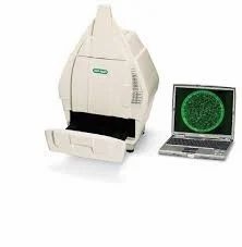
Gel Documentation System (Gel Doc)
Introduction
A Gel Documentation System, also called Gel Doc, Gel Image System or Gel Imager is used in molecular biology labs for imaging and documentation of protein suspended within polyacrylamide or agarose gels and nucleic acid.
For DNA/RNA detection the gel is stained with ethidium bromide or other strains of nucleic acids like SYBR™ Gold,SYBR™ Green,SYBR™ Safe,Gel Star™,Texas Red,Fluorescein and for Protein Detection with Coomassie Brilliant Blue, Silver Staining,Sypro™ Red,Sypro™ Orange,Pro-QDiamond,Deep Purple.
The gel documentation comes with a UV light transilluminator, accompanied by a hood that acts as a darkroom shielding the user from UV exposure. The images are captured in the CCD camera.
Advanced low light capturing cameras are also being incorporated into the system for acute precision. Cooled cameras allow longer exposure preventing sensor overheating. These produce faint bands and spots in gel images that would normally escape the naked eye.
Features like fluorescence and chemiluminescence at cooling camera temperatures of -28 to -60 °C are much preferred. Instant printing and Wi-Fi control for smartphone or tablet operations are also enabled in modern applications.
Principle
A fluorescent substance bound to nucleic acid is triggered by ultraviolet irradiation causing it to emit fluorescent light. Ethidium Bromide binds with the nucleic acid. The molecular weight and the concentration of the nucleic acid determines their bonding displaying a brighter shine if the molecular weight is large, however, if the molecular is smaller the fluorescence will have a weaker shine.
Components
UV Irradiation source
The irradiation source is the transilluminator observing the DNA bands. In the latest technology ethidium bromide acts as a fluorescent sample tag which is obtained from electrophoresis of agarose gel. If there is no action at the source the automatic shut off timer activates. The latest equipment does not require warming uptime of the UV lamps and work on instant switching. The white light LED option provides viable intensity with a safety cut off that prevents damaging the sample and operators. The fluorescent tags should correspond with the irradiation source, it offers cost cutting without compromising on performance.
Base Plate
A sample tray that holds the gel for inspection and is a black non-reactive surface.
Hood
Acts as a darkroom shielding the user. Very useful when performing fluorescence, chemiluminescence, and visible light applications. The auto door lock prevents overexposure to radiation and document’s captured images. The fold-down and side to side doors offer the least obstruction.
Filter
Shielding UV radiation, amber filters block the background light to maintain as much clarity on captured images. The UV radiation is blocked by emission filters that allow safe sample viewing. Depending on the type of application the filters can be accommodated.
The Imaging system
A high-res camera with a camera controller and printer of 104MP-8.3 MP with CCD (charge-coupled device) camera and flatbed scanner. Computer-controlled motor-driven lens, autofocus, tracking upward and downward sample movement automatically. Images are captured with a camera above the hood. The camera is equipped with photon signal conversion offering deep cooling technology with a wide lens aperture maximizing light sensitivity.
Computer-controlled
A touch screen monitor and a normal monitor comes with inbuilt software that enables image optimization, analysis, and acquisition. Image editing is also possible. It can read documenting colourimetry, gels, western blots, plants, TLC plates, colony blots, fluorescent dyes, and infrared dyes. The system can generate simultaneous imaging of blots and gels for gel-to-gel comparison. Large data can be stored in the hard disk drive. Massive data can be shared through Wi-Fi, ethernet connection immediately, it can work on a user-friendly Window version.
Applications
The system is designed for basic use related to academics and research and commercial lab research that require smaller sized equipment that command greater efficiency. Mostly preferred by pharma labs, they are provided with multiple user accounts along with access permission regulating security and controlled work environment.
The Gel Documentation System can support common protein gel strains and common DNA that vary from Coomassie to Bio-Rad Strain-Free to silver stains along with blue excitable stains.
The system detects affinities of Monoclonal and polyclonal antibody binding, blot and gel imaging, colony identification and counting, Immunoassay, compound protein detection, Post-Translational Modification Characterization, 2D Electrophoresis Protein Quantitation.
Preference
The system is available in a compact size consuming minimum lab space. Its efficiency is highly calibrated for the workflow it supports. There is no fear of sample spillage with its image capturing technique and its compactness adds to the ease of work.
The system is provided with intuitive application-driven software which can help scientists to focus on their experiments and technology take care of itself. The digital images can be exported easily through USB drives.
The technology that Geldoc supports is compatible with strain-free technology that detects protein without staining step separation. A complete imaged directory is conducted for Stain-free gels reducing reagents and time allowing greater efficiency in wet workflow.
the cameras are equipped to maintain a high resolution that impacts research quality.


