Sunday, November 26, 2023

Rheumatoid Arthritis

Rheumatoid Arthritis 

Introduction

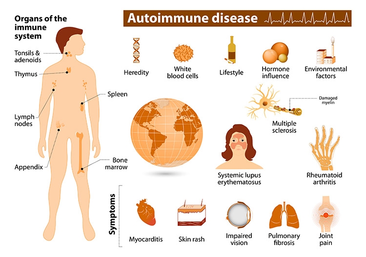
Rheumatoid arthritis, or RA, is an autoimmune and inflammatory disease, which means that your immune system attacks healthy cells in your body by mistake, causing inflammation (painful swelling) in the affected parts of the body.

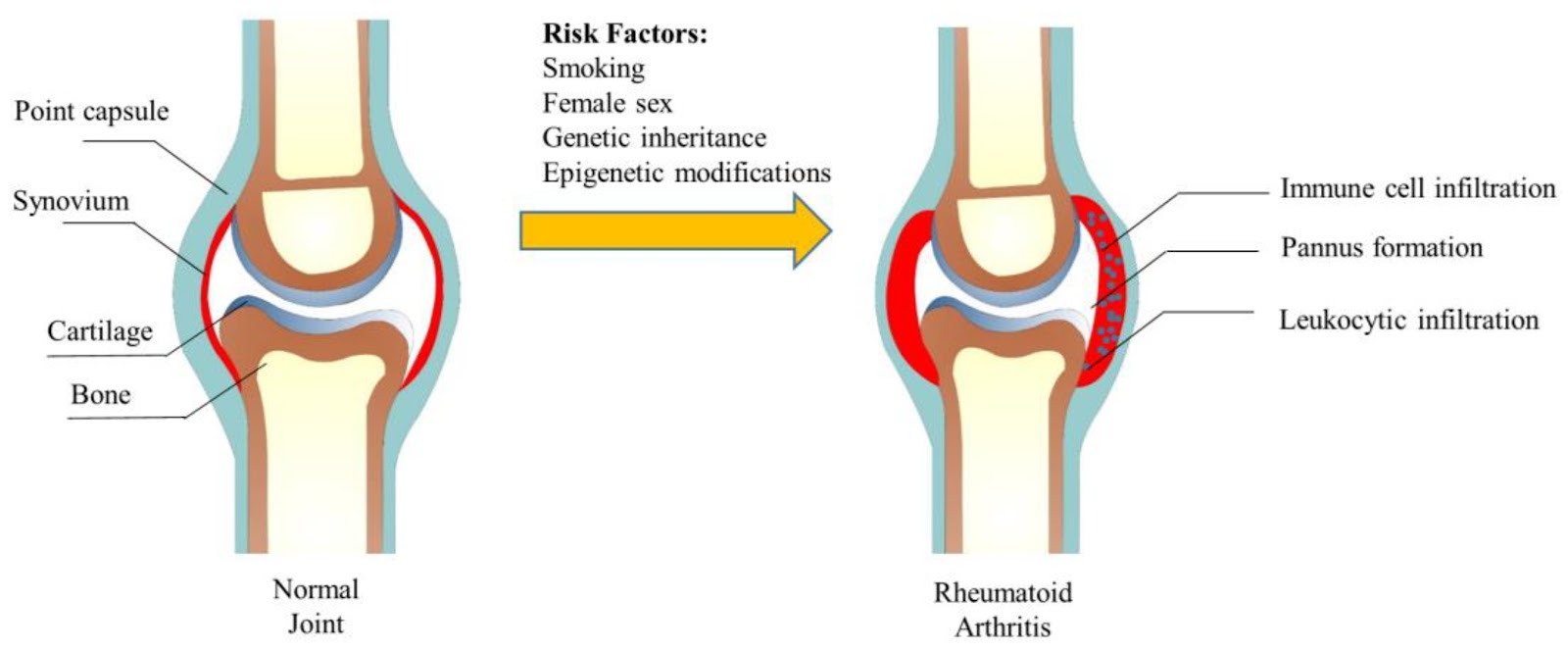
RA mainly attacks the joints, usually many joints at once. RA commonly affects joints in the hands, wrists, and knees. In a joint with RA, the lining of the joint becomes inflamed, causing damage to joint tissue. This tissue damage can cause long-lasting or chronic pain, unsteadiness (lack of balance), and deformity (misshapenness).

RA can also affect other tissues throughout the body and cause problems in organs such as the lungs, heart, and eyes.

Signs and Symptoms

With RA, there are times when symptoms get worse, known as flares, and times when symptoms get better, known as remission.

Signs and symptoms of RA include:

Pain or aching in more than one joint.
Stiffness in more than one joint.
Tenderness and swelling in more than one joint.
The same symptoms on both sides of the body (such as in both hands or both knees).
Weight loss.
Fever.
Fatigue or tiredness.
Weakness.
Causes

RA is the result of an immune response in which the body’s immune system attacks its own healthy cells. The specific causes of RA are unknown, but some factors can increase the risk of developing the disease.

Risk factors

Researchers have studied a number of genetic and environmental factors to determine if they change person’s risk of developing RA.

Characteristics that increase risk

Age. RA can begin at any age, but the likelihood increases with age. The onset of RA is highest among adults in their sixties.

Sex. New cases of RA are typically two-to-three times higher in women than men.

Genetics/inherited traits. People born with specific genes are more likely to develop RA. These genes, called HLA (human leukocyte antigen) class II genotypes, can also make your arthritis worse. The risk of RA may be highest when people with these genes are exposed to environmental factors like smoking or when a person is obese.

Smoking. Multiple studies show that cigarette smoking increases a person’s risk of developing RA and can make the disease worse.

History of live births. Women who have never given birth may be at greater risk of developing RA.

Early Life Exposures. Some early life exposures may increase risk of developing RA in adulthood. For example, one study found that children whose mothers smoked had double the risk of developing RA as adults. Children of lower income parents are at increased risk of developing RA as adults.

Obesity. Being obese can increase the risk of developing RA. Studies examining the role of obesity also found that the more overweight a person was, the higher his or her risk of developing RA became.

Characteristics that can decrease risk

Unlike the risk factors above which may increase risk of developing RA, at least one characteristic may decrease risk of developing RA.

Breastfeeding. Women who have breastfed their infants have a decreased risk of developing RA.

Diagnosis

RA is diagnosed by reviewing symptoms, conducting a physical examination, and doing X-rays and lab tests. It’s best to diagnose RA early—within 6 months of the onset of symptoms—so that people with the disease can begin treatment to slow or stop disease progression (for example, damage to joints). Diagnosis and effective treatments, particularly treatment to suppress or control inflammation, can help reduce the damaging effects of RA.

Treatment 

RA can be effectively treated and managed with medication(s) and self-management strategies. Treatment for RA usually includes the use of medications that slow disease and prevent joint deformity, called disease-modifying antirheumatic drugs (DMARDs); biological response modifiers (biologicals) are medications that are an effective second-line treatment. In addition to medications, people can manage their RA with self-management strategies proven to reduce pain and disability, allowing them to pursue the activities important to them. People with RA can relieve pain and improve joint function by learning to use five simple and effective arthritis management strategies.

Complications

Rheumatoid arthritis (RA) has many physical and social consequences and can lower quality of life. It can cause pain, disability, and premature death.

Premature heart disease. People with RA are also at a higher risk for developing other chronic diseases such as heart disease and diabetes. To prevent people with RA from developing heart disease, treatment of RA also focuses on reducing heart disease risk factors. For example, doctors will advise patients with RA to stop smoking and lose weight.

Obesity. People with RA who are obese have an increased risk of developing heart disease risk factors such as high blood pressure and high cholesterol. Being obese also increases risk of developing chronic conditions such as heart disease and diabetes. Finally, people with RA who are obese experience fewer benefits from their medical treatment compared with those with RA who are not obese.

Employment. RA can make work difficult. Adults with RA are less likely to be employed than those who do not have RA. As the disease gets worse, many people with RA find they cannot do as much as they used to. Work loss among people with RA is highest among people whose jobs are physically demanding. Work loss is lower among those in jobs with few physical demands, or in jobs where they have influence over the job pace and activities.

Manage

RA affects many aspects of daily living including work, leisure and social activities. Fortunately, there are multiple low-cost strategies in the community that are proven to increase quality of life.

Get physically active. Experts recommend that ideally adults be moderately physically active for 150 minutes per week, like walking, swimming, or biking 30 minutes a day for five days a week. You can break these 30 minutes into three separate ten-minute sessions during the day. Regular physical activity can also reduce the risk of developing other chronic diseases such as heart disease, diabetes, and depression. Learn more about physical activity for arthritis.

Go to effective physical activity programs. If you are worried about making arthritis worse or unsure how to safely exercise, participation in physical activity programs can help reduce pain and disability related to RA and improve mood and the ability to move. Classes take place at local Ys, parks, and community centers. These classes can help people with RA feel better. Learn more about the proven physical activity programs that CDC recommends.

Join a self-management education class. Participants with arthritis and (including RA) gain confidence in learning how to control their symptoms, how to live well with arthritis, and how arthritis affects their lives. Learn more about the proven self-management education programs that CDC recommends.

Stop Smoking. Cigarette smoking makes the disease worse and can cause other medical problems. Smoking can also make it more difficult to stay physically active, which is an important part of managing RA. Get help to stop smoking by visiting I’m Ready to Quit on CDC’s Tips From Former Smokers website.

Maintain a Healthy Weight. Obesity can cause numerous problems for people with RA and so it’s important to maintain a healthy weight. 

Type 2 Diabetes

Type 2 Diabetes

Introduction 

Type 2 diabetes is a condition that happens because of a problem in the way the body regulates and uses sugar as a fuel. That sugar also is called glucose. This long-term condition results in too much sugar circulating in the blood. Eventually, high blood sugar levels can lead to disorders of the circulatory, nervous and immune systems.

In type 2 diabetes, there are primarily two problems. The pancreas does not produce enough insulin — a hormone that regulates the movement of sugar into the cells. And cells respond poorly to insulin and take in less sugar.

Type 2 diabetes used to be known as adult-onset diabetes, but both type 1 and type 2 diabetes can begin during childhood and adulthood. Type 2 is more common in older adults. But the increase in the number of children with obesity has led to more cases of type 2 diabetes in younger people.

There's no cure for type 2 diabetes. Losing weight, eating well and exercising can help manage the disease. If diet and exercise aren't enough to control blood sugar, diabetes medications or insulin therapy may be recommended.

Symptoms

Symptoms of type 2 diabetes often develop slowly. In fact, you can be living with type 2 diabetes for years and not know it. When symptoms are present, they may include:

Increased thirst.
Frequent urination.
Increased hunger.
Unintended weight loss.
Fatigue.
Blurred vision.
Slow-healing sores.
Frequent infections.
Numbness or tingling in the hands or feet.
Areas of darkened skin, usually in the armpits and neck.

Causes

Type 2 diabetes is mainly the result of two problems:

1.Cells in muscle, fat and the liver become resistant to insulin As a result, the cells don't take in enough sugar.

2.The pancreas can't make enough insulin to keep blood sugar levels within a healthy range.

Exactly why this happens is not known. Being overweight and inactive are key contributing factors.

How insulin works

Insulin is a hormone that comes from the pancreas — a gland located behind and below the stomach. Insulin controls how the body uses sugar in the following ways:

Sugar in the bloodstream triggers the pancreas to release insulin.
Insulin circulates in the bloodstream, enabling sugar to enter the cells.
The amount of sugar in the bloodstream drops.
In response to this drop, the pancreas releases less insulin.

The role of glucose

Glucose — a sugar — is a main source of energy for the cells that make up muscles and other tissues. The use and regulation of glucose includes the following:

Glucose comes from two major sources: food and the liver.
Glucose is absorbed into the bloodstream, where it enters cells with the help of insulin.
The liver stores and makes glucose.
When glucose levels are low, the liver breaks down stored glycogen into glucose to keep the body's glucose level within a healthy range.

In type 2 diabetes, this process doesn't work well. Instead of moving into the cells, sugar builds up in the blood. As blood sugar levels rise, the pancreas releases more insulin. Eventually the cells in the pancreas that make insulin become damaged and can't make enough insulin to meet the body's needs.

Risk factors

Factors that may increase the risk of type 2 diabetes include:

Weight. Being overweight or obese is a main risk.

Fat distribution. Storing fat mainly in the abdomen — rather than the hips and thighs — indicates a greater risk. The risk of type 2 diabetes is higher in men with a waist circumference above 40 inches (101.6 centimeters) and in women with a waist measurement above 35 inches (88.9 centimeters).

Inactivity. The less active a person is, the greater the risk. Physical activity helps control weight, uses up glucose as energy and makes cells more sensitive to insulin.

Family history. An individual's risk of type 2 diabetes increases if a parent or sibling has type 2 diabetes.

Race and ethnicity. Although it's unclear why, people of certain races and ethnicities — including Black, Hispanic, Native American and Asian people, and Pacific Islanders — are more likely to develop type 2 diabetes than white people are.

Blood lipid levels. An increased risk is associated with low levels of high-density lipoprotein (HDL) cholesterol — the "good" cholesterol — and high levels of triglycerides.

Age. The risk of type 2 diabetes increases with age, especially after age 35.

Prediabetes. Prediabetes is a condition in which the blood sugar level is higher than normal, but not high enough to be classified as diabetes. Left untreated, prediabetes often progresses to type 2 diabetes.

Pregnancy-related risks. The risk of developing type 2 diabetes is higher in people who had gestational diabetes when they were pregnant and in those who gave birth to a baby weighing more than 9 pounds (4 kilograms).

Polycystic ovary syndrome. Having polycystic ovary syndrome — a condition characterized by irregular menstrual periods, excess hair growth and obesity — increases the risk of diabetes.

Complications

Type 2 diabetes affects many major organs, including the heart, blood vessels, nerves, eyes and kidneys. Also, factors that increase the risk of diabetes are risk factors for other serious diseases. Managing diabetes and controlling blood sugar can lower the risk for these complications and other medical conditions, including:

Heart and blood vessel disease. Diabetes is associated with an increased risk of heart disease, stroke, high blood pressure and narrowing of blood vessels, a condition called atherosclerosis.

Nerve damage in limbs. This condition is called neuropathy. High blood sugar over time can damage or destroy nerves. That may result in tingling, numbness, burning, pain or eventual loss of feeling that usually begins at the tips of the toes or fingers and gradually spreads upward.

Other nerve damage. Damage to nerves of the heart can contribute to irregular heart rhythms. Nerve damage in the digestive system can cause problems with nausea, vomiting, diarrhea or constipation. Nerve damage also may cause erectile dysfunction.

Kidney disease. Diabetes may lead to chronic kidney disease or end-stage kidney disease that can't be reversed. That may require dialysis or a kidney transplant.

Eye damage. Diabetes increases the risk of serious eye diseases, such as cataracts and glaucoma, and may damage the blood vessels of the retina, potentially leading to blindness.

Skin conditions. Diabetes may raise the risk of some skin problems, including bacterial and fungal infections.

Slow healing. Left untreated, cuts and blisters can become serious infections, which may heal poorly. Severe damage might require toe, foot or leg amputation.
Hearing impairment. Hearing problems are more common in people with diabetes.

Sleep apnea. Obstructive sleep apnea is common in people living with type 2 diabetes. Obesity may be the main contributing factor to both conditions.

Dementia. Type 2 diabetes seems to increase the risk of Alzheimer's disease and other disorders that cause dementia. Poor control of blood sugar is linked to a more rapid decline in memory and other thinking skills.

Prevention

Healthy lifestyle choices can help prevent type 2 diabetes. If you've received a diagnosis of prediabetes, lifestyle changes may slow or stop the progression to diabetes.

A healthy lifestyle includes:

Eating healthy foods. Choose foods lower in fat and calories and higher in fiber. Focus on fruits, vegetables and whole grains.

Getting active. Aim for 150 or more minutes a week of moderate to vigorous aerobic activity, such as a brisk walk, bicycling, running or swimming.

Losing weight. If you are overweight, losing a modest amount of weight and keeping it off may delay the progression from prediabetes to type 2 diabetes. If you have prediabetes, losing 7% to 10% of your body weight may reduce the risk of diabetes.

Avoiding long stretches of inactivity. Sitting still for long periods of time can increase the risk of type 2 diabetes. Try to get up every 30 minutes and move around for at least a few minutes.

For people with prediabetes, metformin (Fortamet, Glumetza, others), a diabetes medication, may be prescribed to reduce the risk of type 2 diabetes. This is usually prescribed for older adults who are obese and unable to lower blood sugar levels with lifestyle changes.

Type 1 Diabetes


Type 1 Diabetes 

Introduction

Type 1 diabetes, once known as juvenile diabetes or insulin-dependent diabetes, is a chronic condition. In this condition, the pancreas makes little or no insulin. Insulin is a hormone the body uses to allow sugar (glucose) to enter cells to produce energy.

Different factors, such as genetics and some viruses, may cause type 1 diabetes. Although type 1 diabetes usually appears during childhood or adolescence, it can develop in adults.

Even after a lot of research, type 1 diabetes has no cure. Treatment is directed toward managing the amount of sugar in the blood using insulin, diet and lifestyle to prevent complications.

Symptoms

Type 1 diabetes symptoms can appear suddenly and may include:

Feeling more thirsty than usual
Urinating a lot
Bed-wetting in children who have never wet the bed during the night
Feeling very hungry
Losing weight without trying
Feeling irritable or having other mood changes
Feeling tired and weak
Having blurry vision

Causes

The exact cause of type 1 diabetes is unknown. Usually, the body's own immune system — which normally fights harmful bacteria and viruses — destroys the insulin-producing (islet) cells in the pancreas. Other possible causes include:

Genetics

Exposure to viruses and other environmental factors

The Role of Insulin

Once a large number of islet cells are destroyed, the body will produce little or no insulin. Insulin is a hormone that comes from a gland behind and below the stomach (pancreas).
The pancreas puts insulin into the bloodstream.
Insulin travels through the body, allowing sugar to enter the cells.
Insulin lowers the amount of sugar in the bloodstream.
As the blood sugar level drops, the pancreas puts less insulin into the bloodstream.

The Role of Glucose

Glucose — a sugar — is a main source of energy for the cells that make up muscles and other tissues.

Glucose comes from two major sources: food and the liver.
Sugar is absorbed into the bloodstream, where it enters cells with the help of insulin.
The liver stores glucose in the form of glycogen.

When glucose levels are low, such as when you haven't eaten in a while, the liver breaks down the stored glycogen into glucose. This keeps glucose levels within a typical range.

In type 1 diabetes, there's no insulin to let glucose into the cells. Because of this, sugar builds up in the bloodstream. This can cause life-threatening complications.

Risc factors 

Some factors that can raise your risk for type 1 diabetes include:

Family history. Anyone with a parent or sibling with type 1 diabetes has a slightly higher risk of developing the condition.

Genetics. Having certain genes increases the risk of developing type 1 diabetes.

Geography. The number of people who have type 1 diabetes tends to be higher as you travel away from the equator.

Age. Type 1 diabetes can appear at any age, but it appears at two noticeable peaks. The first peak occurs in children between 4 and 7 years old. The second is in children between 10 and 14 years old.

Complications

Over time, type 1 diabetes complications can affect major organs in the body. These organs include the heart, blood vessels, nerves, eyes and kidneys. Having a normal blood sugar level can lower the risk of many complications.

Diabetes complications can lead to disabilities or even threaten your life.

Heart and blood vessel disease. Diabetes increases the risk of some problems with the heart and blood vessels. These include coronary artery disease with chest pain (angina), heart attack, stroke, narrowing of the arteries (atherosclerosis) and high blood pressure.

Nerve damage (neuropathy). Too much sugar in the blood can injure the walls of the tiny blood vessels (capillaries) that feed the nerves. This is especially true in the legs. This can cause tingling, numbness, burning or pain. This usually begins at the tips of the toes or fingers and spreads upward. Poorly controlled blood sugar could cause you to lose all sense of feeling in the affected limbs over time.

Damage to the nerves that affect the digestive system can cause problems with nausea, vomiting, diarrhea or constipation. For men, erectile dysfunction may be an issue.

Kidney damage (nephropathy). The kidneys have millions of tiny blood vessels that keep waste from entering the blood. Diabetes can damage this system. Severe damage can lead to kidney failure or end-stage kidney disease that can't be reversed. End-stage kidney disease needs to be treated with mechanical filtering of the kidneys (dialysis) or a kidney transplant.

Eye damage. Diabetes can damage the blood vessels in the retina (part of the eye that senses light) (diabetic retinopathy). This could cause blindness. Diabetes also increases the risk of other serious vision conditions, such as cataracts and glaucoma.

Foot damage. Nerve damage in the feet or poor blood flow to the feet increases the risk of some foot complications. Left untreated, cuts and blisters can become serious infections. These infections may need to be treated with toe, foot or leg removal (amputation).

Skin and mouth conditions. Diabetes may leave you more prone to infections of the skin and mouth. These include bacterial and fungal infections. Gum disease and dry mouth also are more likely.

Pregnancy complications. High blood sugar levels can be dangerous for both the parent and the baby. The risk of miscarriage, stillbirth and birth defects increases when diabetes isn't well-controlled. For the parent, diabetes increases the risk of diabetic ketoacidosis, diabetic eye problems (retinopathy), pregnancy-induced high blood pressure and preeclampsia.

Prevention

There's no known way to prevent type 1 diabetes. But researchers are working on preventing the disease or further damage of the islet cells in people who are newly diagnosed.

Ask your provider if you might be eligible for one of these clinical trials. It is important to carefully weigh the risks and benefits of any treatment available in a trial.

Saturday, November 25, 2023

Autoimmune Diseases

Autoimmune Disease 

Introduction

Autoimmune diseases are conditions in which your immune system mistakenly damages healthy cells in your body. Types include rheumatoid arthritis, Crohn’s disease, and some thyroid conditions.

Your immune system usually protects you from diseases and infections. When it senses these pathogens, it creates specific cells to target foreign cells.

Usually, your immune system can tell the difference between foreign cells and your cells.

But if you have an autoimmune disease, your immune system mistakes parts of your body, such as your joints or skin, as foreign. It releases proteins called autoantibodies that attack healthy cells.

Some autoimmune diseases target only one organ. Type 1 diabetes damages your pancreas. Other conditions, such as systemic lupus erythematosus, or lupus, can affect your whole body.

Below we provide an overview of some of the most common autoimmune diseases.

Causes

Doctors don’t know exactly what causes the immune system to misfire. Yet some people are more likely to get an autoimmune disease than others.

Some factors that may increaseTrusted Source your risk of developing an autoimmune disease can include:

Your sex: People assigned female at birth between the age of 15 and 44 are more likely to get an autoimmune disease than people assigned male at birth.

Your family history: You may be more likely to develop autoimmune diseases due to inherited genes, though environmental factors may also contribute.

Environmental factors: Exposure to sunlight, mercury, chemicals like solvents or those used in agriculture, cigarette smoke, or certain bacterial and viral infections, including COVID-19Trusted Source, may increase your risk of autoimmune disease.

Ethnicity: Some autoimmune diseases are more common in people in certain groups. For example, White people from Europe and the United States may be more likely to develop autoimmune muscle disease, while lupus tends to occur more in people who are African American, Hispanic, or Latino.

Nutrition: Your diet and nutrients may impact the risk and severity of autoimmune disease.

Other health conditions: Certain health conditions, including obesity and other autoimmune diseases, may make you more likely to develop an autoimmune disease.

Common symptoms

Different autoimmune diseases may have similar early symptoms. These can include:

Fatigue
Dizziness or lightheadedness
Low grade fever
Muscle aches
Swelling
Trouble concentrating
Numbness and tingling in your hands and feet
Hair loss
Skin rash

With some autoimmune diseases, including psoriasis or rheumatoid arthritis (RA), symptoms may come and go. A period of symptoms is called a flare up. A period when the symptoms go away is called remission.

Individual autoimmune diseases can also have their own unique symptoms depending on the body systems affected. For example, with type 1 diabetes, you may experience extreme thirst and weight loss. Inflammatory bowel disease (IBD) may cause bloating and diarrhea.

Most common Autoimmune Diseases

Researchers have identified more than 100 autoimmune diseases. Here are 14 more common ones.

1. Type 1 Diabetes

Your pancreas produces the hormone insulin, which helps regulate blood sugar levels. In type 1 diabetes, the immune system destroys insulin-producing cells in your pancreas.

High blood sugar from type 1 diabetes can damage the blood vessels and organs. This can include your:

Heart
Kidneys
Eyes
Nerves

2. Rheumatoid arthritis (RA)

In RA, your immune system attacks the joints. This causes symptoms affecting the joints such as:

Swelling
Warmth
Soreness
Stiffness

While RA more commonlyTrusted Source affects people as they get older, it can also start as early as your 30s. A related condition, juvenile idiopathic arthritis, can start in childhood.

3. Psoriasis/Psoriatic arthritis

Skin cells grow and then shed when they’re no longer needed. Psoriasis causes skin cells to multiply too quickly. The extra cells build up and form inflamed patches. On lighter skin tones, patches may appear red with silver-white scales of plaque. On darker skin tones, psoriasis may appear purplish or dark brown with gray scales.

Up to 30%Trusted Source of people with psoriasis also develop psoriatic arthritis. This can cause joint symptoms that include:

Swelling
Stiffness
Pain

4. Multiple sclerosis 

Multiple sclerosis (MS) damages the protective coating surrounding nerve cells (myelin sheath) in your central nervous system. Damage to the myelin sheath slows the transmission speed of messages between your brain and spinal cord to and from the rest of your body.

This damage can lead to:

Numbness
Weakness
Balance issues
Trouble walking

Different forms of MS progress at different rates. Difficulties with walking are one of the most common mobility issues with MS.

5. Systemic lupus erythematosus (SLE)

Although doctors in the 1800s first described lupus as a skin disease because of the rash it commonly produces, the systemic form, which is most common, actually affects many organs. This can include your:

Joints
Kidneys
Brain
Heart

Common symptoms can include:

Joint pain
Fatigue
Rashes

6. Inflammatory bowel disease

IBD describes conditions that cause inflammation in the lining of the intestinal wall. Each type of IBD affects a different part of your gastrointestinal (GI) tract.

Crohn’s disease can inflame any part of your GI tract, from the mouth to the anus.
Ulcerative colitis affects the lining of the large intestine (colon) and rectum.
Common symptoms of IBD can include:

Diarrhea
Abdominal pain
Bleeding ulcers

7. Addison’s disease

Addison’s disease affects the adrenal glands, which produce the hormones cortisol and aldosterone as well as androgen hormones. Too little cortisol can affect how your body uses and stores carbohydrates and sugar (glucose). Too little aldosterone can lead to sodium loss and excess potassium in your bloodstream.

Common symptoms of Addison’s disease can include:

Weakness
Fatigue
Weight loss
Low blood sugar

8. Graves’ disease

Graves’ disease attacks the thyroid gland in your neck, causing it to produce too much of its hormones. Thyroid hormones control the body’s energy usage, known as metabolism.

Having too much of these hormones revs up your body’s activities, causing symptoms that may include:

Rapid heart rate (tachycardia)
Heat intolerance
Unintentional weight loss
Swelling of the thyroid gland (goiter)

Some people with Graves’ disease may also experience symptoms affecting the skin (Graves’ dermopathy) or eyes (Graves’ ophthalmopathy).

9. Hashimoto’s thyroiditis

In Hashimoto’s thyroiditis, thyroid hormone production slows to a deficiency. Common symptoms of Hashimoto’s thyroiditis can include:

Weight gain
Sensitivity to cold
Fatigue
Hair loss
Swelling of the thyroid (goiter)

10. Pernicious anemia

Pernicious anemia may happen when an autoimmune disorder causes your body to not produce enough of a substance called intrinsic factor. Having a deficiency in this substance reduces the amount of vitamin B12 your small intestine absorbs from food. It can cause a low red blood cell count.

Without enough of this vitamin, you’ll develop anemia, and your body’s ability for proper DNA synthesis will be altered.

It can cause symptoms that include:

Fatigue
Weakness
Headaches

This rare autoimmune disease typically occurs in people ages 60 to 70 Trusted Source and older.

Human - Nasal Cavity

Human - Nasal Cavity 

Introduction

Your nose is part of your respiratory system. It allows air to enter your body, then filters debris and warms and moistens the air. Your nose gives you a sense of smell and helps shape your appearance. Many common symptoms affect your nose, such as a stuffy nose and nosebleed. Other symptoms may need treatment to keep your nose functioning well.

Your nose, a structure that sticks out from the middle of your face, is part of your respiratory system.

Anatomy


Your nose anatomy includes:

Bone: The hard bridge at the top of your nose is made of bone.

Hair and cilia: Hair and cilia (tiny, hairlike structures) inside your nose trap dirt and particles. Then they move those particles toward your nostrils, where they can be sneezed out or wiped away.

Lateral walls (outer walls): The outer walls of your nose are made of cartilage and covered in skin. The walls form your nasal cavities and your nostrils.

Nasal cavities: Your nose has two nasal cavities, hollow spaces where air flows in and out. They are lined with mucous membranes.

Nerve cells: These cells communicate with your brain to provide a sense of smell.

Nostrils (nares): These are the openings to the nasal cavities that are on the face.

Septum: The septum is made of bone and firm cartilage. It runs down the center of your nose and separates the two nasal cavities.

Sinuses: You have four pairs of sinuses. These air-filled pockets are connected to your nasal cavities. They produce the mucus that keeps your nose moist.

Turbinates (conchae): There are three pairs of turbinates located along the sides of both nasal cavities. These folds inside your nose help warm and moisten air after you breathe it in and help with nasal drainage.

Conditions and Disorders

Health conditions that can affect your nose include:

Allergic rhinitis: Allergic rhinitis (hay fever) can cause irritation, sneezing, runny nose or stuffy nose.

Deviated septum: A deviated septum occurs when your septum is off-center, either at birth or from injury. It can cause breathing problems, nasal congestion and headaches.

Enlarged turbinates: Allergens and irritants can make the turbinates swell, which can block airflow and affect normal breathing.

Injury or trauma: Your nose can be broken or injured, similar to any other external part of your body.

Infection: An infection can cause many of the same symptoms as allergic rhinitis. Examples include sinus infections and the common cold.

Nasopharyngeal cancer: Your nose can be the site of head and neck cancer.

Nasal polyps: Nasal polyps are bumps that can block airflow or prevent your nose from filtering air.

Nasal valve collapse: Often caused by an accident or trauma to your nose, nasal valve collapse is the most common cause of nasal obstruction.

Nosebleed (epistaxis): Nosebleeds occur when a blood vessel in your nose breaks. They are common, and most aren’t serious.

Function

Your nose is involved in several important bodily functions:

Allows air to enter your body.

Contributes to how you look and how you sound when you speak.

Filters and cleans air to remove particles and allergens.

Provides a sense of smell.

Warms and moistens air so it can move comfortably into your respiratory system.

Your nose is also a prominent aspect of your facial appearance and your sense of well-being.

Care

Avoid smoking or breathing in secondhand smoke.

Don’t remove nose hairs, or do it carefully, because they filter dirt and debris.

Drink plenty of water.

Keep your home clean to reduce the amount of dust and other allergens you may breathe in. Wash your bedsheets to remove dust.

Squirt saline into the nasal cavities to keep them clean and moist.

Use a humidifier at home to keep the air moist.

Friday, November 24, 2023

Psychology of Menopause

Psychology of Menopause 

Introduction

The years leading up to menopause and the transition itself can bring changes to your body. But they can also have an effect on your mind, specifically your mental health.

The incidence of depression doubles during this time. Women who have struggled in the past with depression or anxiety might also see a resurgence in symptoms.

Shifts in the levels of female hormones can cause mood changes at other stages of life, so it's not necessarily surprising that they can have some effect on mood during the menopausal transition as well, says Dr. Hadine Joffe, the Paula A. Johnson Associate Professor of Psychiatry in Women's Health at Harvard Medical School and executive director of the Connors Center for Women's Health and Gender Biology at Brigham and Women's Hospital. 

Premenstrual dysphoric disorder (which is a more severe form of premenstrual syndrome, affecting mood) and postpartum depression are other examples of conditions that are driven by hormonal changes inside the body — in these cases, before menstruation or after childbirth.

"These disorders aren't 100% hormone-based," says Dr. Joffe, but female hormones play a major role.

Mood shifts during perimenopause and at menopause are most often mild. "Milder depressive symptoms have clearly been linked with hormone changes," says Dr. Joffe. For example, Joffe was the lead author of a 2019 study in The Journal of Clinical Endocrinology & Metabolism that linked an increase in depression symptoms at perimenopause with fluctuations of two hormones, progesterone and estradiol (the most potent form of estrogen). But when it comes to major depression (the more severe form of clinical depression), the link to female hormone changes is not clear.

The vast majority of women who develop significant mood issues during perimenopause have had them in the past. It's relatively rare for someone with no history of depression or anxiety to suddenly develop a severe case of it at menopause, says Dr. Joffe. In addition, midlife — when menopause occurs — is a time when women sometimes face multiple sources of stress, including caring for children, dealing with aging parents, and navigating life changes, all of which may contribute to the incidence of depression and anxiety at this age.

Anxiety and menopause

While research has clearly linked menopause and depression, the connection is less clear when it comes to anxiety. "We know a lot less about anxiety in menopause," says Dr. Joffe. There is some evidence that women are more likely to experience panic attacks during and after the menopausal transition, she says. (A panic attack is marked by a sudden sense of extreme anxiety, accompanied by symptoms such as sweating, trembling, shortness of breath, or harmless heart rhythm disturbances called palpitations.)

But this apparent connection may reflect the difficulty distinguishing between panic attacks and a common menopausal symptom, hot flashes. These can be similar, says Dr. Joffe. During a panic attack, your heart may race and you may feel sweaty and hot. The same is true of hot flashes. Before a hot flash, some women experience an "aura," which is term doctors use to describe a sensation preceding a brain condition (such as migraine). For these women, the hot flash is preceded by a panicky feeling or a sense of doom. One way to distinguish between hot flashes and panic attacks is that hot flashes don't make you feel short of breath, while panic attacks may, says Dr. Joffe.

Health changes and mood disturbances

Changes in your physical health at the time of menopause may also drive mood changes. For example, anxiety may be triggered by an overactive thyroid gland, which becomes more common with age. In addition, anxiety and depression may be triggered by a lack of sleep, which also becomes more common at the time of menopause, as hormone shifts cause nighttime hot flashes or other sleep disruptions that make it more difficult for women to get the rest they need.

So, what can you do to protect your mental health as you go through menopause?

Be aware that mood changes may accompany other menopausal symptoms.

Monitor your mood and make note of patterns in other factors such as sleep and stress levels. Seek professional help if symptoms become severe and interfere with daily life.

Make lifestyle changes such as increasing exercise, getting adequate sleep, and controlling stress to reduce potential symptoms.

Reach out to others. Don't struggle alone.

Know that it's temporary. Typically, the mood changes that accompany female hormonal changes during the menopausal transition won't last. "Data show that these hormone-related risks ease with increasing time after menopause," says Dr. Joffe. People who opt to treat their condition using antidepressants or other methods won't necessarily have to continue treatment forever, potentially just through this time period, she says. "I see a lot of women who are really fearful that they are descending into a dismal aging experience," says Dr. Joffe. This is not the case, and help is available.

Thursday, November 23, 2023

Human Skin

Human Skin 

Introduction

The skin is the largest organ in the human body and comprises approximately 8% of total body mass.

It is a versatile structure with a wide range of functions; and its exact composition varies across different regions of the body’s surface.

In this article, we will discuss the function, gross structure and ultrastructure of our skin.

Gross Structure

The composition of skin varies across the surface of the body. Skin can be thin, hairy, hirsute, or glabrous. Glabrous skin is the thick skin found over the palms, soles of the feet and flexor surfaces of the fingers that is free from hair.

Throughout the body, skin is composed of three layers; the epidermis, dermis and hypodermis. We shall now examine these layers in more detail.

Ultrastructure

Epidermis

The epidermis is the most superficial layer of the skin, and is largely formed by layers of keratinocytes undergoing terminal maturation. This involves increased keratin production and migration toward the external surface, a process termed cornification.

There are also several non-keratinocyte cells that inhabit the epidermis:

Melanocytes – responsible for melanin production and pigment formation.

(Note – individuals with darker skin have increased melanin production, not an increased number of melanocytes.)

Langerhans cells – antigen-presenting dendritic cells.

Merkel cells – sensory mechanoreceptors.

Layers of the Epidermis

The epidermis can be divided into layers (strata) of keratinocytes – this reflects their change in structure and properties as they migrate towards the surface. From deepest to most superficial, these layers are:

Stratum basale – mitosis of keratinocytes occurs in this layer.

Stratum spinosum – keratinocytes are joined by tight intercellular junctions called desmosomes.

Stratum granulosum – cells secrete lipids and other waterproofing molecules in this layer.

Stratum lucidum – cells lose nuclei and drastically increase keratin production.

Stratum corneum – cells lose all organelles, continue to produce keratin.

A keratinocyte typically takes between 30 – 40 days to travel from the stratum basale to the stratum corneum.

Dermis

The dermis is immediately deep to the epidermis and is tightly connected to it through a highly-corrugated dermo-epidermal junction.

The dermis has only two layers, which are less clearly defined than the layers of the epidermis. They are the superficial papillary layer, and the deeper reticular layer. The reticular layer is considerably thicker, and features thicker bundles of collagen fibres that provide more durability.

The following cell types and structures can be found in the dermis:

Fibroblasts – these cells synthesise the extracellular matrix, which is predominantly composed of collagen and elastin.

Mast cells – these are histamine granule-containing cells of the innate immune system.

Blood vessels and cutaneous sensory nerves
Skin appendages – e.g. hair follicles, nails, sebaceous and sweat glands. Although present in the dermis, these structures are derived from the epidermis which descend into the dermis during development.

Hair Follicles and Sebaceous Glands

The hair follicles and sebaceous glands combine to form a pilosebaceous unit – which is only found on hirsute skin.

Sebaceous glands release their glandular secretions via a holocrine mechanism into the hair follicle shaft. The hair follicle itself is associated with an arrector pili muscle, which contracts to cause the follicle to stand upright.

Sweat Glands

There are two main types of sweat glands:

Eccrine glands – the major sweat glands of the human body. They release a clear, odourless substance, comprised mostly of sodium chloride and water – which is involved in thermoregulation.

Apocrine glands – larger sweat glands, located in the axillary and genital regions. These apocrine glandular products can be broken down by cutaneous microbes, producing body odour.

Hypodermis

The hypodermis, or subcutaneous tissue, is immediately deep to the dermis.

It is a major body store of adipose tissue, and as such can vary in size between individuals depending on the amount of fatty tissue present.

Functions of Skin

The skin provides an essential barrier between the external environment and internal body contents. It protects against mechanical, chemical, osmotic, thermal, and UV damage, and microbial invasion.

Its other functions include:

A role in the synthesis of vitamin D
Regulation of body temperature
Psychosexual communication
A major sensory organ for touch, temperature, pain, and other stimuli.

Disorders of Skin

Alopecia Areata – alopecia is marked by autoimmune destruction of hair follicles, causing hair loss.

Vitiligo – Like alopecia, vitiligo is an autoimmune disease, where melanocytes are targeted and destroyed. Areas of symmetrical depigmentation appear, which are more apparent in darker-skinned individuals.

Psoriasis – In psoriasis, the mitosis of keratinocytes in the stratum basale is drastically increased, producing a thickened stratum spinosum. This is clinically apparent as “scaly” skin, typically on the knees and elbows.

Wednesday, November 22, 2023

Human Ear

Human Ear 

Introduction

Your ears are organs that detect and analyze sound. Located on each side of your head, they help with hearing and balance.

Anatomy

Location

Your ears are on either side of your head, directly over your temporal lobe. This part of your brain is responsible for hearing, speech, memory and some emotion.

Parts

The three main parts of your ear include the outer ear, middle ear and inner ear. Your tympanic membrane (eardrum) separates your outer ear and middle ear.

Outer ear (External ear)

Your outer ear is the part of your ear that’s visible. It’s what most people mean when they say “ear.” Also called the auricle or pinna, your outer ear consists of ridged cartilage and skin, and it contains glands that secrete earwax. Its funnel-shaped canal leads to your eardrum, or tympanic membrane.

Middle Ear

Your middle ear begins on the other side of your tympanic membrane (eardrum). There are three tiny bones in this area — the malleus, incus and stapes. (Healthcare providers refer to these three bones as the ossicles.) They transfer sound vibrations from your eardrum to your inner ear. Your middle ears also house the eustachian tubes, which help equalize the air pressure in your ears.

Inner Ear

Your inner ear contains two main parts: the cochlea and the semicircular canals. Your cochlea is the hearing organ. This snail-shaped structure contains two fluid-filled chambers lined with tiny hairs. When sound enters, the fluid inside of your cochlea causes the tiny hairs to vibrate, sending electrical impulses to your brain.

The semicircular canals, also known as the labyrinthine, are responsible for balance. They tell your brain which direction your head is moving.

Function

Your ears have two main functions: hearing and balance.

Hearing

When sound waves enter your ear canal, your tympanic membrane (eardrum) vibrates. This vibration passes on to three tiny bones (ossicles) in your middle ear. The ossicles amplify and transmit these sound waves to your inner ear. Once the sound waves reach your inner ear, tiny hair cells called stereocilia transform the vibrations into electrical energy and send it along nerve fibers to your brain.

Balance

Your inner ear contains semicircular canals filled with fluid and hair-like sensors. When you move your head, the fluid inside these loop-shaped canals sloshes around and moves the hairs. The hairs transmit this information along the vestibular nerve to your brain. Finally, your brain sends signals to your muscles to help you stay balanced.

Conditions and Disorders

There are many diseases and conditions that can affect your ears, including infection, eustachian tube dysfunction, swimmer’s ear and more.

Ear infection (Otitis media)

Ear infections most commonly occur in your middle ear. Otitis media develops when bacteria and viruses become trapped in your middle ear. This type of infection is more likely to affect children than adults. Ear infection treatment usually involves antibiotics. In severe cases, ear tubes may be necessary.

Eustachian tube dysfunction

Your eustachian tubes connect your middle ears to your throat. When you yawn, sneeze or swallow, your eustachian tubes open to equalize the pressure inside of your ears. If these tubes become clogged, it’s called eustachian tube dysfunction. Symptoms include tinnitus, muffled hearing, sensation of fullness and possible ear pain.

Swimmer’s Ear (Otitis externa)

Swimmer’s ear is an ear canal infection caused by bacteria or fungi. Getting water in your ear can cause this condition. Swimmer’s ear can also occur if you get hair spray or other irritants inside of your ear canal. Additionally, it’s common for people to injure their ear canal with cotton swabs. (Note: When ear cleaning, you should never place cotton swabs inside of your ear canal.)

Ruptured Eardrum

If you get a hole in your tympanic membrane, it’s called a ruptured eardrum. (Your eardrum separates your outer ear from your middle ear.) Infection, trauma, loud sounds or foreign objects in your ears can cause a ruptured eardrum. In most cases, a ruptured eardrum will heal on its own in a few weeks. But sometimes, it requires surgical repair, such as tympanoplasty.

Otosclerosis

Otosclerosis is when abnormal bone remodeling occurs in your middle ear. Bone remodeling is a normal lifelong process in which existing bone tissue replaces itself with new bone tissue. When this process doesn’t go as expected, however, it can cause health problems. With otosclerosis, the tiny bones inside of your middle ear (the malleus, incus and stapes) become hardened and stop vibrating. As a result, sound doesn’t travel properly. Surgery is usually necessary to treat otosclerosis.

Perichondritis

Perichondritis occurs when the skin of your outer ear becomes infected. This condition is usually the result of injury or trauma, such as piercings, contact sports or ear surgery. Antibiotics are necessary to treat perichondritis. In rare cases, you may need surgery to drain any pus from the area.

Vestibular neuritis

Vestibular neuritis occurs when the vestibular nerve in your inner ear becomes inflamed. People with this condition experience a sudden vertigo attack, which is often accompanied by nausea and vomiting. Your healthcare provider will treat vestibular neuritis with medication and possible physical therapy.

Meniere’s disease

This chronic condition affects your inner ear. Common symptoms include dizziness, vertigo and a feeling of fullness in the ear. Most of the time, Meniere’s disease improves on its own over time. However, in severe cases, surgery might be necessary.

Ear Injury

Cuts, fractures and blunt force trauma can cause ear injury. If damage is severe, surgery may be necessary to address the problem. This may include surgery to preserve hearing or cosmetic surgery to improve the appearance of your ear.

Ear Tumors

Ear tumors may be benign (noncancerous) or malignant (cancerous). Types of noncancerous ear tumors include keloids, sebaceous cysts, osteomas and exostoses (bone growths). Noncancerous ear tumors usually require surgical removal.

Cancers that can affect your ears include melanoma, basal cell carcinoma and squamous cell carcinoma. Treatment for these conditions depends on several factors, including the type and stage of cancer, and whether or not it has spread to other parts of your body.

Symptoms

There are a number of symptoms that could indicate a problem with your ears. These warning signs include:

Ear pain.
Ear infection.
Clogged ears.
Muffled hearing.
Itchy ears.
Nausea and vomiting.
A feeling of fullness in your ears.
Ear drainage.

Care

Here are some tips to keep your ears as healthy as possible:

Keep your ears dry by wearing ear plugs when swimming.

Don’t use cotton swabs to clean your ear canal.
Wear protective equipment when playing contact sports.

Turn the volume down when listening to music through headphones.

Wear ear plugs if you’re around loud noises.

Visit your healthcare provider for routine ear examinations.

Human Eye (Photoreceptor)

Human Eye (Photoreceptor)

Introduction

A photoreceptor cell, or photoreceptors, is a type of neuron that can transmit light and is found in the retina of the eye.

The photoreceptor delivers information to other nerve cells via a change in membrane potential when photons are absorbed.

Photoreceptors are specialized cells found in the retina of the eye.

Photoreceptors have such a particular structure and function that they can function in a variety of situations. The eye's receptors are highly specialized neuroepithelial cells.

This is due to the epithelial and neuronal activity of the cells, which enable them to convey visual information. These photoreceptors are vital to life because they have the ability to transform light from the environment.

Types of photoreceptors:

There are three types of photoreceptor cells in mammalian cells.

The three categories are rods, cones, and intrinsically photosensitive retinal ganglion/bipolar cells.

The most well-known and traditional photoreceptors are rods and cones. The information that the visual system utilizes to generate the representation of the visual world that we perceive comes from both of the eye's receptors.

In most cases, the photoreceptors are organized in an uneven pattern.

There are 120 million rod cells and 6 million cone cells in the human retina.

Pigments are known to be present in all types of photoreceptors, making the cells suited for picture visualization.

Photoreceptor cells can attain high photopigment density because they are tightly packed, allowing a large number of photoreceptors to absorb enormous amounts of light photons. This allows the brain to analyze images more efficiently.

Structure and Function of Photoreceptors:

The mechanism by which photoreceptors detect light through the eyes is referred to as photoreception.

Light is absorbed by specialized cells known as photoreceptors, which convert the light information into nerve impulses using this approach.

Rod cells and cone cells are the two primary types of photoreceptors, as previously explained.

Night vision is controlled by the rods, whereas daylight vision is controlled by the cones.

Eye Anatomy: Parts of the Eye Outside the Eyeball

The eye sits in a protective bony socket called the orbit. Six extraocular muscles in the orbit are attached to the eye. These muscles move the eye up and down, side to side, and rotate the eye.

The extraocular muscles are attached to the white part of the eye called the sclera. This is a strong layer of tissue that covers nearly the entire surface of the eyeball.

The Surface of the Eye

The surface of the eye and the inner surface of the eyelids are covered with a clear membrane called the conjunctiva.

Tears lubricate the eye and are made up of three layers. These three layers together are called the tear film. The mucous layer is made by the conjunctiva. The watery part of the tears is made by the lacrimal gland. The eye’s lacrimal gland sits under the outside edge of the eyebrow (away from the nose) in the orbit. The meibomian gland makes the oil that becomes another part of the tear film. Tears drain from the eye through the tear duct.

The Front of the Eye

Light is focused into the eye through the clear, dome-shaped front portion of the eye called the cornea.

Behind the cornea is a fluid-filled space called the anterior chamber. The fluid is called aqueous humor. The eye is always producing aqueous humor. To maintain a constant eye pressure, aqueous humor also drains from the eye in an area called the drainage angle.

Behind the anterior chamber is the eye’s iris (the colored part of the eye) and the dark hole in the middle called the pupil. Muscles in the iris dilate (widen) or constrict (narrow) the pupil to control the amount of light reaching the back of the eye.

Directly behind the pupil sits the lens. The lens focuses light toward the back of the eye. The lens changes shape to help the eye focus on objects up close. Small fibers called zonules are attached to the capsule holding the lens, suspending it from the eye wall. The lens is surrounded by the lens capsule, which is left in place when the lens is removed during cataract surgery. Some types of replacement intraocular lenses go inside the capsule, where the natural lens was.

By helping to focus light as it enters the eye, the cornea and the lens both play important roles in giving us clear vision. In fact, 70% of the eye's focusing power comes from the cornea and 30% from the lens.

The Back of the Eye

The vitreous cavity lies between the lens and the back of the eye. A jellylike substance called vitreous humor fills the cavity.

Light that is focused into the eye by the cornea and lens passes through the vitreous onto the retina — the light-sensitive tissue lining the back of the eye.

A tiny but very specialized area of the retina called the macula is responsible for giving us our detailed, central vision. The other part of the retina, the peripheral retina, provides us with our peripheral (side) vision.

The retina has special cells called photoreceptors. These cells change light into energy that is transmitted to the brain. There are two types of photoreceptors: rods and cones. Rods perceive black and white, and enable night vision. Cones perceive color, and provide central (detail) vision.

The retina sends light as electrical impulses through the optic nerve to the brain. The optic nerve is made up of millions of nerve fibers that transmit these impulses to the visual cortex — the part of the brain responsible for our sight.

Eye Defects and Correction

The eye lens is composed of fibrous, jelly-type material. The curvature of the eye lens can be adjusted to a certain level with the aid of ciliary muscles. 

A change in the curvature of the eye lens can change its focal length. The eye lens becomes thin, and the focal length increases when the muscles of the eyes are relaxed. The objects at a distance can be viewed clearly when the focal length increases. 

To see the objects that are close by, the ciliary muscles contract and increase the curvature of the lens and hence decrease the focal length. The ability of the eye lens to adjust its focal length is called accommodation.

Defects of Vision And Their Correction

Defects in the eye happen due to many reasons. Due to growing age, the vision also decreases, and when the focal length alters, the vision also alters. We know that cataract is a common defect seen in the eye. Cataracts cause partial or sometimes complete vision loss when not treated properly. When the crystalline lens at old age becomes milky and cloudy, it is known as a cataract. When a person undergoes cataract surgery, the vision can be restored.

When the eye loses its ability to adjust its focal length, problems appear like a person cannot see the image correctly (blurring of vision), unable to view nearby objects or far away objects. When the defect in the refractive index occurs, the person cannot see the objects comfortably and distinctly. If not taken timely care of, the eyes might completely lose the power of accommodation. In this article, let us learn about various vision defects and their correction.

Refractive Defects of Vision

Some of the common defects of vision are:

(i) Myopia or near-sightedness

(ii) Hypermetropia or far-sightedness

(iii) Presbyopia

Myopia or Near-Sightedness

Myopia is commonly known as near-sightedness. In this condition, the person can see the objects nearby but cannot see distant objects clearly. 

Faraway objects appear blurry, and a person will not be comfortable seeing them. Myopia condition takes place when the shape of the eyes leads the light rays to bend in a wrong way, focusing images in front of the retina rather than focusing on the retina.

Symptoms:

Blurry vision.
Difficulty in seeing while driving, particularly during night times.
Headaches due to eyestrain.

Correction : When a concave lens of suitable power is used, it assists in focusing the image onto the retina.

Hypermetropia or Far-Sightedness

Hypermetropia is commonly known as far-sightedness. In this condition, the person can see objects at a distance but cannot see nearby objects clearly. Usually, the person with this disorder squints to see nearby objects. Hypermetropia is caused when the light rays from a closeby object are focussed at a point behind the retina.

Symptoms:

Blurry vision.
Headaches due to eyestrain.
Squinting.

Correction : Using spectacles with a converging lens imparts additional focusing power and thus helps form the image on the retina. 

Presbyopia

We know that along with age, the power of the accommodation factor to adjust the focal length also decreases. People have difficulties viewing nearby objects clearly without the assistance of corrective eyeglasses. This condition is referred to as presbyopia. Presbyopia happens when the ciliary muscles weaken and diminish the elasticity of the eye lens. Presbyopia can be seen in people above the age of 40 years.

Symptoms:

Blurred vision due to ageing.
Headaches due to eyestrain.

Correction : This condition can be corrected by using proper eyeglasses or contact lenses. Minor surgery also helps in restoring the vision with better clarity. Advancement in technology has made it easy to correct the refractive defects with contact lenses or through surgical interventions.

Person With Myopia and Hypermetropia Disorders

We can see some people who suffer from myopia and hypermetropia disorders. In such conditions, it is advised to use bifocal lenses. Usually, bi-focal lenses consist of concave as well as convex lenses. The bi-focal lens has a concave lens in the upper portion and a convex lens in the lower portion to facilitate distant vision and near vision.

Tuesday, November 21, 2023

Bacterial Conjugation

Bacterial Conjugation 

Introduction

Conjugation is the transfer of a plasmid or other self-transmissible DNA element and sometimes chromosomal DNA from a donor cell to a recipient cell via direct contact usually mediated by a conjugation pilus or sex pilus.

Recipients of the DNA transferred by conjugation are called transconjugants.

The process of conjugation can transfer DNA regions of hundreds to thousands of kilobases and has the broadest host range for DNA transfer among the methods for bacterial exchange.

Conjugation occurs in and between many species of bacteria, including Gram-negative as well as Gram-positive bacteria, and even occurs between bacteria and plants.

Broad-host-range conjugative plasmids have been used in molecular biology to introduce recombinant genes into bacterial species that are refractory to routine transformation or transduction methods.

Although numerous examples of conjugative plasmids exist, conjugation involving the F plasmid is the most common.

Principle

The process of bacterial conjugation is based on the principle that the plasmid or any other genetic material is transferred from the donor cell to the recipient cell through close physical contact.

Of all the conjugative plasmids, the F (fertility) plasmid of E. coli was the first discovered and is one of the best-studied.

The F plasmid is present in one or two copies per cell and is very large (about 100 kilobases). E. coli harboring the F plasmid are referred to as donor (F+ or male) cells and E. coli lacking the F plasmid are referred to as recipient (F– or female) cells. Only donor cells are capable of transferring the F plasmid to recipient cells.

For transfer of the F plasmid from donor to recipient, intimate contact between cells, resulting in mating-pair formation, is required.

The transfer of genetic material is then brought by membrane fusion of the two cells by the action of different enzymes.

Following the membrane fusion, the replication of donor DNA occurs and is transferred into the recipient cell.

Steps

The following process occurs during the transfer of F plasmid in E. coli by conjugation:

The F plasmid contains tra locus, which includes the pilin This gene, along with some regulatory proteins results in the formation of pilli on the F+ cell surface.

The proteins present in the pilli attach themselves on the F– cell surface. The pilli are responsible for making contact between the cells, but the transfer of plasmid doesn’t occur through the pilli.

The traD enzyme, located at the base of the pilus, initiates membrane fusion.

Once the conjugation is initiated, enzyme relaxase creates a nick in the conjugative plasmid at the oriT
The nicked strand (called the T strand) then unwinds and is transferred to the recipient cell in the 5’-3’ direction.

The complementary strand is synthesized in both cells; thus, both the donor and recipient are F+.

In certain F+ bacterial cells, the F element infrequently (about once in every 10,000 F+ cells) becomes associated with the main bacterial chromosome in such a way that a copy of the chromosome instead is transferred through the conjugation tube from donor to recipient cell.

In the insertion process, the circular F element breaks at a particular point and becomes a linear segment of the bacterial chromosome.

An F+ cell that carries such an integrated F element is known as an Hfr cell (Hfr stands for the high frequency of recombination).

The integrated F element of Hfr cells is ordinarily replicated passively along with the bacterial chromosome and in this way is transmitted from one Hfr generation to the next.

Other conjugative elements

Broad-host-range conjugative plasmids, such as RK2, can be transferred among many bacterial genera and even from bacteria to yeast.

In addition, there exist plasmids that harbor oriT, but that are not self-transmissible because they lack some or all of the necessary tra.

However, if the tra genes are provided on a separate replicon, these plasmids can be mobilized for transfer. Such plasmids are called mobilizable plasmids.

Examples of bacterial conjugation

Agrobacterium tumefaciens causes crown gall tumor in plants by transferring the T DNA element, a part of the Ti (tumor-inducing) plasmid present in this bacterium, into a plant cell where the T element becomes incorporated into the plant cell’s genome.

Conjugative plasmids encoding antimicrobial resistance genes are called R plasmids which are transferred through Shigella spp that might result in a widespread outbreak of antibiotic-resistant Shigella-mediated dysentery.

Bacterial Transduction

Bacterial Transduction 

Introduction

Transduction is a mode of genetic transfer from one bacteria to another through a virus. There is no direct contact between the bacterial cells. The other ways of genetic recombination in bacteria include transformation and conjugation.

In this process, bacteriophages, which infect bacteria, use host cells to multiplicate and while assembling they sometimes pack the bacterial DNA with them. Later, when these viruses infect new bacterial cells, the bacterial genome that they carry may get inserted into the host genome.

Transduction is commonly used in genetic engineering for inserting foreign DNA into the host cell.

Transduction was discovered by Zinder and Lederberg in Salmonella. Hershey and Chase used transduction as a tool to confirm that DNA is the genetic material.

Bacterial Transduction Steps

In transduction, the transfer of bacterial DNA depends on viral infection. The steps involve:

1.Infection of the bacterial cell by bacteriophage.

2.The virus uses the host machinery to make multiple copies either directly by the lytic cycle or first gets incorporated into the bacterial genome by the lysogenic cycle and followed by the lytic stage.

3.During the assembly of bacteriophages, the bacterial genome also gets packed by mistake in the viral head alongside the viral genome. In the lysogenic cycle, during excision of prophage, some parts of the bacterial genome that flank the prophage are also excised and go inside the assembled viral head together with the viral genome.

4.When these viruses infect another bacterial cell, they inject the viral DNA as well as donor DNA into the host cell.

5.The bacterial DNA either forms plasmids or gets inserted into the recipient DNA if it is homologous to the recipient genome. Most of the time it remains as an extrachromosomal DNA. It can also get inserted with the prophage if it is a temperate phage. So the fate depends on the portion of bacterial DNA and also on the nature of bacteriophages.

Types of Transduction

Transduction is common in both virulent and temperate phages, i.e. by lytic or lysogenic cycle. Transduction is of two types:

a.Generalized Transduction – In this, the phage can carry any part of DNA.

b.Specialized Transduction – In this, the phage carries only the specific part of DNA.

Generalized Transduction

Generalized transduction can occur by both lytic or lysogenic cycle. Here, any random part of DNA gets packed in bacteriophages by mistake along with the viral genome. It occurs at the lytic stage of the phage life cycle.

When the virus-containing bacterial DNA infects another cell, it can get inserted into the host genome or if it was a plasmid, then it can reform the plasmid.

Generalized transduction is used to study linkage information, gene mapping, comparing genomes of two different bacteria, mutagenesis, etc.

Example of generalized transduction includes E.coli transduction by P1 phage.

Specialized Transduction

Specialized transduction can occur only through the lysogenic cycle, i.e. by temperate phage. Here, only the specific part of the bacterial DNA is packed into the virus. It occurs when the prophage, i.e. viral DNA, which gets inserted into the bacterial genome in the lysogenic cycle excises.

When prophage excises from bacterial DNA, some parts of bacterial DNA, which are flanked on both sides of the prophage are also excised. Here, the newly packed phage genome consists of both bacterial and viral genome.

Later, when the virus with the recombinant genome infects a new bacterial cell, the bacterial gene also gets inserted into the host genome with the viral genome through lysogeny. The recipient cell now shows the newly acquired characteristics.

Specialized transduction is commonly used for isolation and insertion of genes of choice.

Example of specialized transduction includes E.coli transduction by 𝝀 phage.
Application of Transduction

Transduction is one of the most important tools for genetic engineering.

Transduction is used to insert the genes of choices in animals and plant cells to modify the genetic constituents and get the desired characteristics.

It can be used for gene therapy. It has huge potential to cure genetic diseases.

It is an important tool in genetics and molecular biology research.

Bacterial Transformation

Bacterial Transformation 

Introduction

Bacterial transformation is the transfer of free DNA released from a donor bacterium into the extracellular environment that results in assimilation and usually an expression of the newly acquired trait in a recipient bacterium.

This process doesn’t require a living donor cell and only requires free DNA in the environment.

The recipient that successfully propagates the new DNA is called the transformant.

During extreme environmental conditions, some bacterial genera spontaneously release DNA from the cells into the environment free to be taken up by the competent cells. 

The competent cells also respond to the changes in the environment and control the level of gene acquisition through a natural transformation process.

Transformation is adopted as the most common method of gene transfer as it is the best way for the transfer of artificially altered DNA into recipient cells.

The process of transformation can transfer DNA regions of one to tens of kilobases.

Principle

Bacterial transformation is based on the natural ability of bacteria to release DNA which is then taken up by another competent bacterium.

The success of transformation depends on the competence of the host cell. Competence is the ability of a cell to incorporate naked DNA in the process of transformation.

Organisms that are naturally transformable spontaneously release their DNA in the late stationary phase via autolysis.

Several bacteria, including Escherichia coli, can be artificially treated in the laboratory to increase their transformability by chemicals, such as calcium, or by applying a strong electric field (electroporation) or by using a heat shock.

Electroporation or heat shock increases the competence by increasing the permeability of the cell wall, which allows the entry of the donor DNA.

Similarly, transformants can be selected if the transformed DNA contains a selectable marker, such as antimicrobial resistance, or if the DNA encodes for utilization of a growth factor, such as an amino acid.

In most of the naturally competent bacteria, the free DNA binds to the bacteria, and the DNA is integrated into the chromosomal DNA.

Sometimes, the free DNA is inserted into a plasmid which is capable of replicating autonomously from the chromosome, and thus, the insert doesn’t have to be integrated into the chromosome.

Plasmid encodes some enzymes and antibiotic-resistant markers which are later expressed in the transformant after transformation.

In this process of transformation, the donor DNA is first inserted into the plasmid. The plasmid containing the donor DNA is then inserted into the competent host bacteria.

After the transformation is completed, the bacteria containing the plasmid can be detected either by using a growth media supplemented with a particular antibiotic.

Steps

There are four steps in transformation:

1.Development of competence,
2.Binding of DNA to the cell surface,
3.Processing and uptake of free DNA (usually in a 3’ to 5’ direction), and
4.Integration of the DNA into the chromosome by recombination.

The artificial development of competence can be achieved either through electroporation or through heat shock treatment. The choice depends on the transformation efficiency required, experimental goals, and available resources.

For heat shock, the cell-DNA mixture is kept on ice (0°C) and then exposed to 42°C.

For electroporation, the mixture is transferred to an electroporator and is exposed to a brief pulse of a high-voltage electric field.

The double-stranded DNA released from lysed cells binds noncovalently to cell surface receptors. There is no DNA sequence-specific recognition; thus, these organisms can potentially incorporate DNA from outside their species.

The bound double-stranded DNA is nicked and cleaved into smaller fragments by membrane-bound endonucleases, allowing the single strand to enter the cell through a membrane-spanning DNA translocation channel.

The transformed DNA integrates into the chromosome and replaces the chromosomal DNA fragment by recombination. This integration, however, requires significant nucleotide sequence homology between the donating DNA fragment and the fragment in the chromosome.

In the case of plasmid, the plasmid with the donor DNA is inserted during the heat shock or electroporation. The cells with the plasmid can be detected by growing these cells is a growth media supplemented with a specific antibiotic.

Types of Bacterial Transformation

There are two forms of transformation:

Natural Transformation

In natural transformation, bacteria naturally have the ability to incorporate DNA from the environment directly.

Artificial Transformation

In the case of artificial transformation, the competence of the host cell has to be developed artificially through different techniques.

Examples

The first and most prominent example of bacterial transformation is the transformation of DNA from smooth capsule-positive colonies of Streptococcus pneumonia to the rough capsule-negative colonies. This was the first mechanism of bacterial genetic exchange to be recognized.

Neisseria and H. influenzae take up DNA from their own species which occurs by species-specific recognition.

Natural bacterial transformation is also observed in the case of B. subtilis.

Reverse Transcription

Reverse Transcription Introduction Reverse transcription is the synthesis of DNA from an RNA template. This process is driven by RNA-depende...