Introduction
Phylum Platyhelminthes belongs to the kingdom Animalia. This phylum includes 13,000 species. The organisms are also known as flatworms. These are acoelomates and they include many free-living and parasitic life forms.
Members of this phylum range in size from a single-celled organism to around 2-3 feet long.
Characteristics of Platyhelminthes
Platyhelminthes have the following important characteristics:
1.They are triploblastic, acoelomate, and bilaterally symmetrical.
2.They may be free-living or parasites.
3.The body has a soft covering with or without cilia.
4.Their body is dorsoventrally flattened without any segments and appears like a leaf.
5.They are devoid of the anus and circulatory system but have a mouth.
6.They respire by simple diffusion through the body surface.
7.They have an organ level of organization.
8.They do not have a digestive tract.
9.The space between the body wall and organs is filled with connective tissue parenchyma which helps in transporting the food material.
10.They are hermaphrodites, i.e., both male and female organs are present in the same body.
11.They reproduce sexually by fusion of gametes and asexually by regeneration by fission and regeneration. Fertilization is internal.
12.The life cycle is complicated with one or more larval stages.
13.They possess the quality of regeneration.
14.The flame cells help in excretion and osmoregulation.
15.The nervous system comprises the brain and two longitudinal nerve cords arranged in a ladder-like fashion.
Unique Characteristics of Platyhelminthes
Some of the characteristics that distinguish the organisms belonging to phylum Platyhelminthes from others are:
# Presence of flame cells.
# Ladder-like nervous system.
# Presence of parenchyma in the body cavity.
# Self-fertilization
Classification of Platyhelminthes
The classification of Platyhelminthes are given below:
Turbellaria
Trematoda
Cestoda
1.Turbellaria
These are free-living organisms found mostly in fresh water.
The body is dorsoventrally flattened.
Hooks and suckers are not present.
For eg., Planaria, Otoplana
2.Trematoda
These are mostly parasitic.
Hooks and suckers are usually present.
Eg., Fasciola hepatica, Diplozoon
3.Cestoda
These are exclusively parasitic.
They have hooks and suckers.
Eg., Taenia spp., Convoluta
A few organisms belonging to these species cause severe diseases such as Schistosomiasis, or snail fever. It is one of the most dangerous diseases in tropical countries. Taeniasis is another disease caused by Tapeworms.
Examples of Platyhelminthes
The examples of organisms belonging to phylum Platyhelminthes are:
Dugesia (Planaria)
These are found in freshwater ponds or slow streams. Their body possesses cilia and has the power of regenerating the lost part. The head bears a pair of eyes and two lateral lobes.
Schistosoma
It is found in the mesenteric blood vessels and hepatic portal system of humans and is therefore known as blood fluke. It shows well-marked sexual dimorphism.
Schistosoma causes Schistosomiasis which spreads through contaminated water. The patient suffers from anaemia, pain, fever, liver and spleen enlargement, and diarrhoea.
Fasciola
It is also known as liver fluke since it resides in the liver and bile duct of sheep and goat. It is a hermaphrodite but cross-fertilization takes place.
It causes fascioliasis in animals. In this, the liver of the animal enlarges and the bile ducts are blocked. The infection weakens the muscles of the animals resulting in muscular pain. It might also prove fatal for the animals.
Taenia solium
It is also known as pork tapeworm and is found in all the countries where pork is consumed.
They live as parasites in the small intestine of human beings and their larva are found in the muscles of the pigs. It is a hermaphrodite and undergoes self-fertilization.
Taenia solium causes taeniasis where the patient experiences abdominal pain, anaemia, indigestion, restlessness and false appetite.
There are other organisms such as Taenia saginata that is transferred through beef in the human intestines, and Echinococcus granulosus that lives in the intestine of cats and dogs.
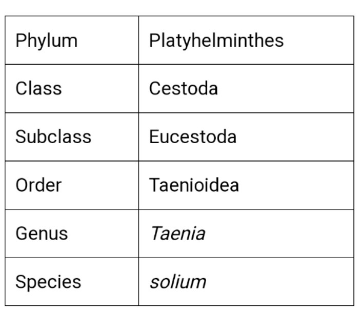
Classification of Taenia solium
Habits and Habitat of Taenia solium
Taenia solium, also known as pork tapeworm, is found worldwide. Thus, its distribution is cosmopolitan.
Its adults dwell in man’s small intestine as an internal parasite, i.e., endoparasitic, where it adheres to the intestinal mucosa by its scolex.
Its life cycle is completed in two hosts, i.e., digenetic, man as the primary host, and pig as a secondary host.
Other animals like goats, cattle, monkeys, and horses also serve as intermediate hosts.
It is especially reported from the European countries where pork is eaten, either raw or improperly cooked.
It absorbs the host’s digestive food through its body wall.
Structure of Taenia solium
1. Shape, size, and coloration
It is usually opaque white, but creamish, yellowish, or greyish coloration is also common.
Its body is long (1-5 meters), dorsoventrally flattened, narrow, ribbon-like.
The two flat surfaces represent the dorsal and ventral surfaces, respectively.
The internal view reveals that the surface closer to the testes is dorsal, and nearer to the female reproductive organs is the ventral surface.
Its body narrows anteriorly and gradually broadens posteriorly.
2. Segmentation
The elongated body of the tapeworm is divided into many segments or parts with about 850, called proglottids.
Segmentation of tapeworms is called pseudometamerism.
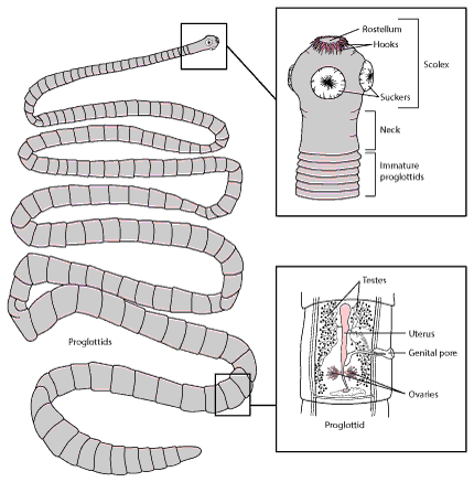
The tapeworm body is divisible into three distinct parts anterior scolex or head, a short-unsegmented neck, and a segmented strobila.
3. Scolex
It is present at the anterior end of the body.
It is knob-like, biradially symmetrical, and 0.6 mm to 1 mm wide.
It appears roughly quadrangular in the en-face view.
It is smaller than the head of a pin, about 1mm in diameter.
It consists of four cup-like muscular suckers having radial muscles.
At its tip is a prominent rounded mobile cone, the rostellum.
Rostellum is armed with 22 to 32 curved and chitinous hooks in 2 circles, the inner circle with larger hooks and the outer ring with smaller ones.
Large hooks each measures 14 to 0.18 mm, and those small each measures 0.11 mm to 0.14 mm.
Each hook consists of a base by which it fixes, a handle directed towards the apex, and a conical blade directed outwardly.
Four hemispherical highly muscular suctorial organs, true suckers or acetabula, are present on the scolex’s broadest part.
It is the organ of attachment to the intestinal mucosa with its suckers and hooks. Thus, it is an organ of adhesion or the holdfast.
4. Neck
Behind, the scolex, well-defined, short, narrow, and unsegmented region present, the neck.
It has been variously termed the budding zone, growth zone, area of proliferation, and segmentation area because it grows continuously and proliferates proglottids by transverse fission or asexual budding.
5. Strobila
The neck is followed by the flattened, ribbon-like body, the strobila.
It forms the main bulk of the body.
It consists of 800 to 1,000 segments or proglottids arranged in a linear series in a chain-like fashion.
The strobila of mature tapeworm measures about 3 meters in length.
A proglottid is a unit parts of the body enclosing a complete set of genitalia and surrounding tissue.
The linear arrangement or repetition of these units is called proglottisation.
The proglottids, internally, remain connected together by muscles, excretory vessels, and nerve cords.
Proglottids are independent, self-contained units, each with a complete set of male and female reproductive organs and a part of the excretory and nervous system.
Proglottids are budded off from the neck region and pushed back due to more proglottids in front.
Anterior proglottids are youngest in strobila, and posterior ones are oldest.
The proglottids are differentiated into three kinds according to the degree of development.
a. Immature proglottids
These are proglottids just behind of neck.
These include nearly 200 anterior proglottids.
They are the youngest, sexually immature, and devoid of reproductive organs.
They are short, broader than long, and rectangular in outline.
b. Mature proglottids
They form the middle part of the strobila.
They are about 450 in number.
These are large and squarish in outline.
The anterior 100 to 150 proglottids contain male reproductive organs only.
The posterior 250 proglottids develop both male and female reproductive organs. Thus, mature proglottids are hermaphrodite.
c. Gravid proglottids
These are the oldest and towards the posterior end of the body.
They include 150 to 350 proglottids.
They are .onger than broad in outline.
They have no reproductive organs.
They contain only branched uterus packed with fertilized eggs.
The proglottids of strobila widen gradually along their length from the anterior to the posterior one. The proglottids bear genital papilla and pore, alternating once to the right and then to the left.
Apolysis
Small groups of gravid proglottids are regularly cut off from the posterior end of strobila and pass out with the host’s feces, called apolysis.
Apolysis serves to transfer the developing embryos to the exterior, where the secondary hosts can ingest them.
It also keeps the body’s size, which may otherwise attain enormous length due to the continued proliferation of new proglottids from the neck region.
Body wall of Taenia solium
The tapeworm lacks a cellular or ciliated epidermis. The body wall of Taenia consists of an Outer tegument and inner basement membrane. The basement membrane includes both the musculature and the packing material called parenchyma.
1. Teguments
It is the outermost, thick, waxy, and enzyme resistant layer clothing the body in the absence of a cellular epidermis.
It is derived from the tegumentary secretory cells.
It is composed of protein impregnated with calcium carbonate and is perforated by numerous fine canals.
It consists of three layers outermost hair-like of finger-like comidial layer, the middle thick homogenous layer, and the innermost basement membrane.
The studies done by Threadgold and others have shown that the outermost cuticle is an intact thick, living, and syncytial layer called tegument.
The tegument is derived from the tegumentary secretory cells.
The tegument is connected to the tegumentary secreting cells with strands of cytoplasm called trabeculae.
It contains mitochondria and lysosomes and gives out microvilli-like processes called microtriches, on its outer surface.
The microvilli help in increasing the surface area of absorption of a nutritive substance from the host and also acts as holdfast organs.
The tegument is helpful in protecting the inner parts of the body.
The tegument is perforated by minute pores t through which substances are absorbed from the host’s intestine.
2. Integumentary musculature
It is situated just below the basement membrane.
It consists of an outer circular and inner longitudianl fibers.
The mesenchymal musculature consists of longitudinal, transverse, or circular, and vertical or dorsoventral muscle fibers.
3. Mesenchyme or parenchyma
The tegument is followed by mesenchyme.
It is a syncytial network formed by branched mesenchymal cells.
It consists of loosely-packed cells with fluid-filled interspaces, forming packing substances around the internal organs.
It does not contain a body cavity.
The parenchyma in young proglottids and neck region is thicker and some free cells which later differentiate to form the reproductive organs.
The turgidity of fluids helps to maintain the form of the body and it also acts as a hydraulic skeleton.
It contains numerous round or oval calcareous bodies composed of concentric layers of calcium carbonate which is secreted by the special mesenchymal lime cells.
The secretion from the lime cells helps to neutralize the acid of the digestive juice of the host.
circular muscle fibers at the margins, divide the mesenchyme into an outer cortex or cortical zone and inner medulla or medullary zone.
Parenchyma also helps in the transport of substances to tissue in absence of a blood vascular system.
Infection and Cysticercosis
Taenia solium infection (taeniasis) is an intestinal infection with adult tapeworms that follows ingestion of contaminated pork. Adult worms may cause mild gastrointestinal symptoms or passage of a motile segment in the stool.
Cysticercosis is infection with larvae of T. solium, which develops after ingestion of ova excreted in human feces. Cysticercosis is usually asymptomatic unless larvae invade the central nervous system, resulting in neurocysticercosis, which can cause seizures and various other neurologic signs.
Neurocysticercosis may be recognized on brain imaging studies. Fewer than half of patients with neurocysticercosis have adult T. solium in their intestines and thus eggs or proglottids in their stool.
Adult worms can be eradicated with praziquantel or niclosamide. Treatment of symptomatic neurocysticercosis is complicated; it includes corticosteroids, antiseizure drugs, and, in some situations, albendazole or praziquantel. Surgery may be required.
Presentation, diagnosis, and management of intestinal infection with the adult T. solium tapeworm are similar to those of T. saginata (beef tapeworm) infection.
However, humans may also act as intermediate hosts for T. solium larvae if they ingest T. solium eggs from human excreta ( see Figure: Taenia solium life cycle).
Some experts postulate that if an adult tapeworm is present in the intestine, gravid proglottids (tapeworm segments) may be passed retrograde from the intestine to the stomach, where oncospheres (immature form of the parasite enclosed in an embryonic envelope) may hatch and migrate to subcutaneous tissue, muscle, viscera, and the central nervous system.
Adult tapeworms may reside in the small bowel for years. They reach 2 to 8 m in length and produce up to 1000 proglottids; each contains about 50,000 eggs.
Humans may develop intestinal infection with adult worms after ingestion of contaminated pork or may develop cysticercosis after ingestion of eggs (making humans intermediate hosts).
1. Humans ingest raw or undercooked pork containing cysticerci (larvae).
2. After ingestion, cysts evaginate, attach to the small intestine by their scolex, and mature into adult worms in about 2 months.
3. Adult tapeworms produce proglottids, which become gravid; they detach from the tapeworm and migrate to the anus.
4. Detached proglottids, eggs, or both are passed from the definitive host (human) in feces.
5. Pigs or humans become infected by ingesting embryonated eggs or gravid proglottids (eg, in fecally contaminated food). Autoinfection may occur in humans if proglottids pass from the intestine to the stomach via reverse peristalsis.
6. After eggs are ingested, they hatch in the intestine and release oncospheres, which penetrate the intestinal wall.
7. Oncospheres travel through the bloodstream to striated muscles and to the brain, liver, and other organs, where they develop into cysticerci. Cysticercosis can result.
Taeniasis and cysticercosis occur worldwide. Cysticercosis is prevalent, and neurocysticercosis is a major cause of seizure disorders in Latin America.
Cysticercosis is rare in countries with low pork consumption (eg, Muslim-predominant countries). Infection in the US or Canada is rare in those who have not traveled abroad, but infection may occur by ingesting ova from people who visited endemic countries and are harboring adult T. solium.
Rarely, zoonotic Taenia species other than T. solium cause neurocysticercosis.
Symptoms and Signs
Intestinal infection
Humans infected with adult T. solium worms are asymptomatic or have mild gastrointestinal complaints. They may see proglottids in their stool.
Cysticercosis
Viable cysticerci (larval form) in most organs cause minimal or no tissue reaction, but dying cysts in the central nervous system, eye, or spinal cord can release antigens that elicit an intense tissue response. Thus, symptoms often do not appear for years after infection.
Infection in the brain (neurocysticercosis) may result in severe symptoms due to mass effect and inflammation induced by degeneration of cysticerci and release of antigens.
Depending on the location and number of cysticerci, patients with neurocysticercosis may present with seizures, signs of increased intracranial pressure, hydrocephalus, focal neurologic signs, altered mental status, or aseptic meningitis.
Cysticerci may also infect the spinal cord, muscles, subcutaneous tissues, and eyes.
Substantial secondary immunity develops after larval infection.


