Cytoskeleton
Introduction
What would happen if someone snuck in during the night and stole your skeleton? Just to be clear, that’s not very likely to happen, biologically speaking. But if it did somehow happen, the loss of your skeleton would cause your body to lose much of its structure. Your external shape would change, some of your internal organs might start moving out of place, and you would probably find it very difficult to walk, talk, or move.
Interestingly enough, the same is true for a cell. We often think about cells as soft, unstructured blobs. But in reality, they are highly structured in much the same way as our own bodies. They have a network of filaments known as the cytoskeleton (literally, “cell skeleton”), which not only supports the plasma membrane and gives the cell an overall shape, but also aids in the correct positioning of organelles, provides tracks for the transport of vesicles, and (in many cell types) allows the cell to move.
In eukaryotes, there are three types of protein fibers in the cytoskeleton: microfilaments, intermediate filaments, and microtubules. Here, we'll examine each type of filament, as well as some specialized structures related to the cytoskeleton.
Microfilaments
Of the three types of protein fibers in the cytoskeleton, microfilaments are the narrowest. They have a diameter of about 7 nm and are made up of many linked monomers of a protein called actin, combined in a structure that resembles a double helix. Because they are made of actin monomers, microfilaments are also known as actin filaments. Actin filaments have directionality, meaning that they have two structurally different ends.
Actin filaments have a number of important roles in the cell. For one, they serve as tracks for the movement of a motor protein called myosin, which can also form filaments. Because of its relationship to myosin, actin is involved in many cellular events requiring motion.
For instance, in animal cell division, a ring made of actin and myosin pinches the cell apart to generate two new daughter cells. Actin and myosin are also plentiful in muscle cells, where they form organized structures of overlapping filaments called sarcomeres. When the actin and myosin filaments of a sarcomere slide past each other in concert, your muscles contract.
Actin filaments may also serve as highways inside the cell for the transport of cargoes, including protein-containing vesicles and even organelles. These cargoes are carried by individual myosin motors, which "walk" along actin filament bundles.
Actin filaments can assemble and disassemble quickly, and this property allows them to play an important role in cell motility (movement), such as the crawling of a white blood cell in your immune system.
Finally, actin filaments play key structural roles in the cell. In most animal cells, a network of actin filaments is found in the region of cytoplasm at the very edge of the cell. This network, which is linked to the plasma membrane by special connector proteins, gives the cell shape and structure.
Intermediate filaments
Intermediate filaments are a type of cytoskeletal element made of multiple strands of fibrous proteins wound together. As their name suggests, intermediate filaments have an average diameter of 8 to 10 nm, in between that of microfilaments and microtubules (discussed below).
Intermediate filaments come in a number of different varieties, each one made up of a different type of protein. One protein that forms intermediate filaments is keratin, a fibrous protein found in hair, nails, and skin. For instance, you may have seen shampoo ads that claim to smooth the keratin in your hair!
Unlike actin filaments, which can grow and disassemble quickly, intermediate filaments are more permanent and play an essentially structural role in the cell. They are specialized to bear tension, and their jobs include maintaining the shape of the cell and anchoring the nucleus and other organelles in place.
Microtubules
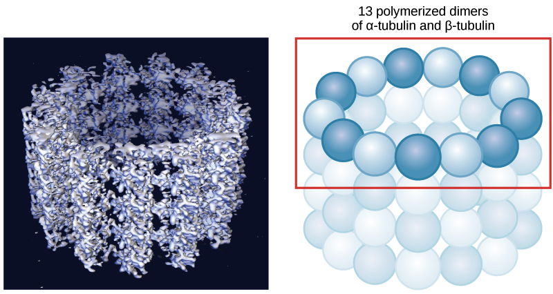
Despite the “micro” in their name, microtubules are the largest of the three types of cytoskeletal fibers, with a diameter of about 25 nm. A microtubule is made up of tubulin proteins arranged to form a hollow, straw-like tube, and each tubulin protein consists of two subunits, α-tubulin and β-tubulin.
Microtubules, like actin filaments, are dynamic structures: they can grow and shrink quickly by the addition or removal of tubulin proteins. Also similar to actin filaments, microtubules have directionality, meaning that they have two ends that are structurally different from one another. In a cell, microtubules play an important structural role, helping the cell resist compression forces.
In addition to providing structural support, microtubules play a variety of more specialized roles in a cell. For instance, they provide tracks for motor proteins called kinesins and dyneins, which transport vesicles and other cargoes around the interior of the cell. During cell division, microtubules assemble into a structure called the spindle, which pulls the chromosomes apart.
Flagella, cilia, and centrosomes
Microtubules are also key components of three more specialized eukaryotic cell structures: flagella, cilia and centrosomes. You may remember that our friends the prokaryotes also have structures that have flagella, which they use to move. Don't get confused—the eukaryotic flagella we're about to discuss have pretty much the same role, but a very different structure.
Flagella (singular, flagellum) are long, hair-like structures that extend from the cell surface and are used to move an entire cell, such as a sperm. If a cell has any flagella, it usually has one or just a few. Motile cilia (singular, cilium) are similar, but are shorter and usually appear in large numbers on the cell surface. When cells with motile cilia form tissues, the beating helps move materials across the surface of the tissue. For example, the cilia of cells in your upper respiratory system help move dust and particles out towards your nostrils.
Despite their difference in length and number, flagella and motile cilia share a common structural pattern. In most flagella and motile cilia, there are 9 pairs of microtubules arranged in a circle, along with an additional two microtubules in the center of the ring. This arrangement is called a 9 + 2 array. You can see the 9 + 2 array in the electron microscopy image at left, which shows two flagella in cross-section.
Upper: Transmission electron micrograph of flagella in cross-section, showing the 9+2 microtubule array organization.
Lower: Cartoon diagram of a motile cililum, showing the singlet microtubules in the center, the outer doublet microtubules arranged in a circle around the singlet microtubules, and the dyneins attached to the doublet microtubules. The whole structure is surrounded by plasma membrane. At the base of the cilium lies a basal body, which is also made up of microtubules.
In flagella and motile cilia, motor proteins called dyneins move along the microtubules, generating a force that causes the flagellum or cilium to beat. The structural connections between the microtubule pairs and the coordination of dynein movement allow the activity of the motors to produce a pattern of regular beating.
You may notice another feature in the diagram above: the cilium or flagellum has a basal body located at its base. The basal body is made of microtubules and plays a key role in assembly of the cilium or flagellum. Once the structure has been assembled, it also regulates which proteins can enter or exit.
The basal body is actually just a modified centriole. A centriole is a cylinder of nine triplets of microtubules, held together by supporting proteins. Centrioles are best known for their role in centrosomes, structures that act as microtubule organizing centers in animal cells. A centrosome consists of two centrioles oriented at right angles to each other, surrounded by a mass of "pericentriolar material," which provides anchoring sites for microtubules.
The centrosome is duplicated before a cell divides, and the paired centrosomes seem to play a role in organizing the microtubules that separate chromosomes during cell division. However, the exact function of the centrioles in this process still isn’t clear. Cells with their centrosome removed can still divide, and plant cells, which lack centrosomes, divide just fine.


