Chromosome Structure
Definition
Chromosomes are thread-like structures present in the nucleus, which carries genetic information from one generation to another. They play a vital role in cell division, heredity, variation, mutation, repair and regeneration.
In Eukaryotic cells, genetic material is present in the nucleus in chromosomes, which is made up of highly organized DNA molecules with histone proteins supporting its structure.
Karl Nägeli in 1842, first observed the rod-like structure present in the nucleus of the plant cell.
W. Waldeyer in 1888 coined the term ‘chromosome’.
Walter Sutton and Theodor Boveri in 1902 suggested that chromosomes are the physical carrier of genes in the eukaryotic cells.
The number of chromosomes in any species is constant for all the cells. The number of chromosomes in gametes (e.g. sperms, egg) is half of the somatic cell and known as a haploid set of chromosomes, which is the result of meiosis during sexual reproduction. Chromosome number is preserved in the mitotic division of somatic cells, which is required for an organism to grow, repair and regenerate.
Chromosome number varies in different species. A nematode species contains only 2 chromosomes in a cell, whereas a protozoan species contains as much as 1600 chromosomes in the cell. Most of the plant and animal species contain 8 to 50 number of chromosomes in its somatic cell. The number of chromosomes does not reflect the complexity of a species. A human cell contains total 23 pair of chromosomes (2n, total 23×2=46), of which 22 are autosomes and 1 sex chromosome.
Karyotyping is a technique to study the structure of chromosomes present in a species. Chromosomes are isolated, stained and photographed. This technique is useful in finding out any chromosomal abnormalities.
Chromosome Structure
The chemical composition of a chromosome is histone proteins and DNA. Each cell has a pair of each kind of chromosome known as a homologous chromosome. Chromosomes are made up of chromatin, which contains a single molecule of DNA and associated proteins. Each chromosome contains hundreds and thousands of genes that can precisely code for several proteins in the cell. Structure of a chromosome can be best seen during cell division.
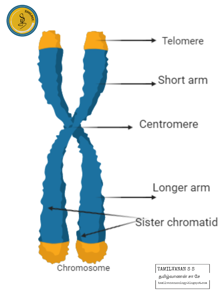
Chromosome structure and organisation
Main parts of chromosomes are:
Chromatid: Each chromosome has two symmetrical structures called chromatids or sister chromatids which is visible in mitotic metaphase.Each chromatid contains a single DNA molecule. At the anaphase of mitotic cell division, sister chromatids separate and migrate to opposite poles.
Centromere and kinetochore: Sister chromatids are joined by the centromere. Spindle fibres during cell division are attached at the centromere. The number and position of the centromere differs in different chromosomes. The centromere is called primary constriction. Centromere divides the chromosome into two parts, the shorter arm is known as ‘p’ arm and the longer arm is known as ‘q’ arm. The centromere contains a disc-shaped kinetochore, which has a specific DNA sequence with special proteins bound to them. The kinetochore provides the center for polymerisation of tubulin proteins and assembly of microtubules
Secondary constriction and nucleolar organisers: Other than centromere, chromosomes possess secondary constrictions. Secondary constrictions can be identified from centromere at anaphase because there is bending only at the centromere (primary constriction). Secondary constrictions, which contain genes to form nucleoli are known as the nucleolar organiser.
Telomere: Terminal part of a chromosome is known as a telomere. Telomeres are polar, which prevents the fusion of chromosomal segments.
Satellite: It is an elongated segment that is sometimes present on a chromosome at the secondary constriction. The chromosomes with satellite are known as sat-chromosome.
Chromatin: Chromosome is made up of chromatin. Chromatin is made up of DNA, RNA and proteins. At interphase, chromosomes are visible as thin chromatin fibres present in the nucleoplasm. During cell division, the chromatin fibres condense and chromosomes are visible with distinct features. The darkly stained, condensed region of chromatin is known as heterochromatin. It contains tightly packed DNA, which is genetically inactive. The light stained, diffused region of chromatin is known as euchromatin. It contains genetically active and loosely packed DNA. At prophase, the chromosomal material is visible as thin filaments known as chromonemata. At interphase, bead-like structures are visible, which are an accumulation of chromatin material called chromomere. Chromatin with chromomere looks like a necklace with beads.
Structural Organisation of Chromatin
Chromatin consists of DNA and associated proteins. DNA is packaged in a highly organised manner in chromosomes.
Nucleosomes are the basic unit of chromatin. It is 10 nm in the diameter.
DNA packing is facilitated by proteins called histones. DNA is wound around histone proteins to form a nucleosome.
There are 5 types of histone proteins in the eukaryotic chromosomes, namely H1, H2A, H2B, H3 and H4.
Histones are positively charged due to the presence of amino acids with basic side chains and it associates with negatively charged DNA due to the presence of phosphate groups.
Histone proteins play an important role in gene regulation.
A typical nucleosome contains 200 bp of DNA helix. The core particle of the nucleosome consists of approximately 146 base pairs (bp) of DNA coiled around a core of eight histone molecules (2 molecules of 4 histone proteins). That is linked by linker DNA of about 80 bp.
Nucleosomes prevent DNA from getting tangled.
Linker DNA and the fifth histone (H1) pack adjacent nucleosomes to a 30 nm compact chromatin fibre.
These fibres form a large coiled loop held together by non-histone proteins (actin, 𝛂 and 𝛃 tubulin, myosin) called scaffolding proteins to form extended chromatin which is 300 nm in diameter.
Chromatin further condenses with the help of protein known as condensin, it binds to DNA and wraps it into coiled loops and we get the compacted chromosome.
Types of Chromosome
Based on the number of centromeres present
Monocentric: having only one centromere.
Holocentric: having diffused centromere and microtubules are attached along the length of a chromosome.
Acentric: chromosome may break and fuse together to form a chromosome without a centromere. It cannot attach to the mitotic spindle.
Dicentric: chromosomal aberration where chromosomes break and fuse together with two centromeres. They are also unstable as two centromeres tend to migrate to opposite poles resulting in fragmentation.
Based on the position of the centromere
Types of chromosomes
Telocentric: rod-like chromosome with centromere present on the proximal end. No ‘p’ arm or short arm of chromosome, is present. Telomeric chromosomes are not found in humans.
Acrocentric: rod-like, centromere present at one end giving rise to one very short arm and an exceptionally long arm.
Submetacentric: L-shaped or J-shaped, with centromere near the centre of the chromosome giving rise to two unequal arms.
Metacentric: V-shaped chromosomes with centromere in the middle giving rise to two equal arms.
Giant Chromosomes
Polytene chromosome
Balbiani first discovered a structure in the nuclei of secretory glands of midges. Painter, Heitz and Bauer, rediscovered them in the salivary gland of Drosophila and recognised them as a chromosome. Also known as Salivary gland chromosome. These are called polytene by Kollar due to the presence of many chromonemata in them. These are present in some cells of the larvae of Dipteran insects. These are very large due to the presence of high DNA content
The polytene chromosome of Drosophila’s salivary gland has 1000 DNA molecules Chironomus has 1600 DNA molecules in its each polytene chromosome. There is a series of alternating dark and clear bands called interband. Chromosome puffs or Balbiani rings are present, which are the swelling of bands due to DNA unfolding into open loops. These are the region of the intense transcription or mRNA formation.
Lamp brush chromosome
First discovered in the oocytes of salamander. The name is given due to its resemblance with a brush that is used for cleaning lamp, glass chimneys, etc. They occur at the diplotene stage of oocytes in vertebrates and invertebrates. Lampbrush chromosomes are also found in the spermatocytes of many animals and also found in the giant nucleus of an algae Acetabularia. They are present as a bivalent with 4 chromatids.
Chromosomal axis is formed from highly condensed chromatin and lateral loops extend from the row of chromomeres. Lateral loops of DNA are always symmetrical and formed due to intense RNA synthesis. In the oocytes of salamander, there are 10,000 loops present per haploid set of chromosomes. The centromere doesn’t bear any loops
Functions of Chromosomes
The main function of chromosomes is to carry the genetic material from one generation to another.
Chromosomes play an important role and act as a guiding force in the growth, reproduction, repair and regeneration process, which is important for their survival.
Chromosomes protect the DNA from getting tangled and damaged.
Histone and non-histone proteins help in the regulation of gene expression.
Spindle fibres attached to the centromere help in the movement of the chromosome during cell division
Each chromosome contains thousands of genes that precisely code for multiple proteins present in the body.


