Wednesday, June 21, 2023

Circulatory system - Human

Circulatory system - Human.

The heart and blood are major parts of the circulatory system. All components contribute to oxygen and nutrient delivery throughout your body.

Combined with the cardiovascular system, the circulatory system helps fight off disease, helps the body maintain a normal body temperature, and provides the right chemical balance for the body to achieve homeostasis, or a state of stability among all its systems.

The circulatory system consists of four major components:

heart
arteries
veins
blood

Heart

The heart is a fist-sized organ that pumps blood throughout your body. It's the primary organ of your circulatory system.

Your heart contains four main sections (chambers) made of muscle and powered by electrical impulses. Your brain and nervous system direct your heart’s function.

The heart’s main function is to move blood throughout your body. Your heart also:

Controls the rhythm and speed of your heart rate. Maintains blood pressure.

The heart works with other body systems to control your heart rate and other body functions. The primary systems are:

Nervous system: Your nervous system helps control your heart rate. It sends signals that tell your heart to beat slower during rest and faster during stress.

Endocrine system: Your endocrine system sends out hormones. These hormones tell your blood vessels to constrict or relax, which affects your blood pressure. Hormones from your thyroid gland can also tell your heart to beat faster or slower.

Parts

The parts of the heart are like the parts of a house. Your heart has:

Walls.
Chambers (rooms).
Valves (doors).
Blood vessels (plumbing).
Electrical conduction system (electricity).

Heart walls

Your heart walls are the muscles that contract (squeeze) and relax to send blood throughout your body. A layer of muscular tissue called the septum divides your heart walls into the left and right sides.

Your heart walls have three layers:

Endocardium: Inner layer.
Myocardium: Muscular middle layer.
Epicardium: Protective outer layer.

The epicardium is one layer of your pericardium. The pericardium is a protective sac that covers your entire heart. It produces fluid to lubricate your heart and keep it from rubbing against other organs.

Heart chambers

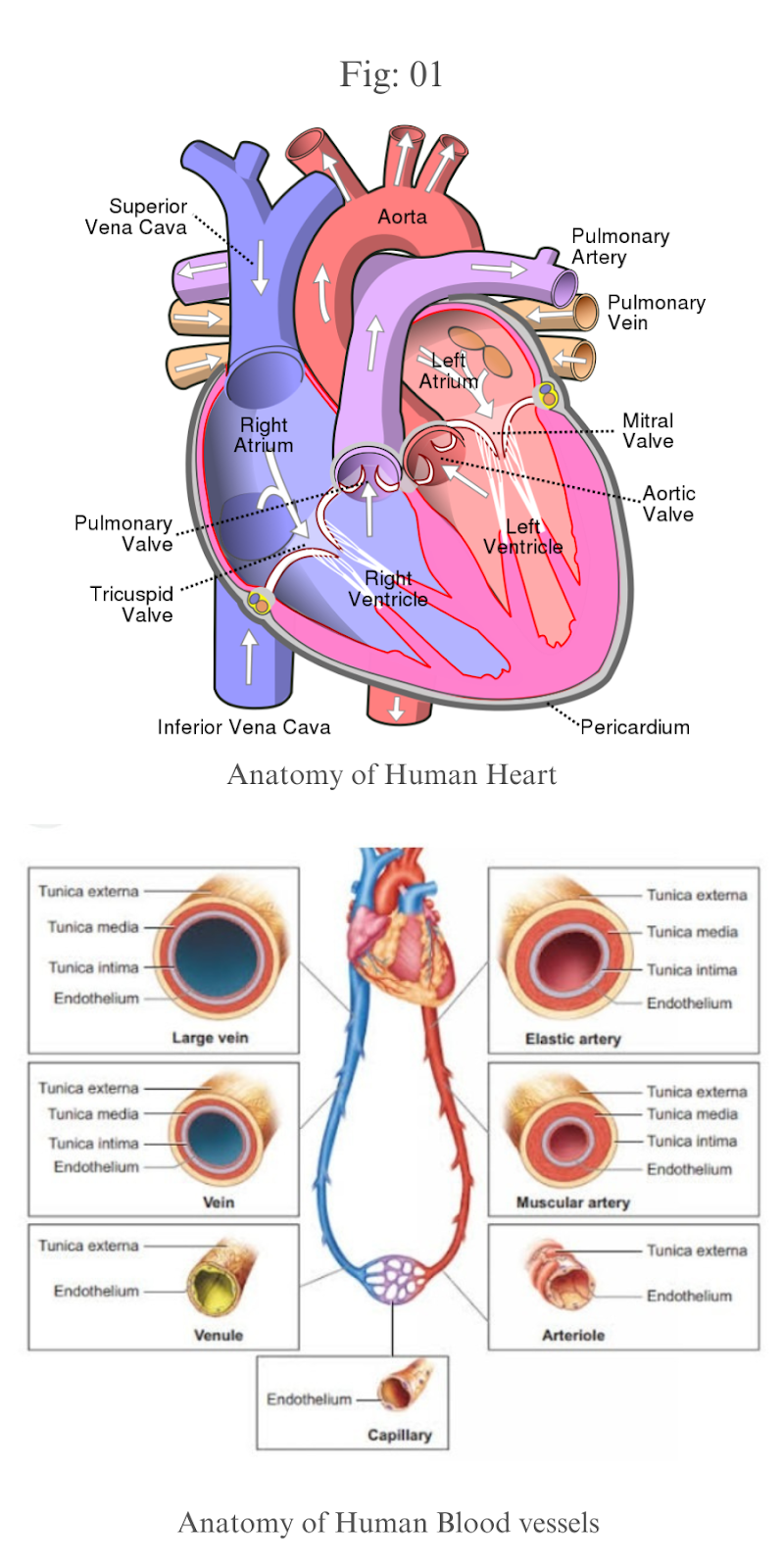
Your heart is divided into four chambers. You have two chambers on the top (atrium, plural atria) and two on the bottom (ventricles), one on each side of the heart.

Right atrium: Two large veins deliver oxygen-poor blood to your right atrium. The superior vena cava carries blood from your upper body. The inferior vena cava brings blood from the lower body. Then the right atrium pumps the blood to your right ventricle.

Right ventricle: The lower right chamber pumps the oxygen-poor blood to your lungs through the pulmonary artery. The lungs reload blood with oxygen.

Left atrium: After the lungs fill blood with oxygen, the pulmonary veins carry the blood to the left atrium. This upper chamber pumps the blood to your left ventricle.

Left ventricle: The left ventricle is slightly larger than the right. It pumps oxygen-rich blood to the rest of your body.

Heart valves

Your heart valves are like doors between your heart chambers. They open and close to allow blood to flow through.

The atrioventricular (AV) valves open between your upper and lower heart chambers. They include:

Tricuspid valve: Door between your right atrium and right ventricle.

Mitral valve: Door between your left atrium and left ventricle.

Semilunar (SL) valves open when blood flows out of your ventricles. They include:

Aortic valve: Opens when blood flows out of your left ventricle to your aorta (artery that carries oxygen-rich blood to your body).

Pulmonary valve: Opens when blood flows from your right ventricle to your pulmonary arteries (the only arteries that carry oxygen-poor blood to your lungs).

Arteries

The arteries and their function

Arteries carry blood away from the heart.

The artery walls have three layers: tunica intima (inner), tunica media (middle), and tunica externa (outer).

The middle layer is usually the thickest. It’s made up of smooth muscle that changes the size of the artery to regulate blood flow.

There are three main types of arteries. They get smaller and smaller the further they are from the heart.

Elastic arteries

The aorta and pulmonary arteries are the elastic arteries. They receive blood directly from the heart and need to be elastic to accommodate the surge and contraction as blood pushes through with each heartbeat.

The aorta is the body’s most important artery.

Pulmonary arteries take deoxygenated blood from the right ventricle to the lungs. They’re the only arteries that carry deoxygenated blood.

Muscular arteries

The muscular arteries move blood from the elastic arteries through the body. They’re made of smooth muscle, which can expand and contract as blood flows. The femoral and coronary arteries are two examples of muscular arteries.

Arterioles

The smallest arteries are the arterioles, which move blood from the muscular arteries to the capillaries. The capillaries connect the arteries, which take blood from the heart, and the veins, which take blood to the heart.

The number of capillaries in a body system depends on the amount of material exchange. Skeletal muscle, the liver, and the kidney all have a large number of capillaries because their body systems need a lot of oxygen and nutrients. The cornea of the eye is one area that has no capillaries.

Veins

The veins and their function

The blood moves back to the heart through veins.

The blood travels from the capillaries into the venules, which are the smallest veins. As the blood moves closer to the heart, the veins get larger and larger.

Like the arteries, veins have walls made up of layers called the tunica intima, tunica media, and tunica externa. There are some important differences between the arteries and veins:

In veins, the walls have less smooth muscle and connective tissue.

The walls of veins are thinner than artery walls.
Veins have less pressure and can hold more blood than arteries.

At any time, about 70 percentTrusted Source of the body’s total blood supply is in the veins.

Valves

The veins include valves, small pieces of tissue which keep blood flowing in the right direction.

The valves in the medium and large veins keep the blood flowing towards the heart. In the arms and legs, these valves make sure gravity doesn’t pull blood in the wrong direction.

There are four valves in the heart.

The tricuspid valve separates the right atrium from the right ventricle. The mitral valve, or bicuspid valve, separates the left atrium from the left ventricle.

The remaining two valves are the semilunar valves. The pulmonic valve, or pulmonary valve, separates the pulmonary artery from the right ventricle. The aortic valve separates the aorta and the left ventricle.

Blood

The blood and its function

Blood is the transport medium of nearly everything within the body. It moves hormones, nutrients, oxygen, antibodies, and other important things needed to keep the body healthy.

The blood has four main components.

Plasma

About 55 percent of the blood is plasma. Plasma is what makes blood liquid.

Plasma moves blood cells through the body by way of the circulatory system. It also carries hormones, nutrients, antibodies, and waste products.

Plasma is made up of:

water
salts
sugar
fat
protein

Red blood cells

Red blood cells, also called erythrocytes, make up about 40 to 45 percent of the blood’s volume. These cells have no nucleus, which means they can easily change shape as they move through the body’s arteries and veins.

Red blood cells contain a protein called hemoglobin. It carries oxygen from the lungs to the rest of the body and returns carbon dioxide to the lungs, where it’s exhaled.

White blood cells

White blood cells, also called leukocytes, make up just 1 percent of the blood. They protect the body from infection.

There are five major types of white blood cells.

Most white blood cells are neutrophils, which live for less than 1 day. Neutrophils are the body’s immediate response team.

Types of lymphocytes include B lymphocytes (B cells) and T lymphocytes (T cells). B lymphocytes make antibodies, while T lymphocytes regulate other immune cells and target infected cells and tumors.

The other major types are basophils, eosinophils, and monocytes.

Platelets

Platelets, also called thrombocytes, are cell fragments.

Platelets are essential for blood clotting. They stick to an injured blood vessel lining to provide the basis for a clot. This stops bleeding and promotes healing.


Circulatory System 

Oxygen enters the bloodstream through tiny membranes in the lungs that absorb oxygen as it’s inhaled. As the body uses oxygen and processes nutrients, it creates carbon dioxide, which your lungs expel as you exhale.

The circulatory system works thanks to constant pressure from the heart and valves throughout the body. This pressure ensures that veins carry blood to the heart and arteries transport it away from the heart. (Hint: To remember which one does which, remember that “artery” and “away” both begin with the letter A.)

There are three different types of circulation that occur regularly in the body:

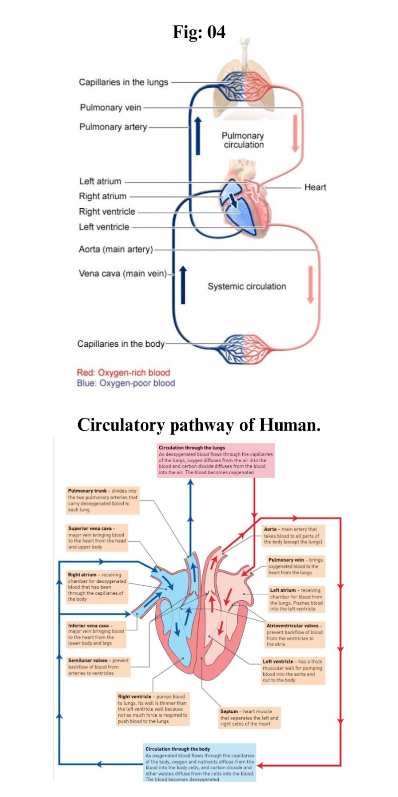
Pulmonary circulation. This part of the cycle carries oxygen-depleted blood away from the heart, to the lungs, and back to the heart.

Systemic circulation. This is the part that carries oxygenated blood away from the heart and to other parts of the body.

Coronary circulation. This type of circulation provides the heart with oxygenated blood so it can function properly.

Pulmonary arteries take blood with low levels of oxygen from the right ventricle to the lungs.

Pulmonary veins move oxygen-rich blood from the lungs to the heart’s left atrium.

Systemic arteries take oxygen-rich blood from the left ventricle to the body’s tissues.

Systemic veins move blood with low levels of oxygen from the body’s tissues to the heart’s right atrium.

Coronary arteries take oxygen-rich blood from the aorta to the heart muscle.

Coronary veins move blood with low levels of oxygen from the heart’s myocardium (middle muscular layer) to its right atrium.

Electrical conduction system

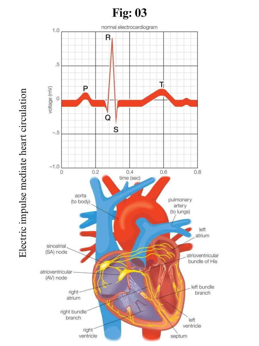
Your heart’s conduction system is like the electrical wiring of a house. It controls the rhythm and pace of your heartbeat. It includes:

Sinoatrial (SA) node: Sends the signals that make your heart beat.

Atrioventricular (AV) node: Carries electrical signals from your heart’s upper chambers to its lower ones.

Your heart also has a network of electrical bundles and fibers. This network includes:

Left bundle branch: Sends electric impulses to your left ventricle.

Right bundle branch: Sends electric impulses to your right ventricle.

Bundle of His: Sends impulses from your AV node to the Purkinje fibers.

Purkinje fibers: Make your heart ventricles contract and pump out blood.

Blood Circulation pathway

The right and left sides of your heart work together to make sure blood flows throughout your whole body. Blood flows through your heart in a series of steps. These steps take place in the space of one heartbeat — just a second or two.

On the right side

1. Oxygen-poor blood from all over your body enters your right atrium through two large veins, your superior vena cava and inferior vena cava. These veins drain blood from your upper body and lower body, respectively, and directly empty it into your right atrium.

2. Your tricuspid valve opens to let blood travel from your right atrium to your right ventricle.

3. When your right ventricle is full it squeezes, which closes your tricuspid valve and opens your pulmonary valve.

4. Blood flows through your main pulmonary artery and its branches to your lungs, where it gets oxygen and releases carbon dioxide.

On the left side

1. Oxygen-rich blood travels from your lungs to your left atrium through large veins called pulmonary veins. These veins directly empty the blood into your left atrium.

2. Your mitral valve opens to send blood from your left atrium to your left ventricle.

3. When your left ventricle is full it squeezes, which closes your mitral valve and opens your aortic valve.

4. Your heart sends blood through your aortic valve to your aorta, where it flows to the rest of your body.

Symptoms of poor circulation

There are many symptoms of poor circulation, including:

chest pain
dizziness or feeling faint
shortness of breath
pain, weakness, or numbness in the limbs
swollen limbs
slow or rapid heartbeat, or palpitations
fatigue

The symptoms depend on the type of circulatory condition. As an example, peripheral arterial disease can cause leg and foot problems like:

leg cramping while walking or resting
cold feet or legs
change of leg color
change in toenail color or thickness
loss of hair on the legs and feet
ulcers (sores) that don’t heal on the legs and feet

Conditions that affect the circulatory system

There are several conditions that can affect the heart and circulatory system, including:

Peripheral arterial disease. In peripheral arterial disease, blood flow in the arteries of the legs is restricted. This is usually due to buildup of plaque in the arteries.

Arteriosclerosis. In arteriosclerosis, plaque buildup in the blood vessels becomes calcified and hard. The arteries are less flexible, leading to higher blood pressure, stroke, heart damage, and kidney damage.

Heart attack. During a heart attack, a blockage of blood flow to the heart muscle leads to death of heart muscle tissue. It’s also known as a myocardial infarction.

Angina. In angina, the heart muscle isn’t getting enough blood. This leads to crushing chest pain, fatigue, nausea, and shortness of breath.

Mitral valve conditions. In mitral valve prolapse, mitral valve stenosis, or mitral valve regurgitation, problems with the mitral valve cause oxygenated blood in the heart to flow backward, or blood flow to be slowed down or constricted.

Arrhythmias or dysrhythmias. These terms are both used to describe a heart rate that’s abnormal.

Ischemia. In ischemia, there’s not enough blood flow in the heart, and muscles don’t get enough oxygen.

Aortic disease. This group of conditions affects the aorta. One example is an aortic aneurysm, where the aorta is weak and bulges out.

Extra notes: 

Your heart pumps about 2,000 gallons of blood each day. That’s enough to fill an 8-by-10-foot swimming pool.

It beats around 100,000 times daily. In an average life span of almost 79 years, your heart beats nearly 2.9 billion times.


GPCR (G-protein-Coupled Receptors)

GPCR (G-protein-Coupled Receptors) 

G-protein–coupled receptors (GPCRs) form a remarkable modular system that allows transmission of a wide variety of signals over the cell membrane, between cells and over long distances in the body. 

G-protein-coupled receptors (GPCRs) are the largest and most diverse group of membrane receptors in eukaryotes. These cell surface receptors act like an inbox for messages in the form of light energy, peptides, lipids, sugars, and proteins. Such messages inform cells about the presence or absence of life-sustaining light or nutrients in their environment, or they convey information sent by other cells.

GPCRs play a role in an incredible array of functions in the human body, and increased understanding of these receptors has greatly affected modern medicine. In fact, researchers estimate that between one-third and one-half of all marketed drugs act by binding to GPCRs.

Background

Every human cell is surrounded by a plasma membrane, a phospholipid bilayer. The membrane makes it possible for the cell to maintain a specific mix of biochemically active species, while preventing unwanted entry of other substances from the outside environment. For proper function, the biochemical machinery inside a cell needs to be able to receive instructions from the outside. 

Changes in hormone levels on the outside of the cell elicit adaptive changes in enzyme activity on the inside. Odour molecules affect cells in the olfactory epithelium and substances in the food influence chemical activities in taste bud cells, which in turn elicit electrical signals that transfer information to the brain. 

Indeed, human cells are constantly communicating with each other and the surrounding environment, which requires a molecular framework and a mechanism for transmission of information across the plasma membrane. 

Moreover, in the body, signal transmission may take place over long distances. To be able to respond promptly, the brain needs rapid information from our senses, for vision, smell, taste and more. Again, this requires a molecular mechanism for transmission of information over the plasma membrane.

The molecular framework consists of G-protein–coupled receptors (GPCRs). Those are proteins located in the plasma membrane. The name GPCR refers to a common mode of receptor signaling via GTP-binding proteins on the inside of the cell. 

Because their polypeptide chain passes seven times through the plasma membrane, the GPCRs are also called seven-transmembrane (7TM) receptors. They mediate a wide range of physiological signals from the outside of the cell.

The signal can be a change in concentration of peptides, hormones, lipids, neurotransmitters, ions, odourants, tastants, etc., or an influx of photons to the eye. GPCRs convey these signals to the inside of the cell and elicit a series of reactions involving other proteins, nucleotides and metal ions, which eventually deliver a message and an appropriate cellular and physiological response.

Many physiological processes in mammals depend on 7TM receptors, which are also the targets for a large portion of all pharmaceutical drugs. About a thousand human genes code for 7TM receptors, and they are involved in sensing a wide range of extracellular stimuli. Examples include the adrenergic receptors, dopamine receptors, histamine receptors, the light receptor rhodopsin, and the many odor and taste receptors.

Discovery  of GPCRs

Most of what we know today about the intriguing molecular properties of 7TM receptors has been discovered over the past 40 years. However, rhodopsin had been identified as a photosensitive pigment as early as the 1870s, and its covalent ligand retinal was reported in 1933 . 

The history of ligand-activated receptors started more than a century ago, when it was noted that reactive cells have a ‘receptive substance’ on their surface. 

Early experiments with tissue preparations measured the responses to stimulators (agonists) and inhibitors (antagonists). During the following half century (about 1920–1970), classical receptor theory was developed, based on the law of mass action and dose-response data.

Some of the signaling components inside the cell were described in molecular detail before the receptors were. These include the second messenger cyclic AMP (cAMP) and the enzyme adenylyl cyclase, cAMP-dependent protein kinase and heterotrimeric G-proteins.

The nature of the ligand-activated receptors remained controversial up to the 1970s. The existence of receptors as separate molecular entities was debated, and it was even speculated that receptors and the enzyme adenylyl cyclase were the same protein.

Structure of GPCRs

Members of the GPCR superfamily share the same basic architecture of 7TM α-helices, an extracellular amino-terminal segment and an intracellular carboxy-terminal tail. 

These plasma membrane-bound receptors have evolved to recognize a diversity of extracellular physical and chemical signals, such as nucleotides, peptides, amines, Ca2+ and photons. On recognition of such signals, the GPCRs act as proximal event in signaling pathways that influence a wide variety of metabolic and differentiated functions.

An extensive analysis of about 200 GPCR sequences revealed that the total length of GPCRs can vary between 311 and ∼1,490 amino acid residues. The largest variations in length are found in the N and C termini with size up to 879 and 371 amino acid residues, respectively. 

GPCRs are not only encoded by eukaryotic genes but also by viral genes. Human GPCRs genomic genes are predominantly intronless.

The 7TM α-helices are connected by three intracellular and three extracellular loops. The extracellular loops of the GPCR can be glycosylated and contain two highly conserved cysteine residues, which build disulfide bonds to stabilize the receptor structure. GPCRs contain extracellular N-terminus domains (ECL1, ECL2 and ECL3) of variable size, ranging from 154 residues (calcitonin receptor) to 36 residues (rhodopsin receptor). 

This domain contains asparagines residues and motifs for N-glycosylation, which influences intracellular trafficking of receptors to the plasma membrane, and cysteine residues in ECL1 and ECL2 loops that can influence protein folding critical for trafficking of a function receptor to the cell surface.19 The N terminus of some GPCRs is involved in ligand binding, activation and downregulation. 

The 7TM α-helices helices of GPCRs are arranged to form a tight, ring-shaped central core that is highly hydrophobic in nature. Similar to most TM proteins, the hydrophobic amino acid residues are presumably arranged to face the lipid bilayer, whereas the more hydrophilic amino acid residues face towards the core. 

Furthermore, helix-helix interaction contributes to the functional tertiary structure of the GPCRs necessary for receptor folding and stability, ligand binding and ligand-induced conformational changes for G protein coupling. Thus, mutations in the TM domain can have an array of deleterious effects.

GPCRs contain intracellular carboxyl-terminal domains (ICL1, ICL2 and ICL3) which are involved in several aspects of GPCR signaling. This domain contains Ser and/or Tyr residues which serve as sites for G protein receptor kinase-mediated phosphorylation and receptor desensitization. 

Some GPCRs contain a cysteine residue in the C-terminal domain, which can serve as a site for palmitoylation. This can create a fourth IL (intracellular loops) because of the ability of the palmitoylated cysteine to insert in the plasma membrane. Also, C terminus may be involved in the interactions with other proteins that mediate GPCR signaling, such as the calcyon, PDZ domain-containing proteins, and Homer/Vesl proteins.19 The GPCR vary not only in sequence, but also in length of amino acid and carboxyl-termini (especially the C3 loop). The serine residue at the carboxy terminus region of GPCRs gets phosphorylated by G protein-coupled receptor kinases (GRKs).

GRKs constitute of six mammalian Ser/Thr protein kinase that phosphorylate agonist-bound, or activated, GPCR as their primary substrates, GRK-mediated receptor phosphorylation rapidly initiates profound impairment of receptor signaling, or desensitization.20 While the X-ray crystal structures of several GRKs have been solved, but the mechanism of GRK interaction with GPCRs was not known. Recently, many proposed a mechanism whereby the N-terminus of GRK2 protein forms an intramolecular interaction that selectively enhances the catalytic activity of the kinase towards GPCR substrates.

Recently, lot reported the 3.2 angstrom (A°) crystal structure of the bovine GPCR (opsin) in its G-protein-interacting conformation. (Ops-GalphaCT peptide complex). Recently report the crystal structure of ligand-free native GPCR (opsin) from bovine retinal rod cells at 2.9 A° resolution. Compared to GPCR rhodopsin, opsin showed some structural changes in the conserved E(D)RY and NPxxY(x)5,6F regions and in TM5–TM7. At the cytoplasmic side, TM6 was found to be tilted outwards by 6–7 A°, whereas the helix structure of TM5 was more elongated and close to TM6. The authors have suggested that the opsin structure sheds new light on ligand binding to GPCRs and on GPCR activation.

Family of GPCRs

These are the largest class of receptors, with more than one thousand GPCRs identified so far. The GPCRs family is the third most abundant family in Caenorhabditis elegans, comprising 5% of its genome with approximately 1,100 members. The Drosophila genome has at least 160 GPCRs. Based on the now entirely known human genome, careful estimation suggests that about 3–4% of the human genes code for GPCRs, about 1,200–1,300 members of the GPCR superfamily in the human genome, many of which are known to homo- and heterodimerize. However, in the case of plants a single GPCR has been isolated from pea and maize, and computational analysis shows their presence in Arabidopsis, Populus and rice. The GPCRs superfamily is divided into 6 major families which share little sequence homology among each other and some functional similarity. The 6 families of GPCRs are as follows:

Family A (rhodopsin receptor family): Family A is commonly known as rhodopsin family. It is the largest family of GPCRs and includes receptors for odorants and small ligands. This family is further divided into three groups. Group 1 contains GPCRs for small ligands including rhodopsin and β-adrenergic receptors. The binding site is localized within the seven TMs. Group 2 contains receptors for peptides whose binding site includes the N-terminal, the extracellular loops and the superior parts of TMs. Group 3 contains GPCRs for glycoprotein hormones. It is characterized by a large extracellular domain and a binding site which is mostly extracellular but at least with contact with extracellular loops e1 and e3.

Family B (secretin receptor family): Family B is commonly known as secretin family. It recruits about 60 members and is characterized not only by the lack of the structural signature present in family A but also by the presence of a large N-terminal ectodomain. Family B GPCRs have a similar morphology to group A3 GPCRs, but they do not share any sequence homology. Their ligands include high molecular weight hormones such as glucagon, secretine, calcitonin, growth hormone-releasing hormone, corticotropin-releasing factor, VIP-PACAP and the Black widow spider toxin, α-latrotoxin.

Family C (metabotropic glutamate receptors family): Family C recruits about two dozens GPCRs such as metabotropic glutamate receptors (mGluR) and the Ca2+ sensing receptors. This family also includes GABA-B receptors, taste receptors, olfactory receptors and a group of putative pheromone receptors coupled to the G protein Go (termed VRs and Go-VN). Like family B, these receptors possess large ectodomains responsible for ligand binding.

Family D (fungus pheromone receptor family): Family D comprises pheromone receptors (VNs) associated with Gi.

Family E (cAMP receptor family): cAMP receptors (cAR) have only seen found in D. discoideum but its possible expression in vertebrate has not yet been reported.

Family F (frizzled/smoothened receptor family): Family F includes the ‘frizzled’ and the ‘smoothened’ (Smo) receptors involved in embryonic development and in particular in cell polarity and segmentation

GPCR look like 

GPCRs bind a tremendous variety of signaling molecules, yet they share a common architecture that has been conserved over the course of evolution. Many present-day eukaryotes — including animals, plants, fungi, and protozoa — rely on these receptors to receive information from their environment. For example, simple eukaryotes such as yeast have GPCRs that sense glucose and mating factors. Not surprisingly, GPCRs are involved in considerably more functions in multicellular organisms. Humans alone have nearly 1,000 different GPCRs, and each one is highly specific to a particular signal.

GPCRs consist of a single polypeptide that is folded into a globular shape and embedded in a cell's plasma membrane. Seven segments of this molecule span the entire width of the membrane — explaining why GPCRs are sometimes called seven-transmembrane receptors — and the intervening portions loop both inside and outside the cell. The extracellular loops form part of the pockets at which signaling molecules bind to the GPCR.


GPCR mechanism

As their name implies, GPCRs interact with G proteins in the plasma membrane. When an external signaling molecule binds to a GPCR, it causes a conformational change in the GPCR. This change then triggers the interaction between the GPCR and a nearby G protein.

G proteins are specialized proteins with the ability to bind the nucleotides guanosine triphosphate (GTP) and guanosine diphosphate (GDP). Some G proteins, such as the signaling protein Ras, are small proteins with a single subunit. However, the G proteins that associate with GPCRs are heterotrimeric, meaning they have three different subunits: an alpha subunit, a beta subunit, and a gamma subunit. Two of these subunits — alpha and gamma — are attached to the plasma membrane by lipid anchors. 

A G protein alpha subunit binds either GTP or GDP depending on whether the protein is active (GTP) or inactive (GDP). In the absence of a signal, GDP attaches to the alpha subunit, and the entire G protein-GDP complex binds to a nearby GPCR. This arrangement persists until a signaling molecule joins with the GPCR. At this point, a change in the conformation of the GPCR activates the G protein, and GTP physically replaces the GDP bound to the alpha subunit. As a result, the G protein subunits dissociate into two parts: the GTP-bound alpha subunit and a beta-gamma dimer. Both parts remain anchored to the plasma membrane, but they are no longer bound to the GPCR, so they can now diffuse laterally to interact with other membrane proteins. G proteins remain active as long as their alpha subunits are joined with GTP. However, when this GTP is hydrolyzed back to GDP, the subunits once again assume the form of an inactive heterotrimer, and the entire G protein reassociates with the now-inactive GPCR. In this way, G proteins work like a switch — turned on or off by signal-receptor interactions on the cell's surface.

Whenever a G protein is active, both its GTP-bound alpha subunit and its beta-gamma dimer can relay messages in the cell by interacting with other membrane proteins involved in signal transduction. Specific targets for activated G proteins include various enzymes that produce second messengers, as well as certain ion channels that allow ions to act as second messengers. Some G proteins stimulate the activity of these targets, whereas others are inhibitory. Vertebrate genomes contain multiple genes that encode the alpha, beta, and gamma subunits of G proteins. The many different subunits encoded by these genes combine in multiple ways to produce a diverse family of G proteins

Secondary Messenger GPCR

Activation of a single G protein can affect the production of hundreds or even thousands of second messenger molecules. (Recall that second messengers — such as cyclic AMP [cAMP], diacylglycerol [DAG], and inositol 1, 4, 5-triphosphate [IP3] — are small molecules that initiate and coordinate intracellular signaling pathways.) One especially common target of activated G proteins is adenylyl cyclase, a membrane-associated enzyme that, when activated by the GTP-bound alpha subunit, catalyzes synthesis of the second messenger cAMP from molecules of ATP. In humans, cAMP is involved in responses to sensory input, hormones, and nerve transmission, among others.

Phospholipase C is another common target of activated G proteins. This membrane-associated enzyme catalyzes the synthesis of not one, but two second messengers — DAG and IP3 — from the membrane lipid phosphatidylinositol. This particular pathway is critical to a wide variety of human bodily processes. For instance, thrombin receptors in platelets use this pathway to promote blood clotting.


Conclusion

GPCRs are a large family of cell surface receptors that respond to a variety of external signals. Binding of a signaling molecule to a GPCR results in G protein activation, which in turn triggers the production of any number of second messengers. Through this sequence of events, GPCRs help regulate an incredible range of bodily functions, from sensation to growth to hormone responses.

Tuesday, June 20, 2023

Entamoeba histolytica

Entamoeba histolytica

1.Introduction: 

Entamoeba histolytica is an anaerobic parasitic amoebozoa, part of the genus Entamoeba. Predominantly infecting humans and other primates causing amoebiasis, E. histolytica is estimated to infect about 35-50 million people worldwide. E. histolytica infection is estimated to kill more than 55,000 people each year. Previously, it was thought that 10% of the world population was infected, but these figures predate the recognition that at least 90% of these infections were due to a second species, E. dispar. Mammals such as dogs and cats can become infected transiently, but are not thought to contribute significantly to transmission.

Binomial nomenclature of Entamoeba histolytica:

Kingdom: Protista
Subkingdom: Protozoa
Phylum: Sarcomastigophora
Subphylum:Sarcodina
Super class: Rhizopoda
Class: Lobosea
Orders: Euamoebida
Genus: Entamoeba
Species: E. histolytica


2.Structure of Entamoeba histolytica:

Life cycle of histolytica is relatively simple and consists of infective cyst and invasive trophozoites stage. Life cycle completes in a single host, i.e. humans get infected with E. histolytica cyst from contaminated food and water. Infection can also be acquired directly by sexual contact.

2.1) Cyst 

Infection occurs by ingestion of mature cysts through fecally contaminated water or food. Due to protection from walls, cysts survive several days and sometimes weeks. They are responsible for transmission.

• It is the infective form of parasite.
• Shape: It is round or round or oval in shape
• Size: 12-15 µm in diameter
• It is surrounded by a highly refractile membrane called cyst wall. The cyst wall is resistant to digestion l by gastric juice in human stomach
• Nucleus: A mature cyst is quadrinucleate.
• Cytoplasm: Cytoplasm shows chromatid bars and glycogen masses but no RBCs or food particles.
• Mature cyst passed out in stool from infected patients and remained without further development in soil for a few days.

2.2) Excystation 

Excystation is the process by which cysts transform into trophozoites. When the cysts enter the ileum of the small intestine of the host, the process of excystation begins. Trophozoites are released in the small intestine and from here they migrate to the large intestine.

It is the intermediate stage between trophozoite and cyst
It is smaller in size; 10-20µ
It is round or slightly ovoid with blunt pseudopodia projecting from periphery
No RBC or food materials are found on its endoplasm.

2.3) Trophozoite 

Trophozoites are unicellular parasites that measure from 14 to 18 mm in diameter. They multiply in the small intestine by binary fission to produce cysts that exit via human stool. Several trophozoites remain inside the lumen of the small intestine. The rest attach themselves to the intestinal mucosa, enter the bloodstream and further grow in the extraintestinal regions of the host like lungs, liver, brain.

• It is the growing and feeding stage of parasite
• Shape; not fixed because of constantly changing position
• Size: ranging from 18-40 µm; average being 20-30 µm
• Cytoplasm: cytoplasm is divided into two portions; a clear transparent ectoplasm and a granular endoplasm. Ingested RBCs, tissue granules and food materials are also found in endoplasm
• Nucleus: It is single, spherical in shape and size ranging from 4-6µ Nucleus contains central karyosome and fine peripheral chromatin.
• Trophozoites are actively motile with the help of pseudopodia.
• Trophozoites are anaerobic parasite, ( present in large intestine)


3.Life Cycle of Entamoeba histolytica: 

•The mature Cyst is resistant to low pH of the stomach, so remains unaffected by the gastric juices.
•The cyst wall is then lysed by intestinal trypsin and when the cyst reaches the caecum or lower part of ilium excystation occurs. The neutral or alkaline environment as well as bile components favor excystation.
•Excystation of a cyst gives 8 trophozoites. Trophozoites are actively carried to the large intestine by peristalsis of the small intestine. Trophozoites then gain maturity and divide by binary fission.
•The trophozoites adhere to mucus lining of the intestine by lectin and secretes proteolytic enzymes which causes tissue destruction and necrosis. Parasite, when gaining access to blood, migrates and causes extra-intestinal diseases.
•When the load of trophozoites increases, some of the trophozoites stop multiplying and revert to cyst form by the process of encystation.
•These cysts are released in feces completing the life cycle.


4.Pathogenesis:

4.1. Mode of infection:

• Faeco-oral route
• Ingestion of cyst contaminated foods and water

4.2. Virulence factors:

• i. Cyst wall: cyst wall is resistant to low pH and gastric juice of the stomach.
• ii. Lectin: Surface of trophozoite contains lectin that is specific to (N-acetyl-galactosamine and galactose sugar) present in the surface of intestinal epithelium.
• iii. Ionophore-like protein: It causes leakage of ions such as Na+, K+, Ca++ from target cells.
• iv. Hydrolytic enzymes: Phosphatase, proteinase, glycosidase and RNase cause tissue destruction and necrosis.
• v. Toxin and haemolysin

4.3. Pathogenesis;

• The parasites express a large number of virulence factors including lectin, lytic peptide, cysteine, proteinases and phospholipase.
• Excystation of cyst in intestine releases 4 trophozoites which then colonizes the large intestine. The binding of trophozoites with the colonic epithelium is a dynamic process in pathogenesis. After adherence trophozoite lyse the target cell by its ionophore-like protein that causes leakage of ions from cytoplasm. The proteolytic enzymes secreted by the amoeba causes tissue destruction giving flask shaped amoebic ulcer, is a
typical feature of intestinal amoebiasis.

Trophozoites penetrates the columnar epithelium of
mucosa causes lysis and moves deep inside till they reach the submucosa layer and multiply rapidly. Ultimately amoeba destroys considerable area of the submucosa leading to an abscess formation which breaks down to form ulcer. The ulcer is flask shaped with a narrow neck and broad base. The ulcer may be localized in the ileo-caecal region or generalized throughout the large intestine.
• From the intestine, the parasites may be carried to other vital organs such as the liver, heart, brain etc through blood circulation. Pulmonary and hepatic amoebic abscesses are frequent and rarely cerebral, cutaneous and splenic amoebic abscesses.


5.Clinical manifestation:

•Infection ranges from asymptomatic to invasive intestinal amoebiasis and extra-intestinal amoebiasis

5.1. Intestinal Amoebiasis

i. Asymptomatic infection: 90% of E. histolytica infection is mild or asymptomatic
ii. Symptomatic infection: 
•Non dysenteric amoebic colitis (mild diarrhea)
•Acute amoebic dysentery: it is more common and characterized by abdominal pain, fever and tenderness. Stool contains RBCs, charcot-leyden crystals and trophozoites. Complications: toxic megacolon, fulminant amoebic colitis, ameboma, amoebic peritonitis, perianal ulceration

5.2. Extra intestinal amoebiasis:

• i. Hepatic infection: non suppurative hepatitis, liver abscesses, other complications
• ii. Pulmonary infection: chest pain, dyspnoea, non-productive cough
• iii. Cerebral infection: it is rare and occurs as a complication of liver of pulmonary amoebiasis
• iv. Genitourinary infection: involves kidney and genital organs
• v. Splenic infarction
• vi. Cutaneous amoebiasis

6.Lab Diagnosis:

Specimen: stool, pus or liver abscesses, sputum and biopsy samples

• i. Stool macroscopy: in amoebic dysentery stool is offensive, semi-solid, dark brown color and acidic in nature, mixed with blood, mucus and fecal materials.
• ii. Microscopy: Normal saline preparation of fresh fecal material reveals trophozoites with RBCs in its cytoplasm and its amoebic motility.
• iii. Stool Ag detection: ELISA to detect 170 KD lectin of E. histolytica
• iv. Stool culture: Robinson’s medium and NH polyxenic culture medium are used to culture E. histolytica
• v. Serology: IHA, IFA etc are used to detect antibody in serum against E. histolytica
• vi. PCR: It is sensitive test , used to differentiate E. histolytica with other Entamoeba species
• vii. Radiological finding: X-rays, MRI, CT scan, ultrasonography etc for extra intestinal amoebiasis..
• viii. Blood test: blood count, Liver function test, Kidney function test
• ix. Intradermal test

7.Treatment:

Amoebicidal drugs used to destroy the parasite inside human body may be grouped under following categories 

7.1. Tissue Amebicides:

These are the drugs which directly act on the trophozoite stage of the parasite residing inside the tissues-
(a) Emetine and dehydro- emetine (DHE) are the drugs of choice to kill trophozoites residing inside intestinal wall, liver and other metastatic lesions.
(b) Chloroquine (4 aminoquinoline) is used specifically for the parasite present in liver and lung.

7.2. Luminal Amebicides:

• These are the drugs which act when they come in contact with the trophozoites as well as cystic forms of E. histolytica present only in the intestinal lumen. That is why, they are also known as contact amoebicides.
• The important luminal amebicides are Di-iodohydroxyquinoline (diodoquin), iodochlor hydroxy quinoline (clioquinol) chlorphenoxamine (mebinol), chlorbetamide (mantomide), acetarsone (stovarsol), carbarsone (milibis), emetine bismuth iodide (EBI), paromomycin (humatin) etc.

7.3. Both luminal and tissue amebicides:

• The new group of drugs administered orally act on parasite residing in tissue as well as the lumen of intestine are 
• Niridazole group (Ambilhar) and Metronidazole group (flagyl, Metrogyl etc.,)

8.Conclusion:

Entamoeba histolytica invasion in human hosts vary from showing zero symptoms to being fatal. This parasite, discovered in 1859 by Lambl. S. Chaudin, showed the difference between pathogenic and non-pathogenic forms of amoeba.


Monday, June 19, 2023

JAK - STAT Pathway

 JAK - STAT Pathway 


Index


S. No.

Content 

1

Introduction 

2

Discovery of JAK - STAT Pathway

3

Components of JAK - STAT Pathway - JAK family & STAT family 

4

Activation of JAK - STAT Pathway

5

Regulation of JAK - STAT Pathway - Positive & Negative 

6

Human Disease 

7

Conclusion 


1.Introduction:


The Janus kinase/signal transducer and activator of transcription (JAK/STAT) signaling pathway is regarded as one of the central communication nodes in the cell function. More than 50 cytokines are produced by the JAK/STAT pathway and it also initiates downstream processes. Mutation in JAK/STAT causes a lot of diseases. JAKs are noncovalently associated with cytokine receptors, mediate tyrosine phosphorylation of receptors, and recruit one or more STAT proteins. Although STATs can be activated by partially overlapping cytokines, different STATs have non redundant biological effects. Thus, inhibiting the JAK/STAT pathway is promising for treating various diseases. 


2.Discovery of JAK/STAT Pathway: 


The JAK/STAT signaling pathway was first discovered when studying how IFNs lead to the activation of a transcription factor.

In 1990, the transcriptional activator interferon-stimulated gene factor 3 (ISGF3), a transcription factor that responds to IFN-α, was discovered to be composed of multiple interacting polypep-tide chains (48, 84, 91, and 113 kDa). 

The discovery of JAKs happened in 1989–1994. In 1989, Wilks et al. identified that a tyrosine kinase has a recognizable kinase domain and a pseudokinase domain.

Thus, in the late 1980s to early 1990s, the components and outlines of the JAK/STAT signaling pathway were completed.

Research on more proteins and functions of the JAK/STAT pathway has continued to the present, making the JAK/STAT landscape more abundant.


3.Components of JAK/STAT Pathway: 


The JAK/STAT signaling pathway is evolutionarily conserved. It is composed of ligand-receptor complexes, JAKs, and STATs. There are 4 members in the JAK family: JAK1, JAK2, JAK3, and TYK2. The STAT family comprises seven members: STAT1, STAT2, STAT3, STAT4, STAT5a, STAT5b, and STAT6. 


3.1.The JAK family:


JAK family consists of non-receptor tyrosine protein kinases. When cytokines bind to their receptors, JAK tyrosine kinases are activated and transmit regulatory signals. The JAK family has four main members, JAK1, JAK2, JAK3, and TYK2. 


JAK3 - Only expressed in the bone marrow and lymphatic system, as well as endothelial cells and vascular smooth muscle cells, Other members - Expressed in almost all tissues. 


JAKs have seven homology domains (the JAK homology domain, JH).

Starting from the carboxyl terminus, JH1 is the first JH, known as the kinase domain, and is composed of approximately 250 amino acid residues. 


JH1 encodes a kinase protein that constitutes the kinase structure domain that phosphorylates a substrate. 

JH2 is a PK domain. Its main function is to regulate the activity of the kinase domain. 

JH3 with one-half of JH4 constitutes the Src-homology 2(SH2) domain.

The combination of one-half of JH4, JH5, JH6, and JH7 constitutes the FERM domain. and 

The SH2 and FERM domains mainly regulate the binding of JAK and cytokine-receptor membrane- proximal box 1/2 regions. 


TABLE 1

S. No., 

Janus Kinase

Cytokine - Receptor signaling 

1

JAK1

Cytokine of the γc receptor subunit (IL-2R, IL-4R, IL-7R, IL-9R, and IL-15R) IL-21R


Class II cytokine receptor (IFNα/βR, IFN-R, and IL-10 family cytokine receptor)

Receptor with gp130 subunit: (IL-6R, IL-11R, CNTF-R,

OSM-R, LIF-R, CT-1 receptor)

2

JAK2

Gp130 receptor family


The class II cytokine-receptor family (3) IL-3 receptor family (IL-3R, IL-5R, and 

IL-3 receptor family (IL-3R, IL-5R, and GM-CSF receptor)

Single-chain receptors (GH-R, EPO-R, TPO-R, PRL-R)

3

JAK3

All of γC receptors: (IL-2R, IL-4R, IL-7R, IL-9R, IL-15R, IL-21R) 

4

TYK2

IFN-α/β, IL-6R family, IL-10R family, IL-12R, Il-13R, IL-23R 


3.2.The STAT family: 


STAT family is composed of STAT1, STAT2, STAT3, STAT4, STAT5a, STAT5b, and STAT6. STAT family members consist of 750–900 amino acids. 


From the N-terminus to the C-terminus, there are the N-terminal domain and coil, helix domain, DNA- binding domain, connection domain, SH2 domain, and transcription-activation domain. Six domains regulate different functions of STAT. 


(1) The N-terminal domain promotes the formation of STAT dimers, which enables their subsequent binding with transcription factors. Studies have also shown that the N-terminus can also promote the interaction of STAT and transcription coactivators, the PIAS family, and receptors and regulate nuclear translocation.

(2) The coiled-coil domain is composed of a potentially dynamic four-helix bundle. This domain is related to regulatory proteins and participates in the control of nuclear import and export processes. It can interact with p48/IRF9, Nmi, c-Jun, StlP, etc. 

(3) The linking domain, as the name implies, structurally connects the DNA-binding domain to the SH2 domain. It is involved in the transcriptional regulation of STAT1. 

(4) The DNA-binding domain can recognize and bind to the DNA sequence in the regulatory region of the target gene. It also participates in the regulation of nuclear import and export. 

(5) The SH2 domain of STAT is very different from other SH2 domains, but this domain is very conserved in the STAT family.68 The primary function of SH2 is to recognize phosphotyrosine motifs of cytokine receptors. Moreover, the SH2 domain cooperates with activated JAK to drive the SH2 domain of STAT to interact with the tail of another STAT monomer after phosphorylation to form a homodimer or heterodimer. 

(6) The transcriptional activation domain is critical for DNA transcription elements and the recruitment of co-activators through a conserved serine phos- phorylation site and regulating the transcription. 


STAT4, STAT5, and STAT6 can be used as targets for ubiquitin-dependent destruction, while STAT1, STAT2, and STAT3 are more stable, indicating that the transcriptional active region also regulates protein stability.


TABLE 2

S. No.

STAT

Cytokine & Growth factor

Biological Functions 

1

STAT1

All interferons, IL-2, IL-6, PDGF, EGF, HGF, TNF, angiotensin II.

Regulate cell growth and differentiation;


Promote cell apoptosis;

Inhibit tumor occurrence;

Regulate immune response.

2

STAT2

Type IIFNs

Type I interferon response mediates the body’s antiviral effect.

3

STAT3

IL-6 family (IL-6、IL-11、IL-31、LIF 、CNTF , CT-1 、OSM 、CLCF1)


IL-10 family (IL-10、IL-19、IL-20、IL-22、IL-24、IL-26)


IL-21、IL-27、G-CSF、Leptin and IFN-Is

Regulates Th17 immune response;


Regulates cell growth, differentiation, and apoptosis.;

Regulate the occurrence of tumors (promote and inhibit).

4

STAT4

Type IIFNs, IL-12, IL-23

Regulate the differentiation and development of Th1-type cells and induce Th1-type immune response.

5

STAT5 a & 

STAT5 b

IL-3, Prolactin, IL-2 cytokine family (IL-2, IL-4, IL-7, IL-9 and IL-15)


EGF, EPO, GM-CSF, TPO, GH and PDGF


IL3, IL-5

Regulate the growth and development of mice;


Regulate cell growth, differentiation, and apoptosis;

Regulate the production of immune cells (NK cells, T cells, etc.);

Related to tumor progression.

6

STAT6

IL-4, IL-13

Regulate the differentiation of Th2 cells;


Regulate the conversion between immunoglobulin isotypes;

Promote the proliferation and maturation of B cells, and induce the

expression of MHC-II and IgE.


4.Activation of JAK/STAT Pathway: 


The classic JAK/STAT signaling is as follows the cell ligand interacts with its receptor to cause receptor dimerization. 

  1. Never-theless, gp130,134 EpoR,135,136 TNF-R1,137 IL-17R,138, IL-10R,139 and GH receptor140 etc., can pre-form inactive receptor dimers before binding to the ligands, which may facilitate rapid receptor complex assembly and signal transduction.

  2. The connection between the ligand and the receptor induces transphosphoryla-tion of JAK.

  3. Activated JAK causes tyrosine phosphorylation of the bound receptor, forming a docking site for STATs. 

  4. At this docking site, JAK phosphorylates STAT, and then STAT dissociates from the receptor and forms homodimers or heterodimers through SH2- domain–phosphotyrosine interactions. 

  5. These dimers translocate to target gene promoters, regulating the transcription of the target genes. 

  6. STAT usually regulates transcription through the following mechanisms: (1) STAT binds to its DNA target site to drive transcription activation. (2) STAT protein may form a transcription complex with non-STAT transcription factors to trigger the transcription mediated by STAT; (3) STAT associates with non-STAT DNA-binding elements to promote STAT- dependent transcription; (4) STAT and non-STAT transcription factors can synergistically activate transcription by binding to clusters of independent DNA-binding sites.


5.Regulation of JAK/STAT Pathway:


Regulation of JAK/STAT signaling pathway was mediated by cascade pathways by downstream and upstream process of DNA, that regulates in two different ways positive as well as negative way of regulation. 


5.1.Positive regulation of JAK/STAT signaling


In addition to the main components of the JAK/STAT signaling pathway, many related proteins play indispensable roles in STAT- dependent transcription and JAK–STAT interactions with other signaling pathways. 


Upon costimulation of glucocorticoids and prolactin, activated STAT5 and glucocorticoid receptor (GR) form a complex.

GR acts as a transcriptional coactivator of STAT5 to promote STAT5-dependent transcription.

Moreover, CBP and p300 act as auxiliary activators of STAT1α to regulate the response of JAK/STAT, but this regulation can be realized by integration of common transcripts of the JAK/STAT and other signaling path- ways. 

Another cytoplasmic protein, Nmi, may promote the activation of STAT1 and STAT5 through the recruitment of STAT1 and STAT5 by CBP.

In vitro GST pull-down assay results showed that STATs except STAT2 could interact with Nmi.66 Some adaptor proteins can also promote the JAK/STAT signaling pathway. 

The SH2 protein subfamily composed of lymphocyte adaptor protein (Lnk), SH2-B, and APS has potential adaptor functions. SH2-2B can promote the activation of JAK2 induced by GH, while APS is a negative regulator of the JAK/STAT signaling pathway.

The signal transducing adapter molecule is a transduction adapter molecule containing an SH3 domain and one ITAM domain. It can interact with JAK2 and JAK3 through its ITAM domain to enhance IL-2 and GM-CSF-mediated C-myc transcription. 


5.2Negative regulation of JAK/STAT signaling 


Many negative regulators are involved in the regulation of JAK/ STAT signal transduction. They maintain the balance and steady state of the JAK/STAT pathway. There are three main types of negative regulation of the JAK/STAT signaling pathway: protein inhibitor of activated STAT (PIAS), SOCS/CIS family members, and PTPs (protein tyrosine phosphatases).


6.Human Disease: 


The JAK/STAT pathway is a highly conserved pathway of signal transduction. It regulates multiple cellular mechanisms associated with varieties of disease development. Dysregulation of the JAK/STAT pathway is associated with various diseases. For example, JAK2V617F mutation frequently occurs in myeloproliferative neo-plasms (MPN). 


TABLE 3

S. No., 

Gene

Mutation 

Overexpression 

Disease

1

JAK1

JAK1

___

Primary mediastinal B-cell lymphoma


JAK1

___

Hepatocellular carcinoma


___

JAK1

Hair loss 


___

JAK1

Atopic dermatitis 


___

JAK1

Age-related frailty


___

JAK1

Colorectal cancer

2

JAK2

JAK2

(JAK2 V617F)


Myeloproliferative neoplasms


___

JAK2

Hodgkin lymphoma


___

JAK2

Rheumatoid arthritis


___

JAK2

Atopic dermatitis


JAK2 (V615L

and M532V)

___

Lung tumor

3

JAK3

JAK3 (L156P,

E183G, R172Q)

___

T-cell leukemia/lymphoma


JAK3

___

Natural killer T-cell lymphoma


JAK3 (A572V

and A573)

___

Severe combined immunodeficiency syndromes


JAK3 (A1090S)

___

Lung Tumor 

4

STAT3

STAT3

___

Job’s syndrome


___

STAT3

Rheumatoid arthritis


___

STAT3

Cervical Cancer


___

STAT3

Bladder cancer

5

STAT6

STAT6


Primary mediastinal B-cell lymphoma


7.Conclusion:


The JAK/STAT pathway is central to extracellular cytokine-activated receptor-mediated signal transduction, which is involved in cellular proliferation and differentiation, organ development, and immune homeostasis. In this review, we discussed the composition and function of the JAK/STAT pathway and discussed the role of JAK/STAT in various diseases. Dysregulation of the JAK/STAT signaling pathway is recognized as a major contributor to various diseases, especially malignant tumors, and autoimmune diseases.


Reverse Transcription

Reverse Transcription Introduction Reverse transcription is the synthesis of DNA from an RNA template. This process is driven by RNA-depende...