Wednesday, September 13, 2023

Osteoporosis


Osteoporosis

Introduction:

Osteoporosis is a disease that weakens your bones. It makes your bones thinner and less dense than they should be. People with osteoporosis are much more likely to experience broken bones (bone fractures).

Your bones are usually dense and strong enough to support your weight and absorb most kinds of impacts. As you age, your bones naturally lose some of their density and their ability to regrow (remodel) themselves. If you have osteoporosis your bones are much more fragile than they should be, and are much weaker.

Most people don’t know they have osteoporosis until it causes them to break a bone. Osteoporosis can make any of your bones more likely to break, but the most commonly affected bones include your:

Hips (hip fractures).
Wrists.
Spine (fractured vertebrae).

The sooner a healthcare provider diagnoses osteoporosis, the less likely you are to experience bone fractures. Ask a healthcare provider about checking your bone density, especially if you’re over 65, have had a bone fracture after age 50, or someone in your biological family has osteoporosis.

Osteoporosis is common in people over 50. Experts estimate that half of all people assigned female at birth and 1 in 4 people assigned male at birth over 50 have osteoporosis.

Studies have found that 1 in 3 adults over 50 who don’t have osteoporosis yet have some degree of reduced bone density (osteopenia). People with osteopenia have early signs of osteoporosis. If it’s not treated, osteopenia can become osteoporosis.


Symptoms:

Osteoporosis doesn’t have symptoms the way lots of other health conditions do. That’s why healthcare providers sometimes call it a silent disease.

You won’t feel or notice anything that signals you might have osteoporosis. You won’t have a headache, fever or stomachache that lets you know something in your body is wrong.

The most common “symptom” is suddenly breaking a bone, especially after a small fall or minor accident that usually wouldn’t hurt you.

Even though osteoporosis doesn’t directly cause symptoms, you might notice a few changes in your body that can mean your bones are losing strength or density. These warning signs of osteoporosis can include:

1.Losing an inch or more of your height.
2.Changes in your natural posture (stooping or bending forward more).
3.Shortness of breath (if disks in your spine are compressed enough to reduce your lung capacity).
4.Lower back pain (pain in your lumbar spine).

It might be hard to notice changes in your own physical appearance. A loved one may be more likely to see changes in your body (especially your height or posture). People sometimes joke about older adults “shrinking” as they age, but this can be a sign that you should visit a healthcare provider for a bone density test.


Causes:

Osteoporosis happens as you get older and your bones lose their ability to regrow and reform themselves.

Your bones are living tissue like any other part of your body. It might not seem like it, but they’re constantly replacing their own cells and tissue throughout your life. Up until about age 30, your body naturally builds more bone than you lose. After age 35, bone breakdown happens faster than your body can replace it, which causes a gradual loss of bone mass.

If you have osteoporosis, you lose bone mass at a greater rate. People in postmenopause lose bone mass even faster.

Osteoporosis risk factors:

Anyone can develop osteoporosis. Some groups of people are more likely to experience it, including:

.Anyone over 50.
.People assigned female at birth (AFAB), especially people AFAB in postmenopause.
.People with a family history (if someone in your biological family has osteoporosis).
.People who are naturally thin or who have “smaller frames.” People with thinner statures often have less natural bone mass, so any losses can affect them more.
.People who smoke or use tobacco products.

Some health conditions can make you more likely to develop osteoporosis, including:

.Endocrine disorders — any condition that affects your parathyroid glands, thyroid gland and hormones (like thyroid disease and diabetes).
.Gastrointestinal diseases (like celiac disease and inflammatory bowel disease [IBD]).
.Autoimmune disorders that affect your bones (like rheumatoid arthritis or ankylosing spondylitis — arthritis that affects your spine).
.Blood disorders (or cancers that affect your blood like multiple myeloma).

Some medications or surgical procedures can increase your risk of osteoporosis:

.Diuretics (medications that lower your blood pressure and clear extra fluid from your body.
.Corticosteroids (medications that treat inflammation).
Medications used to treat seizures.
.Bariatric (weight loss) surgery.
.Hormone therapy for cancer (including to treat breast cancer or prostate cancer).
.Anticoagulants.
.Proton pump inhibitors (like those that treat acid reflux, which can affect your calcium absorption).

Certain aspects of your diet and exercise routine can make you more likely to develop osteoporosis, including:

.Not getting enough calcium or vitamin D in your diet.
.Not getting enough physical exercise.
.Regularly drinking alcohol (more than two drinks per day).

Diagnosis: 

A healthcare provider will diagnose osteoporosis with a bone density test. A bone density test is an imaging test that measures the strength of your bones. It uses X-rays to measure how much calcium and other minerals are in your bones.

Healthcare providers sometimes refer to bone density tests as DEXA scans, DXA scans or bone density scans. All of these are different names that refer to the same test.

A bone density test uses low levels of X-rays to measure the density and mineral content of your bones. It’s similar to a typical X-ray. It’s an outpatient procedure, which means you won’t have to stay in the hospital. You can go home as soon as you finish your test. There are no needles or injections in this test.

Checking for changes in your bone density is the best way to catch osteoporosis before it causes a bone fracture. Your provider might suggest you get regular bone density tests if you have a family history of osteoporosis, if you’re over 50, or you have osteopenia.

Treatment: 

Your healthcare provider will suggest a combination of treatments that slow down your bone loss and strengthen your existing bone tissue. The most important part of treating osteoporosis is preventing bone fractures.

The most common osteoporosis treatments include:

Exercise: Regular exercise can strengthen your bones (and all the tissue connected to them, like your muscles, tendons and ligaments). Your provider might suggest weight-bearing exercise to strengthen your muscles and train your balance. Exercises that make your body work against gravity like walking, yoga, Pilates and tai chi can improve your strength and balance without putting too much stress on your bones. You might need to work with a physical therapist to find exercises and movements that are right for you.

Vitamin and mineral supplements: You might need over-the-counter or prescription calcium or vitamin D supplements. Your provider will tell you which type you need, how often you should take them and which dosage you’ll need.

Medications for osteoporosis: Your provider will tell you which prescriptions will work best for you and your body. Some of the most common medications providers use to treat osteoporosis include hormone therapies like replacement estrogen or testosterone and bisphosphonates. People with severe osteoporosis or a high risk of fractures might need medications, including parathyroid hormone (PTH) analogs, denosumab and romosozumab. These medications are usually given as injections.

Prevention: 

Exercise and making sure you get enough calcium and vitamin D in your diet are usually all you’ll need to prevent osteoporosis. Your provider will help you find a combination of treatments that’s best for you and your bone health.

Follow these general safety tips to reduce your risk of an injury:

.Always wear your seatbelt.
.Wear the right protective equipment for all activities and sports.
.Make sure your home and workspace are free from clutter that could trip you or others.
.Always use the proper tools or equipment at home to reach things. Never stand on chairs, tables or countertops.
.Follow a diet and exercise plan that’s healthy for you.
.Use a cane or walker if you have difficulty walking or have an increased risk for falls.

Sunday, September 10, 2023

Bioluminescence in Animals (15)


Bioluminescence in Animals 

Introduction 

Bioluminescence is the natural emission of light by living organisms. This light is produced as a result of a chemical reaction that takes place in the cells of bioluminescent organisms. In most instances, reactions involving the pigment luciferin, the enzyme luciferase, and oxygen are responsible for the emission of light. Some organisms have specialized glands or organs called photophores that produce light. Photophores house light-generating chemicals or sometimes bacteria that emit light. A number of organisms are capable of bioluminescence including some types of fungi, marine animals, some insects, and a few bacteria.

Dark light 

There are a variety of uses for bioluminescence in nature. Some organisms use it as a defense mechanism to surprise or distract predators. The emission of light also serves as a means of camouflage for some animals and as a means to make potential predators more visible. Other organisms use bioluminescence to attract mates, to lure potential prey, or as a means of communication.

Bioluminescent Organisms

Bioluminescence is observed among a number of marine organisms. This includes jellyfish, crustaceans, algae, fish, and bacteria. The color of the light emitted by marine organism is most commonly blue or green and in some cases red. Among land dwelling animals, bioluminescence occurs in invertebrates such as insects (fireflies, glow worms, millipedes), insect larvae, worms, and spiders. Below are examples of organisms, terrestrial and marine, that are bioluminescent.

1.Jellyfish


Jellyfish are invertebrates that consist of a jelly-like material. They are found in both marine and freshwater habitats. Jellyfish typically feed on dinoflagellates and other microscopic algae, fish eggs, and even other jellyfish.

Jellyfish have the ability to emit blue or green light. A number of different species use bioluminescence primarily for defense purposes. The light emission is typically activated by touch, which serves to startle predators. 

The light also makes predators more visible and may attract other organisms that prey on jellyfish predators. Comb jellies have been known to secrete luminescent ink that serves to distract predators providing time for the comb jelly to escape. Additionally, bioluminescence is used by jellyfish to warn other organisms that a particular area is occupied.

2.Dragonfish


Black dragonfish are monstrous-looking, scaleless fish with very sharp, fang-like teeth. They are typically found in deep sea aquatic habitats. These fish have specialized organs known as photophores that produce light. 

Tiny photophores are located along its body and larger photophores are found below its eyes and in a structure that hangs below its jaw known as barbel. Dragonfish use the glowing barbel to lure fish and other prey. 

In addition to the production of blue-green light, dragonfish are also capable of emitting red light. Red light helps the dragon fish locate prey in the dark.

3.Dinoflagellates


Dinoflagellates are a type of unicellular algae known as fire algae. They are found in both marine and freshwater environments. Some dinoflagellates are capable of bioluminescence due to the production of chemical compounds that produce light when they react. 

Bioluminescence is triggered by contact with other organisms, objects, or by the movement of the surface of waves. Drops in temperature may also cause some dinoflagellates to glow. Dinoflagellates use bioluminescence to ward off predators. When these organisms light up, they give the water a beautiful blue, glowing hue.

4.Anglerfish


Anglerfish are strange looking deep sea fish with sharp teeth. Protruding from the dorsal spine of the females is a bulb of flesh that contains photophores (light-producing glands or organs). This appendage resembles a fishing pole and lure that hangs above the animal's mouth. 

The luminescent bulb lights up and attracts prey in the dark aquatic environment to the large open mouth of the anglerfish. The lure also serves as a means to attract male anglerfish. 


Bioluminescence seen in anglerfish is due to the presence of bioluminescent bacteria. These bacteria reside in the glowing bulb and produce the necessary chemicals to emit light. 

In this mutualistic symbiotic relationship, the bacteria receive protection and a place to live and grow. The anglerfish benefits from the relationship by gaining a means of attracting food.

5.Firefly


Fireflies are winged beetles with light producing organs located in their abdomen. Light is created by the reaction of the chemical luciferin with oxygen, calcium, ATP, and the bioluminescent enzyme luciferase within the light organ. 

Bioluminescence in fireflies serves several purposes. In adults, it is primarily a means to attract mates and to lure prey. The flashing light patterns are used to identify members of the same species and to distinguish male fireflies from female fireflies. 

In firefly larvae, the glowing light serves as a warning to predators not to eat them because they contain distasteful toxic chemicals. Some fireflies are capable of synchronizing their light emission in a phenomenon known as simultaneous bioluminescence.

6.Glow Worm


A glow worm is not actually a worm at all but the larvae of various groups of insects or adult females that resemble larvae. 

Adult female glow worms do not have wings, but have light producing organs along their thoracic and abdominal areas. 

Like fireflies, glow worms use chemical bioluminescence to attract mates and lure prey. Glow worms produce and hang suspended from long silky fibers that are covered in a sticky substance. 

They emit light to attract prey, such as bugs, that become entrapped in the sticky fibers. Glow worm larvae emit light to warn predators that they are toxic and would not make a good meal.

7.Fungi


Bioluminescent fungi emit a green glowing light. It has been estimated that there are over 70 species of fungi that are bioluminescent. 

Scientists believe that fungi, such as mushrooms, glow in order to attract insects. Insects are drawn to the mushrooms and crawl around on them, picking up spores. 

The spores are spread as the insect leaves the mushroom and travels to other locations. Bioluminescence in fungi is controlled by a circadian clock that is regulated by temperature. 

As the temperature drops when the sun sets, the fungi begin to glow and are easily visible to insects in the dark.

8.Squid


There are a number of species of bioluminescent squid that make their home in the deep sea. These cephalopods contain light producing photophores over large portions of their bodies. 

This enables the squid to emit a blue or green light along the length of its body. Other species use symbiotic bacteria to produce light.

Squid use bioluminescence to attract prey as they migrate to the surface of the waters undercover of night. Bioluminescence is also used as a type of defense mechanism known as counter-illumination. 

Squids emit light to camouflage themselves from predators that typically hunt by using light variations to detect prey. Due to bioluminescence, the squid don't cast a shadow in the moonlight making it difficult for predators to detect them.

9.Octopus


While common in other cephalopods such as squid, bioluminescence does not typically occur in octopuses. The bioluminescent octopus is a deep sea creature with light-producing organs called photophores on its tentacles. 

The light is emitted from organs that resemble suckers. The blue-green light serves to attract prey and potential mates. The light is also a defense mechanism used to startle predators providing time for the octopus to escape.

10.Sea Salp


Salps are marine animals that resemble jellyfish, but they are actually chordates or animals with a dorsal nerve chord. Shaped like a barrel, these tiny free-swimming animals drift in the ocean individually or form colonies that stretch several feet in length. 

Salps are filter feeders that feed primarily on phytoplankton, such as diatoms and dinoflagellates. They play an important role in marine ecosystems by controlling phytoplankton blooms. 

Some salp species are bioluminescent and use light to communicate between individuals when linked in vast chains. Individual salps also use bioluminescence to attract prey and potential mates.

11.Millipedes


The Motyxia millipede, also commonly known as the Sierra luminous millipede, is another bioluminescent invertebrate. 

In a paper published in Current Biology, researchers report that this millipede's bright light is a warning to predators that it is highly toxic. 

Motyxia defends itself by oozing cyanide, but the light tells predators to stop before they take a bite.

After a 50-year absence, the millipede Xystocheir bistipita was rediscovered. This species, which is also bioluminescent, is considered an evolutionary sister of Motyxia.

12.Comb Jelly


Most bioluminescent creatures are found in the ocean, often at depths below the reach of sunbeams. Some species of comb jellies, or Ctenophora, are examples of this. 

The comb jelly produces blue or green light, but the movement of its combs can scatter the light, producing a rainbow effect. The light produced by comb jellies can be used to both confuse and attract predators.

13.Bobtail Squid


The bobtail squid has formed a symbiotic relationship with bioluminescent bacteria known as Vibrio fischeri. 

In exchange for food, the glowing bacteria help the squid camouflage itself at night. 

The bacteria live under the surface of the squid's mantle, which can act as a filter to control the brightness of the light.

14.Lanternfish


The name lanternfish may be given to any number of fish species belonging to the Myctophidae family. Lanternfish are abundant deep-sea creatures, with over 250 species. 

Each species has a specific pattern of light organs. They utilize their bioluminescence to observe prey and predators, for camouflage, and to attract mates.

15.Krill


Most types of krill, tiny shrimp-like creatures, are bioluminescent. Their light-emitting organs are driven by an enzyme reaction. Near the bottom of the food chain, krill feed on plankton and are the primary food source for many ocean animals. 

Krill, which travel in great numbers, may use bioluminescence to communicate. These creatures are responsible for the amazing effect of glowing waves that can be seen in the video below.

Mollusk Construct a shell - Mathematical Perspective


Mollusk Construct a shell - Mathematical Perspective 

Introduction 

Mollusks are fabulous architects. They build houses that protect their soft bodies from predators and the elements—shells of uncommon strength, durability and beauty. Many of these shells have spectacularly complex shapes—logarithmic spirals bedecked with fractal spines or other ornaments, all executed with near-perfect mathematical regularity. Yet mollusks, of course, know nothing of math. How, researchers have wondered, do these humble creatures produce such intricate patterns so precisely?

For more than 100 years scientists have recognized that cells, tissues and organs must respond to the same physical forces that govern other kinds of matter. 

But for most of the 20th century biologists focused on understanding how the genetic code directs the formation of biological patterns and on figuring out how those patterns function. 

In recent decades, however, investigators have begun to apply physics-based mathematical modeling to questions about biological form. Our own work along these lines over the past few years has yielded intriguing insights into how shells acquire their ornate structures.

Using the tools of differential geometry, a mathematical discipline that studies curves and surfaces, we have determined that the elaborate shapes of shells arise from a few simple rules that the mollusks follow when constructing their homes. 

These rules interact with mechanical forces produced during shell growth to generate myriad pattern variations. Our findings help to explain how Byzantine features such as spines have evolved independently in so many lineages of gastropods, which make up the largest mollusk group. 

These creatures need not undergo the same genetic changes to acquire similar ornaments, because the laws of physics do most of the work. 

Rules of Construction

The business of building the shell falls to the mollusk's mantle. This thin, soft organ secretes layer on layer of a substance rich in calcium carbonate at the opening, or aperture, of the shell. It only needs to follow three basic rules to form the characteristic spiral seen in the shells of snails and their relatives, the gastropods. 

The first rule is expand: by uniformly depositing more material than it did on the previous pass, the mollusk creates a slightly larger opening at each iteration. This process generates a cone from an initial circle. 

The second rule is rotate: by depositing slightly more material on one side of the aperture, the mollusk achieves a full rotation of that aperture, building a doughnut shape, or torus, from an initial circle. 

The third rule is twist: the mollusk rotates the points of deposition. Follow just the expand and rotate operations, and you get a planospiral shell like that of the chambered nautilus. Add the twist step, and the result is what mathematicians describe as a nonplanar, helicospiral shell.

For some shell builders, that is the end of the story, as sleek and elegant a home as one could want. For others, some embellishment is in order. To understand how ornaments such as spines form, we must examine the forces produced during shell growth. 

The shell secretion process revolves around an interesting mechanical system. The mantle is attached to the shell via the so-called generative zone, a region of secreted but not yet calcified material. 

It is in this interaction between mantle and shell that the potential for pattern formation exists. Any mismatch between mantle and aperture will physically stress the mantle tissue. 

If the mantle is too small for the opening, it will have to stretch to attach to it. If the mantle is too large, it will have to compress to fit. 

And if the generative zone becomes deformed because of these stresses, the new material the mantle secretes at that stage will assume the deformed shape and permanently solidify in the shell, further influencing the mantle at the next growth step. 

Essentially, if the shell does not grow at the exact same rate as the growing mollusk, deformations will arise, generating features we recognize as ornaments.


Spines constitute the most prominent ornamentation, typically protruding at a right angle to the shell aperture and often extending centimeters beyond the shell surface. 

These projections form at regular periods in which the mantle undergoes a growth spurt. During a growth spurt, the mantle develops so quickly that it has an excess of length and cannot align with the aperture. 

This mismatch causes the mantle to buckle slightly. The material it secretes assumes the buckled shape. By the next increment, the mantle has grown further and has again exceeded the aperture, which has the effect of amplifying the buckled pattern. 

We reasoned that this repeated process of growth and mechanical interaction gives rise to a row of spines, the precise pattern of which is determined primarily by the rate of the growth spurt and the stiffness of the mantle.

To test this idea, we developed a mathematical model of a mantle growing on a foundation that evolves at each iteration. 

As we experimented with typical growth and material properties in the model, a wide variety of spine patterns emerged, similar to the forms that are observed in real shells, confirming our hypothesis.

This Old House

Spines are not the only flourish that mollusks may add to their shells. Another type of pattern is found on the shells of ammonites, a group of extinct mollusks related to today's cephalopods (nautiluses, octopuses and their cousins). 

Ammonites ruled the seas for 335 million years before disappearing around 65 million years ago. The abundance of their fossilized remains, along with their great diversity of forms and apparently high rate of evolution, has made them one of the most studied groups of fossil invertebrates.

The most striking characteristic of the ammonite shell beyond its planar-logarithmic spiral form is the regular ribbing that runs parallel to the shell edge. 

This ornamentation probably stems from the same mechanical conflict that produces spines, yet it is a completely different pattern. The forces are the same, but the magnitude and the geometry on which they operate are not.

The aperture of the ammonite is basically circular. If the mantle radius is larger than the current aperture radius, the mantle will be compressed but not enough to generate the degree of elastic instability needed to produce spines. 

Rather the compressed mantle pushes outward, and the shell radius at the next increment is larger. But this outward motion is opposed by the calcifying generative zone, which acts as a torque spring trying to maintain the current shell orientation.

We surmised that the effect of these two opposing forces is an oscillatory system: the shell radius increases, reducing compression, but overshoots to a state of tension; the “stretched” mantle then pulls inward to decrease its tensile force, again overshooting to a state of compression. 

A mathematical description of this “morphomechanical oscillator” confirmed our hypothesis, producing regular ribs with a wavelength and amplitude that increased during the growth and development of the mollusk. These mathematical predictions closely resemble the known forms of ammonites.

Mathematical modeling also predicts that the greater the expansion rate of the growing mollusk—the rate at which the diameter of the shell opening increases—the less pronounced its ribs are. These findings help to explain the observation that increased aperture curvature correlates with increased ribbing pattern, an evolutionary trend that has been noted by paleontologists for more than a century.

This relation between expansion rate and ribbing also provides a simple mechanical and geometric explanation for a long-standing puzzle of mollusk evolution: the shells of the chambered nautilus and its relatives—a group known as the nautilids—have remained essentially smooth since at least 200 million years ago, leading some observers to suggest that the group has apparently not evolved in that time. Indeed, today's few surviving nautilid species are often described as “living fossils.” 

Our biophysical growth model, however, shows that the smoothness of nautilid shells is merely a mechanical consequence of rapid aperture expansion. 

The nautilids' lineage may have evolved more than their shell morphology suggests, but lacking the distinctive ornamental patterns that paleontologists use to distinguish species, their actual evolution remains largely hidden.

We still have much to learn about how mollusks make their marvelous abodes. A short stroll through any good shell collection reveals a number of patterns scientists have yet to explain. 

For example, roughly 90 percent of gastropods are “right-handed,” building their shells such that they coil in a clockwise direction. Only 10 percent wind to the left. Scientists have only just begun to probe the mechanisms that lead to this prevalence of right-handedness. 

The origins of some exquisite ornamentations are likewise unknown, such as the fractallike spine pattern found in a number of species in the muricid family of mollusks. Also, although we know that environmental factors influence shell growth rate, the impact of these variables on shell form is less clear.

With these and other mysteries still surrounding seashells—which are model organisms for exploring broader questions about pattern formation in nature—we have our work cut out for us. But an understanding of the physical forces that govern their development only heightens their allure.

Wednesday, September 6, 2023

Development of Heart in Chick


Development of Heart in Chick 

Introduction

Two types of heart are present in the ver­tebrate groups – lymph hearts and the heart of the arteriovenous system. The latter one is a centralized, well-muscularized organ, pre­sent ventral to the oesophageal segment of the gut. Its function is to receive blood from the veins and to propel it forward via the arteries. 

Fundamentally the embryonic heart is composed of the following structures:

i. A thin walled sinus venosus, that con­nects with a series of converging veins.

ii. The atrium lying anterior to the sinus.

iii. The ventricle lying anterior to the atrium.

iv. The bulbus cordis that contains a series of diverging arteries.

The bulbus cordis (to some extent) and the ventricle of the embryonic heart forms the main propulsive mechanism of the heart, while the atrium and sinus comprises the blood receiving areas.

Origin of Heart (Formation of Endocardial Tube) in Chick 

Although the adult heart is an unpaired structure, it has a bilateral origin in most vertebrates. It develops from a pair of primordia. In birds prospective heart cells can be localized in pregastrular stages.

During gastrulation, these pre-cardiac cells pass through the primitive streak and are inclu­ded in the primary mesenchyme. The heart, thus, arises from the splanchnic mesoderm and it develops precociously before the body of the embryo becomes separated from the yolk sac.

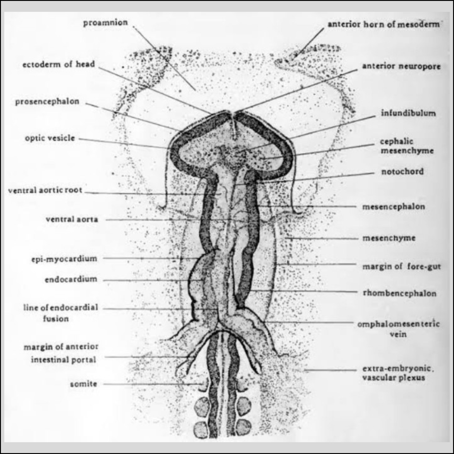
At about 24 hours of incubation in chick embryo, the coelom on each side of the ante­rior intestinal portion undergoes dilation to form the amniocardiac vesicles that gradu­ally push in from each side towards the mid­line. The splanchnic mesoderm bounding the amniocardiac vesicles on each side under­goes thickening.

These thickenings are the primordia of the epimyocardium. They are destined to form the external coat of the heart (epicardium) and the muscular layer of the heart (myocardium). Thereafter, cells detach from the epimyocardium and aggregate loosely between the splanchnic mesoderm and the adjacent endoderm of the intestine.

These cells form the primordium of the endocardium, (endothelial lining of the heart). Shortly, thereafter, the endocardial cells arrange themselves to produce a pair of thin-walled tubes.


While the above changes have been tak­ing place, the closure of the foregut at the level of the heart was also progressing. As a result the paired endocardial tubes were brought progressively close together. The two endocardial tubes finally fuse into a single tube lying in the midline.

At the time of fusion of the two primor­dia the tubes unite sequentially starting from the union of the truncus arteriosus, followed by the ventricle, atrium and finally the sinus venosus. Thus, in a chick embryo of 30 hours of incubation, fusion of the trunco-ventricular region is complete, while the atrium and sinus venosus are still double.

The entire fusion gets completed at about the 48 hour. If the cardia primordia are pre­vented from fusing by either chemical or sur­gical means, then separate independently beating hearts are formed on each side of the body.


Progressive Fusion of the Paired Primordia

While the two endocardial tubes were fusing into one, the epimyocardial layers were bending towards the midline to form a single tube around the endocardium. Ventral to the endocardial tubes the epimyocardial layers of opposite sides come into contact with each other.

At the region of contact the limbs of the mesodermal folds next to the endo­cardium fuse with each other to form ventral mesocardium. The ventral mesentery ruptures and disappears almost as rapidly as it is formed. Thus, the original right and left coelomic chambers become con­fluent to form a median unpaired pericardial cavity.

Dorsally the right and left epimyocardial layers also come close to each other. But here they do not fuse immediately as happens ventral to the heart. They persist for a time as a double-layered supporting membrane called the dorsal mesocardium, by which the primitive heart remains sus­pended from the floor of the pharynx.

Bending of the Tubular Primordial Heart and Establishment of its Regional Divisions

At the end of the 2nd day and during the 3rd day of incubation, the early heart under­goes a change in shape from a straight tubu­lar form to an S-shaped configuration. The primitive ventricle bends to the right and the atrium travels upward above the ventricle.


By the end of the 3rd day the heart of chick attains the S- shaped condition. This is considered to be the first step in the development of heart in all vertebrates.

The primary factor that brings about the regional differentiation of the heart is the rapid elongation of the primitive cardiac tube, resulting in its bending into a S-shape. The factors that cause the heart to bend and also that leading to the subdivision of the heart into atrial and ventricular regions has still remained obscure.

The ventricle is formed by the bent mid- portion of the original cardiac tube. As this ventricular loop becomes progressively more extensive, it at first projects ventrally but later is brought to its characteristic adult position, caudal to the atrium. The narrow connecting portion between the ventricle and the atrium is called the atrioventricular canal.

Right from its earliest appearance, the atrium and the ventricle show external indi­cations of their division into right and left side. A prominent median furrow (ventri­cular septum) appears at the apex of the ventricular loop. The atrium meanwhile has undergone rapid dilation and bulges out as a bilobed configuration on either side of the midline.

Beginning of Cardiac Function in Chick

Due to its metabolic requirements, the heart of chick embryo is required to com­mence beating early in its development. Thus, in chick at about 29 hours of incuba­tion, the earliest tentative beats begin while the paired cardiac primordia are still fusing. The intensity and regularity of the heart beat increases rapidly as the cardiac primordia continues to fuse, along with the laying down of the major divisions of the heart.

The first contraction appears in the ven­tricle before the atrium is fully established. The initial contraction rate of the ventricle is very slow but when the atrium starts pul­sating, the heart rate increases. The right heart begins to beat at a more rapid rate than the left at about the time when the sinus venosus would normally form. This is sug­gestive that the origin of the cardiac pace­maker is in the right cardiac primordium.

 Partitioning of the Heart in Chick

At the end of the 4th day, an interatrial septum between the two atrial lobes, begin to develop from the dorsoanterior area, slightly to the left of the sinus venosus opening. The interatrial septum continues to form posterior ward towards the narrow atrioventricular canal.

At the same time in the atrioventricular canal, two endocardial thickenings arise called the endocardial cushions. These are formed one dorsally and the other ventrally. At the apex of the ventricle another septum appears called the inter-ventricular septum. It grows forward towards the atrioventricular canal.


The two endocardial cushions grow towards each other,and about the sixth day of incubation, they fuse with one another. They thus, separate the atrioventricular canal into two channels (right and left atrio­ventricular canals) by the formation of a cushion septum.

The interatrial septum grows towards the endocardial cush­ion and finally unites with the cushion sep­tum. The interatrial septum, however, is never completely formed during the embry­onic life of chick.


Partitioning of the Atrio-Ventricular Canal

A small septum or fenestrae appear in the atrial septum permitting blood to pass through it. During the last week of incubation the fenestral opening become smaller and smaller and completely closes shortly after hatching.

Meanwhile the inter-ventricular septum grows forward to unite with the cushion sep­tum. Till the 4th day only one passage way leaves the heart via the developing bulbus cordis and ventral aorta. During the 5th day, a spiral septum begins to form within the caudal portion of the ventral aortal sac and the bulbus cordis.

Within the bulbus, this septum grows caudalward in a spiral man­ner, separating the pulmonary trunk ventral­ly and the root of the systemic aorta dorsally.

This septum continues to grow backwards also towards the inter-ventricular septum where it unites with a similar septum at the caudal end of the bulbus. At about the seventh day, the original bulbus cordis thus becomes divided into two separate vessels, a pulmonary trunk, which unites with the right ventricle and a systemic trunk, which is continuous with the left ventricle.

Valves of the Heart in Chick

As the above progress is taking place, the valves of the heart also start forming. When the spiral septum was developing in the bul­bus cordis, three semilunar or cup-shaped valves appear at the base of the systemic trunk and also at the base of the pulmonary trunk. These valves prevent the backflow of blood from the systemic trunk into the left ventricle and from the pulmonary trunk into the right ventricle.

When the cushion septum was forming, the atrioventricular or cuspid valves were formed at the two atrioventricular openings. The right auriculoventricular valve appears as a single strong muscular fold between the right chambers, while two valves make up the bicuspid valve between the left cham­bers. These valves prevent the back flow of blood into the atrium from the ventricles.

At the opening of the sinus into the right atrium, the right and left sides of the opening enlarge and produce folds that form the sinuatrial (sinuauricular) valves. A third valve, the Eustachian valve or sinus septum arises during the last Week of incubation, as a fold from the dorsal side of the sinus that projects into the right atrium dividing the sinuatrial opening.

Final Development of the Heart in Chick

As the time approaches for hatching, the sinus venosus becomes incorporated almost completely into the wall of the right atrium, while a small portion is probably incorpora­ted into the cardiac end of the left precaval vein (superior vena cava). The sinuatrial valves also disappear. At the time of hatching the fenestrae at the interatrial septum closes and the oxygenated and deoxygenated blood are completely separated.

Role of Organizer in the Development of Heart in Chick

It has been demonstrated in case of amphibian embryos that if the endoderm is removed at the neurula stage of develop­ment, the heart fails to form. Even when frag­ments of pre-cardiac mesoderm are cultivated in a culture medium, normal heart tissue does not result. Thus, the endoderm exercis­es an essential inductive influence on the pre­-cardiac myoderm.

However, in chick embryos, pre-cardiac mesodermal cells, iso­lated before the endoderm has formed, are capable of differentiating into cardiac muscle in vitro (Litvin et al., 1992), thus suggesting that the endodermal cells do not exercise any essential inductive influence on the forma­tion of heart.

*****_____*****

Tuesday, September 5, 2023

Development of Brain in Chick


Development of Brain in Chick 

Introduction 

The neural tube formed from the neural ectoderm is located dorsally in the median plain of the embryo and forms the basis for the central nervous system. Before going into the development of the brain a brief idea about the formation of neural tube in chick is dealt with.

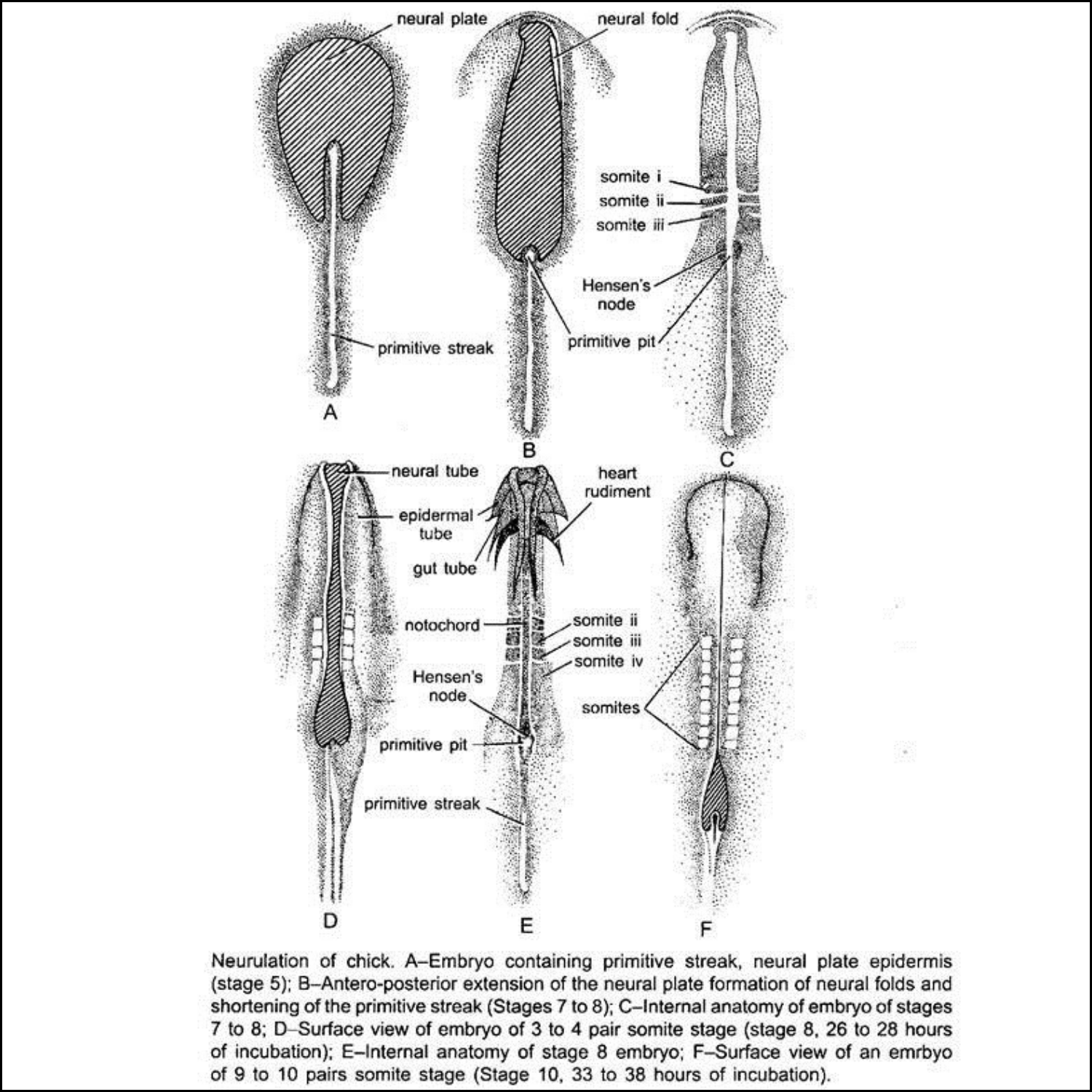
During gastrulation of chick a primitive streak with Hensen’s node is formed. In the 24 hours chick-embryo, as epiboly of ectodermal cells is taking place at the surface of the embryo, it brings about an elongation of the presumptive neural ecto­dermal cells above the notochordal area. 


As gastrulation comes to an end, the single-layered neural ectoderm rapidly becomes thick and stratified to form the neu­ral plate. Starting from the anterior towards the posterior side, the neural plate gradually sinks down and its margins becomes elevated resulting in the formation of a neural groove bounded by the neural folds. 


The groove continues to deepen and the neural folds meet above it, converting the original plate into a neural tube. The meeting of the neural folds is accompanied by the meeting of the epidermal ectoderm, which results in detaching the neural tube from the overlying ectoderm.

The primitive neural tube is composed of two major regions — the future brain region at its anterior end and the rudiments of the spinal cord at the posterior end. The brain, thus, can be defined simply as the anterior part of the neural tube lying within the crani­um. However, its morphogenetic history is one of increasing complexity of both gross anatomical form and internal organization.

The brain of Amphioxus hardly differs architecturally from the spinal cord. However, in amphibians to mammals, the prospective brain differs from the spinal cord behind. Therefore, in chick the fore part of the neural tube is considerably wider than that of the spinal tube.

As the neural folds are about to meet (at the time of formation of the neural tube), it is possible to distinguish two subdivisions in the forming brain. The pri­mordial brain tube is initially divided into an anterior pros-encephalon (archencephalon) and posterior deuteroencephalon. 


The regionally specific induction mecha­nisms influence the formation of the neural tube. Thus, the pros encephalon is induced by prechordal plate mesoderm and the deuteroencephalon by the anterior portion of the notochordal mesoderm, both lying beneath the primordial brain.

Formation of three parts in Brain

The deuteroencephalon undergoes a con­striction termed the isthmus, that divides it into a mesencephalon (mid brain) behind the pros encephalon (fore brain), and a rhomb encephalon (hind brain) in front of the spinal tube. Thus, the brain is said to pass through a three-part stage.

The distinction between these three divisions rests more on functional than anatomical ground. Accordingly, the pros encephalon is associated with the sense of smell, the mesencephalon with vision and the rhomb encephalon with hearing.

The anterior end of the neural tube during its early stages of development, tends to form primitive segments called neuromeres. These neuromeres fuse as they contribute to the primitive brain regions. 


Formation of five parts in Brain


Further embryonic modifications result in a subdivision of two of the three primary brain regions at about 75 to 80 hours of incu­bation. The pros encephalon is divided into anterior telencephalon and a posterior diencephalon. The mesencephalon remains intact. The rhomb encephalon is also divided into two, forming an anterior metencephalon and a posterior medulla or myelencephalon. 

The five divisions of the brain are arranged generally in a straight line in case of lower vertebrates. However, in case of chick and other higher vertebrates, the brain tube tends to bend upon itself at an early period of its embryogeny, by formation of flexures.


Formation of Flexures in Chick

Flexures are formed at three regions to create sharp angles in the long axis of the embryonic brain. These are accompanied by a more general flexing of the entire head. At the time of the primordial brain tube formation a primary neural flexure occurs at the mesencephalon region called the cepha­lic flexure.

The cephalic flexure is more pro­nounced in birds. It sharply bends the fore- brain to the ventral side. Soon thereafter, a second flexure called cervical flexure develops at the caudal portion of the myelencephalon and the anterior part of the spinal cord.

It bends the entire brain region of chick ventrally and is pro­nounced in birds like that of other amniotes. A third flexure or pontine flexure appears later at the mid-region of the rhomb encephalon, in the area between myelen­cephalon and metencephalon.

It counter acts the effect of the other two flexures by bending the brain in the opposite direction, that is dorsally. The pontine flex­ure is found only in the higher vertebrates.

Out-pocketing’s of the Brain in Chick

As the five primary subdivisions of the brain are established, its wall exhibits a num­ber of local out-pocketing’s. The telen­cephalon gives rise to two lateral pouches, the telencephalic vesicles. The telencephalic vesicles represent the rudi­ments of the cerebral lobes. The dien­cephalon on the other hand forms four or five evaginations.

These are — a mid-dorsal evagination, the epiphysis (rudiments of the pineal body); a second mid-dorsal evagina­tion occurs in front of the epiphysis called paraphysis; two ventro-lateral outgrowths, the optic vesicles and the infundibulum arising from a mid-ventral evagination.

The Rathke’s pouch arising from the stomodaeum unites with the infundibulum and ultimately differentiates into the anterior lobe of the pituitary body.

From the mesencephalic roof or tectum dorsal swellings appear. In birds like other amniotes, four swellings arise in the tectum, the corpora quadrigemina. From the roof of the metencephalon there arise two cerebellar outpushings.

These out-pocketing’s are accompanied by continued cell multiplications. The differentiating neuro-blasts undergo localized aggregations that bring about thickening of gray and white matter and a variety of folds, fissures and invaginations. The roof of the diencephalon and myelencephalon are thin, highly vascular and are devoid of nervous tissue.

Cavity of the Primitive Five-Part Brain in Chick

The brain as well as the spinal cord are hollow structures and this cavity is generally called the neural cavity or neurocoel. The neurocoel are fluid-filled cavities. The cavities of the telencephalic vesicles extend into the cerebral hemispheres (cere­bral lobes) and are known as the lateral ventricles or the first and second ventricles.

These ventricles communicate with the lumen of the middle of the telencephalon and that of the diencephalon, which together form the third ventricle.

The cavity of the mesencephalon is designated as the cerebral aqueduct or aqueduct of Sylvius. It becomes a narrow passage way and greatly reduced in chick as well as in other amniotes. The cavity of the rhomb encephalon is termed as the fourth ventricle. The cerebral aqueduct con­nects the third ventricle in front with that of the fourth.

In the roof of the third and fourth ventri­cles, small blood vessels from the pia mater form freely branching groups of vessels and are called the anterior and posterior choroid plexus. These plexus, plus those that form the walls of the lateral ventricles secrete cere­brospinal fluid that bathes the tissues of the brain and spinal cord.

Embryonic Induction in Brain Development of Chick

Activation phase of induction results primarily in the formation of neural tube. The normal prospect of the neural tube is its differentiation into four general regions. The most anterior of it will become the fore brain.

Subsequent regions will provide the mid brain and hind brain. This is then followed by the prospective spinal cord. Finally there is an area that fails to produce neural struc­tures at all but contributes to the mesodermal composition of the tail.

The question that may arise is, how the pattern of regions appearing in the neural tube may be related to a pattern present in the inducing mesoderm? This is indicated by two sets of facts. First, in the anatomical fact the notochord terminates anteriorly at approximately the junction between the fore- and mid-brain, the remaining forward dis­tance being occupied by the prechordal mesoderm or plate.

This prechordal plate may thus be considered the inductor of the fore brain and the notochord the inductor of the regions behind. The second fact is that different levels of the mesoderm have unlike inductive capacities. If different regions of the prospective chorda mesoderm from ante­rior to posterior are cut out and grafted into young gastrula, it is found that they induce characteristically different regions of the neu­ral tube.

Immediately after the neural plate is established, this neural primordium itself acts as a secondary inductor. Some examples are induction of sensory structures (lens of the eye etc.), the otic vesicle (inner ear) and the series of ectodermal placodes. The brain and the spinal cord later, also induces the for­mation of bony structure that protects them.

Sunday, September 3, 2023

Neurulation (Formation of neural tube)


Neurulation (Formation of neural tube) 

Introduction:

There are two major ways of forming a neural tube. In primary neurulation, the cells surrounding the neural plate direct the neural plate cells to proliferate, invaginate, and pinch off from the surface to form a hollow tube. In secondary neurulation, the neural tube arises from a solid cord of cells that sinks into the embryo and subsequently hollows out (cavitates) to form a hollow tube. 

The extent to which these modes of construction are used varies among vertebrate classes. Neurulation in fishes is exclusively secondary. In birds, the anterior portions of the neural tube are constructed by primary neurulation, while the neural tube caudal to the twenty-seventh somite pair (i.e., everything posterior to the hindlimbs) is made by secondary neurulation (Pasteels 1937; Catala et al. 1996). 

In amphibians, such as Xenopus, most of the tadpole neural tube is made by primary neurulation, but the tail neural tube is derived from secondary neurulation (Gont et al. 1993). In mice (and probably humans, too), secondary neurulation begins at or around the level of somite 35 (Schoenwolf 1984; Nievelstein et al. 1993).

Primary neurulation:


The events of primary neurulation in the chick and the frog are illustrated in Figures 01 and 02, respectively. During primary neurulation, the original ectoderm is divided into three sets of cells: 

(1) the internally positioned neural tube, which will form the brain and spinal cord, 

(2) the externally positioned epidermis of the skin, and 

(3) the neural crest cells. 

The neural crest cells form in the region that connects the neural tube and epidermis, but then migrate elsewhere; they will generate the peripheral neurons and glia, the pigment cells of the skin, and several other cell types.

Chick Neurulation. By 33 hours of incubation, neurulation in the chick embryo is well underway. Both whole mounts and a complete set of serial cross sections through a 33-hour chick embryo are included in this segment so that you can see this amazing event. The serial sections can be displayed either as a continuum in movie format or individually, along with labels and color-coding that designates germ layers.

The process of primary neurulation appears to be similar in amphibians, reptiles, birds, and mammals (Gallera 1971). Shortly after the neural plate has formed, its edges thicken and move upward to form the neural folds, while a U-shaped neural groove appears in the center of the plate, dividing the future right and left sides of the embryo. The neural folds migrate toward the midline of the embryo, eventually fusing to form the neural tube beneath the overlying ectoderm. The cells at the dorsalmost portion of the neural tube become the neural crest cells.

Neurulation occurs in somewhat different ways in different regions of the body. The head, trunk, and tail each form their region of the neural tube in ways that reflect the inductive relationship of the pharyngeal endoderm, prechordal plate, and notochord to its overlying ectoderm. The head and trunk regions both undergo variants of primary neurulation, and this process can be divided into four distinct but spatially and temporally overlapping stages: 

(1) formation of the neural plate; 

(2) shaping of the neural plate; 

(3) bending of the neural plate to form the neural groove; and 

(4) closure of the neural groove to form the neural tube (Smith and Schoenwolf 1997).

Formation and shaping of the neural plate: 

The process of neurulation begins when the underlying dorsal mesoderm (and pharyngeal endoderm in the head region) signals the ectodermal cells above it to elongate into columnar neural plate cells (Smith and Schoenwolf 1989; Keller et al. 1992 ). 

Their elongated shape distinguishes the cells of the prospective neural plate from the flatter pre-epidermal cells surrounding them. As much as 50% of the ectoderm is included in the neural plate. 

The neural plate is shaped by the intrinsic movements of the epidermal and neural plate regions. The neural plate lengthens along the anterior-posterior axis, narrowing itself so that subsequent bending will form a tube (instead of a spherical capsule).

In both amphibians and amniotes, the neural plate lengthens and narrows by convergent extension, intercalating several layers of cells into a few layers. In addition, the cell divisions of the neural plate cells are preferentially in the rostral-caudal (beak-tail; anterior-posterior) direction (Jacobson and Sater 1988; Schoenwolf and Alvarez 1989; Sausedo et al. 1997. 

These events will occur even if the tissues involved are isolated. If the neural plate is isolated, its cells converge and extend to make a thinner plate, but fail to roll up into a neural tube. 

However, if the “border region” containing both presumptive epidermis and neural plate tissue is isolated, it will form small neural folds in culture (Jacobson and Moury 1995; Moury and Schoenwolf 1995).

Bending of the neural plate

The bending of the neural plate involves the formation of hinge regions where the neural tube contacts surrounding tissues. In these regions, the presumptive epidermal cells adhere to the lateral edges of the neural plate and move them toward the midline. 

In birds and mammals, the cells at the midline of the neural plate are called the medial hinge point (MHP) cells. They are derived from the portion of the neural plate just anterior to Hensen's node and from the anterior midline of Hensen's node (Schoenwolf 1991a,b; Catala et al. 1996). 

The MHP cells become anchored to the notochord beneath them and form a hinge, which forms a furrow at the dorsal midline. The notochord induces the MHP cells to decrease their height and to become wedge-shaped (van Straaten et al. 1988; Smith and Schoenwolf 1989). 

The cells lateral to the MHP do not undergo such a change. Shortly thereafter, two other hinge regions form furrows near the connection of the neural plate with the remainder of the ectoderm. 

These regions are called the dorsolateral hinge points (DLHPs), and they are anchored to the surface ectoderm of the neural folds. These cells, too, increase their height and become wedge-shaped.

Cell wedging is intimately linked to changes in cell shape. In the DLHPs, microtubules and microfilaments are both involved in these changes. Colchicine, an inhibitor of microtubule polymerization, inhibits the elongation of these cells, while cytochalasin B, an inhibitor of microfilament formation, prevents the apical constriction of these cells, thereby inhibiting wedge formation (Burnside 1973; Karfunkel 1972; Nagele and Lee 1987). 

After the initial furrowing of the neural plate, the plate bends around these hinge regions. Each hinge acts as a pivot that directs the rotation of the cells around it (Smith and Schoenwolf 1991).

Meanwhile, extrinsic forces are also at work. The surface ectoderm of the chick embryo pushes toward the midline of the embryo, providing another motive force for the bending of the neural plate (Alvarez and Schoenwolf 1992). 

This movement of the presumptive epidermis and the anchoring of the neural plate to the underlying mesoderm may also be important for ensuring that the neural tube invaginates into the embryo and not outward. 

If small pieces of neural plate are isolated from the rest of the embryo (including the mesoderm), they tend to roll inside out (Schoenwolf 1991a). The pushing of the presumptive epidermis toward the center and the furrowing of the neural tube creates the neural folds.

Closure of the neural tube: 


The neural tube closes as the paired neural folds are brought together at the dorsal midline. The folds adhere to each other, and the cells from the two folds merge. In some species, the cells at this junction form the neural crest cells. In birds, the neural crest cells do not migrate from the dorsal region until after the neural tube has been closed at that site. In mammals, however, the cranial neural crest cells (which form facial and neck structures) migrate while the neural folds are elevating (i.e., prior to neural tube closure), whereas in the spinal cord region, the crest cells wait until closure has occurred (Nichols 1981; Erickson and Weston 1983).

The closure of the neural tube does not occur simultaneously throughout the ectoderm. This is best seen in those vertebrates (such as birds and mammals) whose body axis is elongated prior to neurulation. 

Depicts neurulation in a 24-hour chick embryo. Neurulation in the cephalic (head) region is well advanced, while the caudal (tail) region of the embryo is still undergoing gastrulation. Regionalization of the neural tube also occurs as a result of changes in the shape of the tube. 

In the cephalic end (where the brain will form), the wall of the tube is broad and thick. Here, a series of swellings and constrictions define the various brain compartments. 

Caudal to the head region, however, the neural tube remains a simple tube that tapers off toward the tail. The two open ends of the neural tube are called the anterior neuropore and the posterior neuropore.

Unlike neurulation in chicks (in which neural tube closure is initiated at the level of the future midbrain and “zips up” in both directions), neural tube closure in mammals is initiated at several places along the anterior-posterior axis (Golden and Chernoff 1993; Van Allen et al. 1993). 

Different neural tube defects are caused when various parts of the neural tube fail to close.

Failure to close the human posterior neural tube regions at day 27 (or the subsequent rupture of the posterior neuropore shortly thereafter) results in a condition called spina bifida, the severity of which depends on how much of the spinal cord remains exposed. 

Failure to close the anterior neural tube regions results in a lethal condition, anencephaly. Here, the forebrain remains in contact with the amniotic fluid and subsequently degenerates. Fetal forebrain development ceases, and the vault of the skull fails to form. 

The failure of the entire neural tube to close over the entire body axis is called craniorachischisis. Collectively, neural tube defects are not rare in humans, as they are seen in about 1 in every 500 live births. Neural tube closure defects can often be detected during pregnancy by various physical and chemical tests.

Human neural tube closure requires a complex interplay between genetic and environmental factors. Certain genes, such as Pax3, sonic hedgehog, and openbrain, are essential for the formation of the mammalian neural tube, but dietary factors, such as cholesterol and folic acid, also appear to be critical. 

It has been estimated that 50% of human neural tube defects could be prevented by a pregnant woman's taking supplemental folic acid (vitamin B12), and the U.S. Public Health Service recommends that all women of childbearing age take 0.4 mg of folate daily to reduce the risk of neural tube defects during pregnancy (Milunsky et al. 1989; Czeizel and Dudas 1992; Centers for Disease Control 1992).

The neural tube eventually forms a closed cylinder that separates from the surface ectoderm. This separation is thought to be mediated by the expression of different cell adhesion molecules. Although the cells that will become the neural tube originally express E-cadherin, they stop producing this protein as the neural tube forms, and instead synthesize N-cadherin and N-CAM. 

As a result, the surface ectoderm and neural tube tissues no longer adhere to each other. If the surface ectoderm is experimentally made to express N-cadherin (by injecting N-cadherin mRNA into one cell of a 2-cell Xenopus embryo), the separation of the neural tube from the presumptive epidermis is dramatically impeded (Detrick et al. 1990; Fujimori et al. 1990).

Secondary neurulation


Secondary neurulation involves the making of a medullary cord and its subsequent hollowing into a neural tube. Knowledge of the mechanisms of secondary neurulation may be important in medicine, given the prevalence of human posterior spinal cord malformations.

In frogs and chicks, secondary neurulation is usually seen in the neural tube of the lumbar (abdominal) and tail vertebrae. In both cases, it can be seen as a continuation of gastrulation. In the frog, instead of involuting into the embryo, the cells of the dorsal blastopore lip keep growing ventrally. 

The growing region at the tip of the lip is called the chordoneural hinge (Pasteels 1937), and it contains precursors for both the posteriormost portion of the neural plate and the posterior portion of the notochord. 

The growth of this region converts the roughly spherical gastrula, 1.2 mm in diameter, into a linear tadpole some 9 mm long. The tip of the tail is the direct descendant of the dorsal blastopore lip, and the cells lining the blastopore form the neurenteric canal. 

The proximal part of the neurenteric canal fuses with the anus, while the distal portion becomes the ependymal canal (i.e., the lumen of the neural tube) ( Gont et al. 1993).


Reverse Transcription

Reverse Transcription Introduction Reverse transcription is the synthesis of DNA from an RNA template. This process is driven by RNA-depende...