Wednesday, January 31, 2024

Haemophilia

Haemophilia 

Key facts

#Haemophilia is an inherited bleeding disorder, which means it can be passed on from birth parents to their children.

#If you have haemophilia, your blood doesn’t clot properly, which makes it difficult to control bleeding.

#Some people have mild haemophilia, while others are more severely affected.

#The main symptoms of haemophilia are easy bruising, having large bruises, and greater than normal bleeding from surgery or menstruation.

#There is no cure for haemophilia, but heavy bleeding can be controlled with medical treatment.

Introduction

Haemophilia is an inherited bleeding disorder, which means it can be passed on from birth parents to their children. If you have haemophilia, your blood doesn’t clot properly. This can lead to bleeding that is difficult to control.

When a blood vessel is injured, special proteins in the blood called ‘clotting factors’ act to control blood loss by plugging or patching up the injury. People with haemophilia have lower than normal levels of a clotting factor.

Types of Haemophilia

There are 2 types of haemophilia.

1.Haemophilia A (also called classic haemophilia) is the most common type. It is caused by a lack of clotting factor 8.

2.Haemophilia B (sometimes called Christmas disease) is caused by a lack of clotting factor 9.

It is important to know the type of haemophilia you have, as each type requires different clotting factor treatment.

Some people have mild haemophilia, while others are more severely affected.

Symptoms of Haemophilia

The main signs of haemophilia are:

*Easy bruising from an early age.

*Internal bleeding for no obvious reason, especially in the joints and muscles.

*Greater than normal bleeding following injury or surgery.

*Abnormally heavy bleeding during menstruation or after giving birth.

Although bleeding problems often start from a very young age, some children don’t have symptoms until they begin walking or running. People with mild haemophilia may not bleed excessively until they get an injury or have surgery.

Causes of Haemophilia

Haemophilia is an inherited condition and occurs in families, but 1 in 3 people with haemophilia will have it even without a family history of the disorder.

Haemophilia is caused by a mutation in a gene that is located on the X chromosome.

Females have 2 copies of the X chromosome in body cells, while males have one X chromosome and one Y chromosome. For this reason, haemophilia is much more common in males. Females who inherit a gene for haemophilia often have an unaffected gene on their other X chromosome. This means that they while they can pass on the gene for haemophilia to their children (be a genetic carrier), they may not be affected by haemophilia themselves.


Diagnosis

If your doctor suspects that you have haemophilia, they may refer you for blood tests to measure the levels of clotting factors in your blood. These tests can show the type and severity of the disease.

Genetic testing can often confirm a diagnosis of haemophilia, but in some cases, doctors won’t know if you have a gene mutation.

Treatment

There is currently no cure for haemophilia. It is possible to manage haemophilia effectively, although it can be complex.

When someone with haemophilia has a bleeding episode, they will need treatment to help their blood clot and stop the bleeding. This usually involves giving clotting factors by infusion or injection.

If you have haemophilia, you need to be very careful not to injure yourself. Be sure to learn how to recognise a bleed, as there may be no visible signs of bleeding, for example, if your bleed inside your body.

Australian guidelines recommend that people with haemophilia get care from a team of healthcare professionals. This team may include doctors (GPs and/or specialists) nurses, medical scientists, physiotherapists, social workers and psychologists.

If you are diagnosed with haemophilia, you should ask your doctor about the benefits of referral to a haemophilia treatment centre, where a health team can provide you with comprehensive care.

Complications of Haemophilia

The most common complication of the haemophilia is damage to joints and muscles that result from bleeding into these areas. These complications are best managed by a multidisciplinary health care team.

Saturday, January 27, 2024

Phobias


Phobias

Introduction

Phobias are an intense fear of something, like heights or certain animals, that causes significant distress. They’re typically treated with therapy, medication, or a combination of both.

A phobia is an irrational fear of something that’s unlikely to cause harm. The word itself comes from the Greek word“phobos,” which means “fear” or “horror.”

Hydrophobia, for example, literally translates to fear of water.

When someone has a phobia, they experience intense fear of a certain object or situation. Phobias are different from regular fears because they cause significant distress, possibly interfering with life at home, work, or school.

People with phobias actively avoid the phobic object or situation, or they endure it with intense fear or anxiety.

Agoraphobia, a fear of places or situations that trigger fear or helplessness, is singled out as a particularly common fear with its own unique diagnosis. Social phobias, which are fears related to social situations, are also singled out with a unique diagnosis.

Phobias come in all shapes and sizes. Because there are an infinite number of objects and situations, the list of specific phobias is quite long.

Types of phobias

According to the DSM-5, specific phobias typically fall within five general categories:

*Fears related to animals (spiders, dogs, insects)

*Fears related to the natural environment (heights, thunder, darkness)

*Fears related to blood, injury, or medical issues (injections, broken bones, falls)

*Fears related to specific situations (flying, riding an elevator, driving)

*Other (fear of choking, loud noises, drowning)
These categories encompass an infinite number of specific objects and situations.

There’s no official list of phobias beyond what’s outlined in the DSM-5, so clinicians and researchers create names for them as the need arises. This is typically done by combining a Greek (or sometimes Latin) prefix that describes the phobia with the –phobia suffix.

For example, a fear of water would be named by combining “hydro” (water) and “phobia” (fear).

There’s also such a thing as a fear of fears (phobophobia). This is actually more common than you might imagine.

People with anxiety disorders sometimes experience panic attacks when they’re in certain situations. These panic attacks can be so uncomfortable that people do everything they can to avoid them in the future.

For example, if you have a panic attack while sailing, you may fear sailing in the future, but you may also fear panic attacks or fear developing hydrophobia.

List of phobias

Studying specific phobias is a complicated process. Most people don’t seek treatment for these conditions, so cases largely go unreported.

These phobias also vary based on cultural experiences, gender, and age.

Here’s a look at some phobias that have been identified.

1.Achluophobia - Fear of darkness
2.Acrophobia - Fear of heights
3.Aerophobia - Fear of flying
4.Agoraphobia - Fear of public spaces or crowds
5.Aichmophobia - Fear of needles or pointed objects
6.Ailurophobia - Fear of cats
7.Alektorophobia - Fear of chickens
8.Algophobia - Fear of pain
9.Amaxophobia - Fear of riding in a car
10.Androphobia - Fear of men
11.Anginophobia - Fear of angina or choking
12.Anthophobia - Fear of flowers
13.Anthropophobia - Fear of people or society
14.Aphenphosmphobia - Fear of being touched
15.Arachnophobia - Fear of spiders
16.Arithmophobia - Fear of numbers
17.Astraphobia - Fear of thunder and lightning
18.Ataxophobia - Fear of disorder or untidiness
19.Atelophobia - Fear of imperfection
20.Atychiphobia - Fear of failure
21.Autophobia - Fear of being alone
22.Bacteriophobia - Fear of bacteria
23.Barophobia - Fear of gravity
24.Bathmophobia - Fear of stairs or steep slopes
25.Batrachophobia - Fear of amphibians
26.Belonephobia - Fear of pins and needles
27.Bibliophobia - Fear of books
28.Botanophobia - Fear of plants
29.Cacophobia - Fear of ugliness
30.Catagelophobia - Fear of being ridiculed
31.Catoptrophobia - Fear of mirrors
32.Chionophobia - Fear of snow
33.Chromophobia - Fear of colors
34.Chronomentrophobia - Fear of clocks
35.Cibophobia - Fear of food
36.Claustrophobia - Fear of confined spaces
37.Coulrophobia - Fear of clowns
38.Cyberphobia - Fear of computers
39.Cynophobia - Fear of dogs
40.Dendrophobia - Fear of trees
41.Dentophobia - Fear of dentists
42.Domatophobia - Fear of houses
43.Dystychiphobia - Fear of accidents
44.Emetophobia - Fear of vomiting
45.Entomophobia - Fear of insects
46.Ephebiphobia - Fear of teenagers
47.Equinophobia - Fear of horses
48.Gamophobia - Fear of marriage or commitment
49.Genuphobia - Fear of knees
50.Glossophobia - Fear of speaking in public
51.Gynophobia - Fear of women
52.Heliophobia - Fear of the sun
53.Hemophobia - Fear of blood
54.Herpetophobia - Fear of reptiles
55.Hydrophobia - Fear of water
56.Hypochondria - Fear of illness
57.Iatrophobia - Fear of doctors
58.Insectophobia - Fear of insects
59.Koinoniphobia - Fear of rooms full of people
60.Leukophobia - Fear of the color white
61.Lilapsophobia - Fear of tornadoes and hurricanes
62.Lockiophobia - Fear of childbirth
63.Mageirocophobia - Fear of cooking
64.Megalophobia - Fear of large things
65.Melanophobia - Fear of the color black
66.Microphobia - Fear of small things
67.Mysophobia - Fear of dirt and germs
68.Necrophobia - Fear of death or dead things
69.Noctiphobia - Fear of the night
70.Nosocomephobia - Fear of hospitals
71.Nyctophobia - Fear of the dark
72.Obesophobia - Fear of gaining weight
73.Octophobia - Fear of the number 8
74.Ombrophobia - Fear of rain
75.Ophidiophobia - Fear of snakes
76.Ornithophobia - Fear of birds
77.Papyrophobia - Fear of paper
78.Paruresis - Fear of urinating in public places or in the presence of others
79.Pathophobia - Fear of disease
80.Pedophobia - Fear of children
81.Philophobia - Fear of love
82.Phobophobia - Fear of phobias
83.Podophobia - Fear of feet
84.Pogonophobia - Fear of beards
85.Porphyrophobia - Fear of the color purple
86.Pteridophobia - Fear of ferns
87.Pteromerhanophobia - Fear of flying
88.Pyrophobia - Fear of fire
89.Samhainophobia - Fear of Halloween
90.Scolionophobia - Fear of school
91.Selenophobia - Fear of the moon
92.Sociophobia - Fear of social evaluation
93.Somniphobia - Fear of sleep
94.Tachophobia - Fear of speed
95.Technophobia - Fear of technology
96.Thalassophobia - Fear of deep water
97.Tonitrophobia - Fear of thunder
98.Trypanophobia - Fear of needles or injections
99.Trypophobia - Fear of clustered patterns of holes
100.Venustraphobia - Fear of beautiful women
101.Verminophobia - Fear of germs
102.Wiccaphobia - Fear of witches and witchcraft
103.Xenophobia - Fear of strangers or foreigners
104.Zoophobia - Fear of animals

Treating a phobia

Phobias are typically treated with therapy, medication, or a combination of both:

Exposure therapy. During exposure therapy, which is a type of cognitive behavioral therapy, you work with a psychologist to learn how to desensitize yourself to the object or situation that you fear. The goal is to improve your quality of life so that you’re no longer hindered or distressed by your fear.

Antianxiety medication. Your doctor may recommend certain anxiety-reducing medications that can help you through exposure therapy. While these medications aren’t exactly a treatment for phobias, they can help make exposure therapy less distressing.

Other medications. Your clinician might also prescribe beta-blockers and benzodiazepines to manage feelings of anxiety or panic.

The Takeaway

Phobias are persistent, intense, and unrealistic fears of a certain object or situation. Specific phobias are related to certain objects and situations. They typically involve fears related to animals, natural environments, medical issues, or specific situations.

While phobias can be extremely uncomfortable and challenging, therapy and medication can help. If you think you may have a phobia that’s causing a disruption in your life, speak with your doctor for an evaluation and treatment options.

Friday, January 26, 2024

Trephination

Trephination

Introduction:

Trephination, also known as trepanning, trepanation or burr holing, is one of the oldest surgical procedures known to humanity and refers to a surgical procedure in which a circular piece of bone is drilled and excised, most commonly from the human skull. While the term trephination typically refers to the creation of a hole in the skull, there are other uses of trephination, such as in the fingernail or toenail. 

Trephination of the skull can be performed while the person is living or dead (i.e., postmortem). It has been performed in many areas of the world and throughout history, spanning from Mesolithic (i.e., prehistoric) to modern times. 

Today, there are four techniques that can be used for trephination: drilling using a trephine (i.e., surgical instrument with a cylindrical blade) or drill, scraping using an abrasive instrument, cutting using an incisive instrument, or boring and cutting using a drill and an incisive instrument. Complications that may arise during trephination include brain injury, hemorrhage, and infection. 

Trephination Uses:

Throughout history, trephination has been a medical technique used to relieve intracranial pressure and headaches. In the fifth century BCE, physicians practicing under the guidance of Hippocrates believed that stagnant blood, like stagnant water, could decay and turn into pus (i.e., liquid produced in infected tissue). For that reason, trephining allowed blood to flow out before it spoiled. This eventually fell out of practice due to low survival rates and infection. 

At the end of the 19th century, physicians learned about the importance of modern antisepsis and infection prophylaxis, and understood the importance of intracerebral pressure in head injury. 

This allowed trephination to return as a surgical therapy for the treatment and management of head trauma, including fractured skulls, head wounds, intracranial hemorrhage, intracranial disorders, chronic headache, brain tumors, and other painful skull disorders. 

Trephining is no longer used for therapeutic purposes in itself, but is typically used for exploratory diagnosis, relieving intracranial pressure due to epidural or subdural hematoma, debriding a penetrating head wound, and gaining access to the brain. 

In modern practice, the technique of drilling a hole in one’s skull is more commonly known as a craniotomy. In this procedure, a surgeon removes a piece of the skull to access the brain in order to treat conditions such as brain lesions and brain tumors. 

After treatment, the piece of skull is then replaced as soon as possible. It is important that, in the process of trephination, surgeons are careful to avoid penetration of the dura mater, which is the outer layer of connective tissue that protects the brain. If the dura mater is damaged, this could result in infection and physical damage to the underlying blood vessels, meninges, and brain tissue.

In 1884, frontal sinus trephination for the treatment of frontal sinus disease was described by physician Alexander Ogston. The technique had a high failure rate from complications of nasofrontal duct stenosis, or blockage of a duct that drains the sinuses. 

In the 1980s, endoscopic sinus surgery became the standard of care for the surgical management of frontal sinus disease. During this procedure, an endoscope (i.e., a camera on the end of a thin tool) is inserted into the nose to open the drainage pathways of the sinuses and restore their function. 

While endoscopy is still the standard of care to date, trephination may be used in cases of frontal sinus disease that are unable to be treated by endoscopy. For example, trephination is used if the surgeon needs an additional port for dissection, if the surgeon needs to rapidly decompress the frontal sinus, or if there is a tumor that has to be resected but cannot be done by endoscopy. 

Frontal sinus trephination is performed under local or general anesthesia. In the case of frontal sinus disease, trephination is used to relieve the pressure of the frontal sinus and drain the inflammatory cells and fluid. Following trephination, intravenous antibiotics are then administered to limit potential infection. 

In addition to treating frontal sinus disease, trephination has been used in the eye for corneal transplant and to treat glaucoma. Glaucoma is a condition in which the outflow of aqueous fluid of the eye is blocked, and intraocular pressure, or pressure within the eye, builds up. 

A procedure known as corneoscleral trephination has been documented in the treatment of glaucoma and consists of using a 1-2mm blade and topical anesthetic to relieve pressure of the eye. 

Lastly, while trephination mainly refers to the process of creating a hole in the human skull, the process of creating a hole in the fingernail or toenail is also referred to as trephination. Trephination of the nail is used as a procedure for subungual hematomas (i.e., blood under the nail). 

Subungual hematomas can occur from trauma, such as crushing one’s finger in a door or stubbing one's toe. Typically, an electric cautery tool is used to melt a hole through the nail and drain the blood from under the nail. Clinicians may also choose to use a needle and rotate it back and forth with slight downward pressure until dark blood is noted, being careful to not damage the nail bed. 

Important facts:

Trephination refers to the process of creating a hole in the skull and, in specific cases, the nail and is one of the oldest surgical procedures documented to date. It has been used throughout history with the goal of relieving pressure; managing symptoms of head trauma, such as subdural and epidural hematomas; and accessing brain tumors. 

Trephination can be performed using a trephine, a drill, by scraping with an abrasive instrument, or cutting with an incisive instrument. Common complications that may arise with trephination include infection and, if the dura mater surrounding the brain is damaged in the process, damage to the underlying blood vessels, meninges, and brain. Modern medicine has used trephination as part of treatments for frontal sinus disease, glaucoma, and subungual hematomas. 

Monday, January 15, 2024

Myopia

Myopia

Introduction

The structure of your eye is to blame. When your eyeball is too long or the cornea -- the protective outer layer of your eye -- is too curved, the light that enters your eye won’t focus correctly. Images focus in front of the retina, the light-sensitive part of your eye, instead of directly on the retina. This causes blurred vision. Doctors call this a refractive error.

High myopia: It’s a more serious form of the condition, where the eyeball grows more than it is supposed to and becomes very long front to back. Besides making it hard to see things at a distance, it can also raise your chance of having other conditions like a detached retina, cataracts, and glaucoma.

Degenerative myopia: Also called pathological or malignant myopia, it is a rare type you usually inherit from your parents. Your eyeball gets longer very quickly and causes severe myopia, usually by the teenage or early adult years. This type of myopia can get worse far into adulthood. Besides making it hard to see things at a distance, you may have a higher chance of having a detached retina, abnormal blood vessel growth in the eye (choroid neovascularization), and glaucoma.

Symptoms

Chances are the only symptom is that more distant objects are blurred. You may also notice:

Headaches
Squinting
Eye strain
Eye fatigue when you try to see objects more than a few Feet away
Children with myopia often have trouble reading the blackboard at school.

Diagnosis and Treatment

An eye exam can show you if you’re myopic. Glasses, contacts, or refractive surgery can usually correct the problem.

When you have myopia, your prescription for glasses or contact lenses will be a negative number. The more negative the number, the stronger your lenses will be. For example, -3.00 is stronger than -2.50.

Your prescription helps the eye focus light on your retina. That clears up your vision.

Eye surgery can improve your vision so much you may no longer need to wear glasses or contacts. The most common procedures for myopia are:

Photorefractive keratectomy: Also called PRK, this surgery uses a laser to sculpt the middle layer of your cornea. That flattens the cornea’s curve and lets light rays focus closer to or on your retina.

LASIK: This is the most common surgery for myopia. The surgeon uses a laser or another tool to create a thin flap on the top layer of your cornea. They sculpt the cornea with another laser and move the flap back into place.

EVO Implantable Collamer Lens (ICL): Using a microscopic incision, a contact lens made of a soft, polmeric materialis implanted into your eye between you natural lens and your iris. It helps refract light on the retina, producing clearer vision.

In the case of high myopia, special contacts or atropine eyedrops have been found to be effective in slowing the progression. In some cases, your doctor may suggest cataract or clear lens replacement surgery. 

Does It Get Better Over Time?

Myopia runs in families and will probably start in childhood. Multifocal lens (glasses or contacts) and eye drops such as atropine, pirenzepine gel, or cyclopentolate can help slow the progression. Your eyes usually stops changing after your teenage years, but not always. The incidents of myopia have been rising at an alarming rate in recent years. If you notice changes in your vision, get your eyes checked. See your eye doctor every year.

Optics of Myopia

Before discussing Optics of Myopia we should ratify parishioner phrase in Myopia. Briefly, the uncorrected myopia or nearsighted person. Usually can see up close better than far away. Hence, the term “nearsighted” make sense.

Derived from two Greek root words from myein “to shut” – ops “eye”.

Myopia is the refractive state of the eye in which with normal tonic accommodation, the parallel rays of light are brought to a focus on a point in front of the retinal plane when the eye is at rest.

In myopia the eyeball is usually deformed ; the deformation occurs at the posterior part of the globe only ,the anterior part is normal. The eyeball is usually large and looks prominent. When adducted the equator can be seen.

Refractive Error Definition 

A refractive error is a very common eye disorder . It exists when the rays of light do not focus on the retina means the eye cannot clearly focus the images from the outside world .

The eye’s optical system is to powerful over converging light in front of the retina . ( Myopia/Nearsighted)

The result of refractive errors is blurred vision, double vision , haziness or eye strain . which is sometimes so severe that it causes visual impairment .

Optics of Myopia: In myopes a near object may be focused without any effort of accommodation if it is situated at the punctum remotum . The image on the retina of an object at infinity is made up of circles of diffusion formed by divergent beam. 

In view of the absence of sharp focus the distant objects appear blurred. The nodal point in myopes is farther away from the retina and the image formed is larger in size which partly compensates for the blur due to absence of sharp focus . 

Accommodation is of little value to myopes, as any exercise of accommodation would only accentuate his visual problem . It would neither increase his vision nor reduce his myopia . In higher errors of refraction the amplitude of accommodation is small. 

The patient has no incentive to improve it and convergence does not get an accommodative influence . All these factors result in fatigue of accommodation and eye strain. Due to dissociation of convergence and accommodation exophoria commonly occurs and this may ultimately break into exotropia.

The eye of simple myopia is large and prominent. The Anterior chamber is deeper than normal and the pupil reacts sluggishly to light. The macula is slightly nearer to the disc than in a normal eye . 

The visual axis gets so altered that in some cases the angle alpha becomes slightly negative which gives an impression of a convergent squint . In myopia more commonly one sees a divergent strabismus.

Optical System 

The optical system is too powerful for it’s axial length , over converging light in front of the retina ( myopia / nearsighted).

Image of Distant Object 

Image of distant object on retina is made up of circle of diffusion formed by divergent beam since the parallel rays of light coming from the infinity are focused in front of the retina.

Far Point of the Myopic Eye : Far point is finite point in front of eye . The far point of myopic eye is 70 cm.

Nodal Point : Nodal point is further away from retina . The nodal point of human eye is located 17 mm in front of the retina.

Accommodation : Accommodation in uncorrected myopes is not developed normally , they may suffer from convergence insufficiency , exophoria and early presbyopia as they grow.

Image Formation: In myopia image formed in front of eye which is corrected by placing the negative lenses .

Optical Treatment : Include prescription of appropriate concave lens minimum acceptance providing maximum vision should be prescribed .

Never Overcorrected myopia

Guidelines For Correcting Low Degree Of Myopia Upto-6D :

Children younger than 8yrs should be fully corrected and instructed to use their glasses constantly.

Adult younger than 30 years :

Usually accept their full correction .

Older than 30 years :

Not able to tolerate a full correction with which the patient feels comfortable .

Prescribe less than full correction with which the patient feels comfortable.

Fossil Formation


Fossil Formation 

Introduction

A fossil is the mineralized partial or complete form of an organism, or of an organism’s activity, that has been preserved as a cast, impression or mold. A fossil gives tangible, physical evidence of ancient life and has provided the basis of the theory of evolution in the absence of preserved soft tissues.

The root of the word fossil derives from the Latin verb ‘to dig’ (fodere). A preserved inorganic object is not a fossil. The same applies to the mummification, desiccation and freezing of organisms, as mineralization is not a characteristic feature of these processes.Fossil form depends on the material in which it is preserved, and the ancient environment. Fossils can also represent organism movement and activity in the form of footprints and boring holes. An insect trapped in amber is a fossil subtype known as an inclusion; the originally soft, gum-like coniferous tree resin has fossilized into stone-like amber with the chitinous skeleton of the insect inside preserved, as shown below.

The oldest human fossil, where human refers to Homo erectus, Homo ergaster, and Homo georgicus, was a set of five skulls found in Dmanisi in Georgia between 1999 and 2005. These date back to approximately 1.8 million years ago. The oldest fossil remains depict five different species of microbe, preserved in a 3.5-billion-year-old rock in Australia. These microbes were carbon-dated by researchers at UCLA and the University of Wisconsin-Madison.

Types of Fossils

Fossil types are grouped according to the process in which they are formed, or to the evidence they have left behind. The method by which fossils are formed is termed as fossilization. Optimal conditions for fossilization are that an organism is buried very soon after its death and in the absence of bacterial or fungal decay, that mineral-rich waters and sediments surround the site, and the immediate environment is cool and hypoxic.

Compressions

Compressions are the most common fossil form, especially in plants, where some or the entire original organism is left behind as an imprint as the organism is slowly compressed between sediment layers. This means compression fossils are often distorted. Coal, a fossil fuel, is an example of a compression, where the combination of fallen vegetation on hypoxic swampland formed a sludge which was slowly compressed under the vertical pressures of swampland silts, and over a course of approximately 300 million years. Coal, a source of fossil fuel energy, is therefore far from a renewable source.

A sub-genre of the compression fossil is the compaction fossil. Compactions have less flattening and distortion, and a three-dimensional form is partially observable. Compactions are rare among crustaceans, as the hardest tissue (the shell or exoskeleton) is not supported by internal structures, leading to collapse and a compressed fossil. In larger, more complex organisms, soft tissues are usually unable to support the skeleton, and therefore the fossil forms are more likely to be compressions than compactions. Compactions of plant forms are more commonly found, but the famous Archaeopteryx bavarica shown below is a good example where some slight three-dimensional structure can still be observed.

Petrifactions

Petrified remains are the result of the replacement of the original remains with very specific minerals, which must be present in sufficient quantities dissolved in the water source. Petrification is an older term and rarely used except at certain tourist sites. The process by which the organism’s components are replaced by water-soluble minerals is called mineralization. These minerals are most commonly calcium carbonate, silicon dioxide, iron sulfide, iron carbonate and calcium phosphate. As the tissue of the dead, buried organism dissolves, the gaps left behind allow these minerals to seep in. Soft tissues are generally less well preserved than the petrifaction (or petrification) of hard tissues, depending on the environment and the rate of the replacement process. Petrified forests, like the one in Arizona shown below, contain stone-like tree stumps, the result of original tissue being replaced with crystalized minerals.

Casts and Molds

When dead organisms from earlier geologic eras are rapidly buried in sand, clay or silt sediments, the soft tissues decay, but harder tissues such as shells, carapaces, teeth and bones require a much longer period to dissolve.

A mold fossil is the equivalent of a plaster cast mold of a wax model. If an organism becomes trapped in sediment, decomposition takes place at an extremely slow rate as the sediment dries out and becomes rock. When the rock is cracked open millions of years later, the impression of the organism can be seen outlined in the rock, as in the shell featured below.

A cast fossil is the equivalent of liquid porcelain poured into a plaster-cast mold. Once dry, the mold can be removed and a porcelain version of the original wax model is the result. To become a cast, mineralization of the slowly decomposing organism is necessary. This process is the same as petrification, but in this case is named according to the fossil form. A cast always has a mold, although these can become lost over millennia. This cast of a giant whelk clearly shows its morphology in three dimensions.

Chemical Fossils or Chemofossils

Sometimes it is only chemicals that are left behind, as in the case of carbonization where all other chemical traces slowly disappear, leaving a thin layer of carbon. This phenomenon is known as a carbon film fossil or phytoleim and looks like a careful black or brown tracing of the original organism in two dimensions. Carbon films usually occur at the same time as compression, leaving a fine carbon print on the surface of a rock. In fact, any organic molecules left behind that prove the existence of past life are considered to be chemical fossils.

Traces, Tracks and Trails

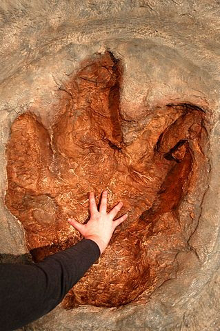
Trace fossils, also called ichnofossils, tell us about an organism’s behavior rather than representing its anatomical form. Traces are split into four sub-groups – tracks, trails, coprolites, and gastroliths. Tracks are footprints, paw prints or claw prints which become covered with sediment before they are washed away by rain or wave. Trails are not usually made by feet, but by tentacles, the crawling patterns of snakes and worms, or the boreholes of prehistoric beetles. The picture below features a Tyrannosaurus rex track.

There are two other classes of trace fossils; coprolites and gastroliths. The first represents fossilized feces which usually contain difficult to digest remnants of a meal. Coprolites are usually petrified, or cast and mold fossils. Gastroliths are stones swallowed by certain animals to aid digestion.

How are Fossils Formed?

As already mentioned, the ideal conditions for fossilization are immediate burial in hypoxic, mineral-rich sediments in a low temperature environment. These elements make the existence of fossils rare. Four types of process contribute to the formation of a fossil. These are mineralization, carbonization, encrustation and distillation. They occur once an organism has become trapped within the surrounding sediment, and primarily depend on the mineral composition of silt and water.

Mineralization

Mineralization is the most common process in fossilization. Water within the sediment contains minerals that, over time, slowly replace the tissues of the original living organism. The most prevalent minerals that contribute to the fossilization process are calcium carbonate, iron sulfide, iron carbonate, calcium phosphate, and silica (silicon, silicon dioxide or quartz).

Carbonization and Distillation

The initiation of carbonization requires a certain type of bacteria that creates an anaerobic environment devoid of any oxygen or nitrogen. This bacteria turns plant life into carbon. Under millions of years of vertical compression, these ancient carbon fields become coal deposits. Distillation describes the process by which carbon film or phytoleim fossils are formed. This is an extremely rare occurrence, but can give a fine image of soft tissues. Fossils obtained by distillation are usually badly preserved, although significant external detail is possible.

Encrustation

Encrustation occurs in the presence of minerals – usually calcium carbonate – deposited in various layers on top of the remains of an organism. As tides rise and fall or water levels fluctuate, minerals slowly form a mold. While mineralization replaces organic matter, encrustation covers it. Mineralization will form a cast, encrustation the mold.

Female orgasms

Female orgasms A female orgasm can be a highly pleasurable experience during masturbation or sexual activity. While orgasms may ...