Saturday, September 30, 2023

Camouflaged Animals

Camouflaged Animals

Introduction

An animal that uses camouflage is one that has adapted itself to look exactly like its surroundings or to blend in with it - it makes them very difficult to spot! For example, if an animal lives in woodland with lots of leaves, trees and branches, it might have adapted to have a brown coat which blends in with the rest of the environment.

For lots of animals, it’s a defense mechanism or a hunting tactic.

Camouflage can also be called cryptic colouration.

There are four main types of camouflage:

Concealing colouration - This is when an animal disguises itself against its background by being the same colour as it.

Disruptive colouration - This is when stripes, spots or other patterns make it hard for other animals to distinguish the outline of their bodies.

Disguise - This is similar to concealing colouration; however, instead of using colour, animals will imitate the texture or shape of their surroundings instead.

Mimicry - These animals pretend to look like other animals and are sometimes called 'imposters'. This is common in prey animals who pretend to be a different animal which is toxic or poisonous to their predator.

Reasons 

Animals can use camouflage for lots of different reasons, but the overarching reason is to survive. They use camouflage to hide their location and identity, particularly from predators if they’re a prey species. On the other hand, some predator species also use camouflage to sneak up on their prey. If the prey has no idea they’re there, that makes it much easier to hunt!

Camouflage varies from species to species. If a species has feathers or scales, they can shed and camouflage themselves much more regularly and easily than an animal with fur. This is why most animals with fur camouflage themselves by season.

Animals have adapted to use camouflage as a result of natural selection: those who have used camouflage are the ones who have survived in the wild.

Examples

There are hundreds of creatures in the animal kingdom that camouflage themselves, and perhaps some are still undiscovered because they do it so well. Here are a few examples of animals that use camouflage.

Chameleon

When you think of camouflage, more often than not, you think of the chameleon. But actually, when chameleons change the colours of their scales, the primary function isn’t to camouflage themselves against their surroundings (though this is a handy side-effect).

In fact, chameleons change colour to regulate their body temperature and to send messages and signals to their fellow chameleons. That being said, they’re still a fantastic example of camouflage. Their scales can completely change colour to match their surroundings (usually the rainforest or a desert), which makes it impossible for predators to find them.

Did you know that some cells in a chameleon’s skin have crystals called guanine crystals? To change the colour of their skin, chameleons adjust the space between these crystals, which affects how light reflects off their skin.

Arctic Fox

The Arctic fox has adapted its white fur to blend in with the vast, snowy Arctic tundra perfectly. But did you know that when the season changes and the ice melts away to reveal grassland, the fox changes colour too? While we instinctively think of the Arctic fox as white, it sheds its fur to make way for a brown coat in the summer months. This allows it to blend in once again and hunt effectively for its prey.

Stick Insect

The clue is in the name - this insect looks exactly like your average stick or branch. This creature has adapted to have a bark-like appearance, which lets it completely camouflage itself against trees. Sometimes, they even sway back and forth in the wind just like a branch to convince predators that they aren’t there at all.
Sea Turtle

Sea turtles use the patterns on their shells to camouflage themselves against the seafloor. The pattern on their shells is similar to the patterns that sunlight creates when it reflects into the water and onto the seafloor. This helps them to hide from their predators, such as tiger sharks and orcas.

Sea Urchin

While the sea urchin’s appearance hasn’t been adapted to mimic its surroundings, it still uses camouflage to protect itself. On the seafloor, it gathers shells, rocks and anything else it can find to help it blend in and hide from predators.

Viceroy Butterfly

To protect itself from predators, the Viceroy butterfly completely mimics the appearance of a Monarch butterfly. Viceroy butterflies are non-poisonous, but the Monarch is poisonous. Therefore, if its predators think its poisonous, they won't try to eat it!

Zebra

To us humans, zebras don’t appear to be camouflaged at all. Their distinctive black and white stripes stand out against the brown savanna where they live and make them easy to spot. However, their main predator, lions, are colour blind. Zebras also live in herds, and their stripes make it difficult for lions to pick out a single zebra to hunt. Therefore, a zebra’s stripes help them to blend in with the herd, rather than their surroundings.

Leopard and Cheetah

Leopards and cheetahs are both well-known for their spots and they play a huge role in these species’ camouflage. The spots help to disguise the outline of the animal’s body, allowing it to blend in with the brown savanna landscape. Using this camouflage tactic, leopards and cheetahs can easily sneak up on their prey and hunt.

Endosymbiotic Theory

Endosymbiotic Theory 

Definition

Endosymbiotic theory is the unified and widely accepted theory of how organelles arose in organisms, differing prokaryotic organisms from eukaryotic organisms. In endosymbiotic theory, consistent with general evolutionary theory, all organisms arose from a single common ancestor. This ancestor probably resembled a bacteria, or prokaryote with a single strand of DNA surrounded by a plasma membrane. 

Throughout time, these bacteria diverged in form and function. Some bacteria acquired the ability to process energy from the environment in novel ways. Photosynthetic bacteria developed the pathways that enabled the production of sugar from sunlight. Other organisms developed novel ways to use this sugar is oxidative phosphorylation, which produced ATP from the breakdown of sugar with oxygen. ATP can then be used to supply energy to other reactions in the cell.

Both of these novel pathways led to organisms that could reproduce at a higher rate than standard bacteria. Other species, not being able to photosynthesis sugars or break them down through oxidative phosphorylation, decreased in abundance until they developed a novel adaptation of their own. 

The ability of endocytosis, or to capture other cells through the enfolding of the plasma membrane, is thought to have evolved around this time. These cells now had the ability to phagocytize, or eat, other cells. In some cells, the bacteria that were ingested were not eaten, but utilized. 

By providing the bacteria with the right conditions, the cells could benefit from their excessive production of sugar and ATP. One cell living inside of another is called endosymbiosis if both organisms benefit, hence the name of the theory. Endosymbiotic theory continues further, stating that genes can be transferred between the host and the symbiont throughout time.

This gives rise to the final part of endosymbiotic theory, which explains the variable DNA and double membranes found in various organelles in eukaryotes. While the majority of cell products start in the nucleus, the mitochondria and chloroplast make many of their own genetic products. 

The nucleus, chloroplasts, and mitochondria of cells all contain DNA of different types and are also surrounded by double membranes, while other organelles are surrounded by only one membrane. Endosymbiotic theory postulates that these membranes are the residual membranes from the ancestral bacterial endosymbiont. 

If a bacteria was engulfed via endocytosis, it would be surrounded by two membranes. The theory states that these membranes survived evolutionary time because each organism retained the maintenance of its membrane, even while losing other genes entirely or transferring them to the nucleus. Endosymbiotic theory is supported by a large body of evidence. The general process can be seen in the following graphic.

Evidence

The most convincing evidence supporting endosymbiotic theory has been obtained relatively recently, with the invention of DNA sequencing. DNA sequencing allows us to directly compare two molecules of DNA, and look at their exact sequences of amino acids. 

Logically, if two organism share a sequence of DNA exactly, it is more likely that the sequence was inherited through common descent than the sequence arose independently. If two unrelated organisms need to complete the same function, the enzyme they evolve does not have to look the same or be from the same DNA to fill the same role. Thus, it is much more likely that organisms who share sequences of DNA inherited them from an ancestor who found them useful.

This can be seen when analyzing the mitochondrial DNA (mtDNA) and chloroplast DNA of different organisms. When compared to known bacteria, the mtDNA from a wide variety of organisms contains a number of sequences also found in Rickettsiaceae bacteria. Fitting with endosymbiotic theory, these bacteria are obligate intracellular parasites. 

This means they must live within a vesicle of an organism that engulfs them through endocytosis. Like bacterial DNA, mtDNA and the DNA in chloroplasts is circular. Eukaryotic DNA is typically linear. The only genes missing from the mtDNA and those of the bacteria are for nucleotide, lipid, and amino acid biosynthesis. An endosymbiotic organism would lose these functions over time, because they are provided for by the host cell.

Further analysis of the proteins, RNA and DNA left in organelles reveals that some of it is too hydrophobic to cross the external membrane of the organelle, meaning the gene could never get transferred to the nucleus, as the cell would have no way of importing certain hydrophobic proteins into the organelle. 

In fact, chloroplasts and mitochondria have their own genetic code, and their own ribosomes to produce proteins. These proteins are not exported from the mitochondria or chloroplasts, but are needed for their functions. 

The ribosomes of mitochondria and chloroplasts also resemble the smaller ribosomes of bacteria, and not the large eukaryotic ribosomes. This is more evidence that the DNA originated inside of the organelles, and is separate completely from the eukaryotic DNA. This is consistent with endosymbiotic theory.

Lastly, the position and structure of these organelles lends to the endosymbiotic theory. The mitochondria, chloroplasts, and nuclei of cells are all surrounded in double membranes. All three contain their DNA in the center of the cytoplasm, much like bacterial cells. 

Although less evidence exists linking the nucleus to any kind of extant species, both chloroplasts and mitochondria greatly resemble several species of intracellular bacteria, existing in much the same manner. The nucleus is thought to have arisen through enfolding of the cell membrane, as seen in the graphic above. 

Throughout the world, there are various endosymbiont bacteria, all of which live inside other organisms. Bacteria exist almost everywhere, from the soil to inside our gut. Many have found unique niches within the cells of other organisms, and this is the basis of endosymbiotic theory.

Related Biology Terms

Endosymbiont – An organism that lives with another organism, cause both organisms to receive benefits.

Cyanobacteria – Still extant, cyanobacteria are photosynthetic bacteria whose ancestors probably became the chloroplasts of plant cells.

Proteobacteria – The bacterial ancestor to the mitochondria organelle.

Eukaryote – An organism with membrane bound organelles, thought to have evolved from endosymbiotic interactions.

Mitochondria


Mitochondria - Structure and Function & Mitochondrial DNA 

Introduction 

We all eat our breakfast, dinner, and lunch, but have you ever wondered how we get energy from food? How can we use this energy? Well, it is due to mitochondria. Mitochondria are important cell organelles in our body and are known as the powerhouse of cells. Different types of cells have different numbers of mitochondria. For example, simple cells have one or two mitochondria, whereas complex cells, such as plant or animal cells, have numerous mitochondria as they require more energy.

Plant cells have hundreds of mitochondria, while the number goes to thousands and lakhs for animal cells. Interestingly, the human body has 1,00,000 to 6,00,000 mitochondria in each cell which can produce 90% of the energy in the body to perform daily tasks. In this article, we will discuss the mitochondria diagram, function of mitochondria, and structure of mitochondria.

Mitochondria

Mitochondria are the powerhouse of cells as they generate energy for cell functioning. The structure of mitochondria is unique. The mitochondrion is a rod or sausage-shaped structure found in animal and plant cells. It is a small organelle whose size is between 0.5 to 1 micrometre in diameter. Hence, it cannot be seen under a microscope unless stained. Unlike other organelles, it has two layers; inner and outer. Each layer performs different functions. 

Let us understand the structure with the mitochondria diagram. 

Mitochondria Structure 

Outer Membrane - It is made of proteins. The membrane allows small protein-like molecules to pass through it. 

Intermembrane Space - It is the space between outer and inner membranes. 

Inner Membrane - This membrane is made of phospholipids and does not allow molecules to pass through it. Special transporters (carrier molecules) are required to transport substances. Here, ATP production takes place. 

Cristae - These are the irregular folds of the inner membrane. They increase the space for chemical reactions to take place by increasing the surface area of the membrane. 

Matrix - It is fluid within the inner membrane. This fluid has several enzymes required for ATP production. It also contains ribosomes, mitochondrial DNA, inorganic and organic molecules, etc.

Mitochondrial DNA 

Mitochondrial DNA is a double stranded circular molecule, which is inherited from the mother in all multi-cellular organisms, though some recent evidence suggests that in rare instances mitochondria may also be inherited via a paternal route. 

Typically, a sperm carries mitochondria in its tail as an energy source for its long journey to the egg. When the sperm attaches to the egg during fertilization, the tail falls off. Consequently, the only mitochondria the new organism usually gets are from the egg its mother provided. 

There are about 2 to 10 transcripts of the mt-DNA in each mitochondrion. Compared to chromosomes, it is relatively smaller, and contains the genes in a limited number.

The size of mitochondrial genomes varies greatly among different organisms, with the largest found among plants, including that of the plant Arabidopsis, with a genome of 200 kbp in size and 57 protein-encoding genes. 

The smallest mtDNA genomes include that of the protist Plasmodium falciparum, which has a genome of only 6 kbp and just 2 protein- encoding genomes. Humans and other animals have a mitochondrial genome size of 17 kbp and 13 protein genes.

Mitochondrial DNA consists of 5-10 rings of DNA and appears to carry 16,569 base pairs with 37 genes (13 proteins, 22 t-RNAs and two r-RNA) which are concerned with the pro­duction of proteins involved in respiration. 

Out of the 37 genes, 13 are responsible for mak­ing enzymes, involved in oxidative phosphorylation, a process that uses oxygen and sugar to produce adenosine tri-phosphate. 

The other 14 genes are responsible for making molecules, called transfer RNA (t-RNA) and ribosomal RNA (r-RNA). In some metazoans, there are about 100 – 10,000 separate copies of mt-DNA present in each cell.

Unlike nuclear DNA, mitochondrial DNA doesn’t get shuffled every generation, so it is presumed to change at a slower rate, which is useful for the study of human evolution. 

Mito­chondrial DNA is also used in forensic science as a tool for identifying corpses or body parts and has been implicated in a number of genetic diseases, such as Alzheimer’s disease and diabetes. 

Changes in mt-DNA can cause maternally inherited diseases, which leads to faster aging process and genetic disorders.

Mitochondria convert the potential energy of food molecules into ATP by the Krebs cy­cle, electron transport and oxidative phosphorylation in presence of oxygen. 

The energy from food molecules (e.g., glucose) is used to produce NADH and FADH2 molecules, via glycolysis and the Krebs cycle. The protein complexes in the inner membrane (NADH de­hydrogenase, cytochrome c reductase, cytochrome c oxidase) use the released energy to pump protons (FT) against a gradient.

Mitochondrial marker enzymes

Mitochondrial marker enzymes are enzymes that are specifically present in mitochondria, in the mt-matrix, the inner mt-membrane, the inter-membrane space, or the outer mt-membrane.

Citrate synthase (mt-matrix)

NAD+ malate dehydrogenase (mt-matrix)

NAD+ glutamate dehydrogenase (mt-matrix)

Succinate cytochrome c reductase (inner mt-membrane)

Rotenone-sensitive NADH cytochrome c reductase (inner mt-membrane)

Adenylate kinase (intermembrane space)

Rotenone-insensitive NADH cytochrome c reductase (outer mt-membrane)

Monoamine oxidase (outer mt-membrane)

Kynurenine hydroxylase (outer mt-membrane)

Function of Mitochondria

The most common function of Mitochondria is energy generation. However, it performs several other vital functions of the body. These include the following.

Energy Generation

Mitochondria help produce ATP molecules which are the energy units of cells. Most energy production takes place in the cristae or folds of the inner membrane. It generates energy by converting chemical energy from food. 

Cell Death

Apoptosis or cell death is an essential part of the regeneration of new cells. As cells damage or become old, they are destroyed by the mitochondria, and new cells are formed. It releases enzymes like Cytochrome C, which helps in cell degeneration. 

Heat Production 

In extreme colds, the body generates its heat by utilising tissue fat. Mitochondria release energy in the form of heat in cold climatic conditions. 

Storing Calcium 

Calcium is involved in several cellular processes. For example, releasing neurotransmitters for nerve conduction and muscle movement, blood clotting, fertilisation, hormone signalling, steroid synthesis, and cellular metabolism. As calcium is so important for the body, cells regulate it tightly. Mitochondria help in the absorption of calcium ions and store them until they are used. 

New Things About Mitochondria

1.Mitochondria are the powerhouse of cells and produce energy. 

2.It has a complex structure, and each performs distinct functions. 

3.Like a nucleus, it has its DNA. 

4.The outer membrane of the mitochondria has a protein called porins which forms protein channels to facilitate molecule transport.  

5.Mature red blood cells have no mitochondria. 

6.Liver cells have more than 2,000 mitochondria. 

7.It has dividing and multiplying abilities. 

8.In human sperm, mitochondria are spiral-shaped and provide energy for motion. 

Summary

Mitochondria are popularly known for their ability to generate energy. They are a double membrane organelle and have a typical structure and are called the powerhouse of a cell. They carry numerous bodily functions and metabolic activities, including heat generation, apoptosis, neurotransmitter regulation, calcium uptake, etc. Interestingly, they have their DNA while other cell organelles do not possess DNA.

Changes in the mitochondrial DNA can lead to insufficient ATP or energy production and other critical diseases. The number of mitochondria varies from cell to cell or organism to organism. For example, human liver cells have more than 2000 mitochondria, whereas human red blood cells have no mitochondria. In the end, we have seen some facts about mitochondria. If there are any doubts related to the topic please ask in the comments.

Antigen - Antibody Reaction

Antigen - Antibody Reaction

Introduction 

Antigen-antibody reaction or antigen-antibody interaction is a particular chemical interaction between antibodies generated by B cells of the white blood cells and antigens during the immune reaction. The process of agglutination combines antigens and antibodies.

It is the basic biological process that the body uses to defend itself against various foreign particles like viruses and their toxic chemicals. An antigen-antibody complex is formed in the blood when antibodies specifically and strongly bind to antigens. The immunological complex is subsequently transferred to cellular systems where it can be eliminated or deactivated.

Antigen (Ag)

(Anti = opposite; gen = anything that causes)

Immunogens are any foreign substances that, once entering our bodies, frequently cause a sequence of immunological responses. While others, known as haptens, require the assistance of other molecules (carrier proteins) to activate an immunological response. All of the immunogens and haptens are referred to as antigens.

They could be polysaccharides, lipids, proteins, or peptides.

An epitope is an antibody-binding location.

Antibody (Ab)

An antibody is a component that the immune system produces in response to antigens. Thus, antigens result in the production of antibodies. They act together to exhibit an immunological response. The general characteristics of an antibody are as follows:

An antibody is also known as an immunoglobulin (Ig)

They are Y-shaped

Glycoproteins

Generated by plasma B-cells.

Paratope is the name of the antigen binding site.

Five types: IgG, IgA, IgM, IgE, and IgD.

Antigen-Antibody Reaction

Antigens and antibodies combine specifically with each other. Antigen-Antibody reaction is the term used to describe this interaction between them. Ag-Ab reaction is a common acronym for it. These serve as the building blocks of humoral or antibody-mediated immunity.

These reactions serve as the foundation for the detection of both specific and non-specific Ags, such as enzymes that cause non-specific diseases. Serological reactions are referred to as Ag-Ab reactions when they occur in vitro.

Stages of Antigen-Antibody Reaction

There are three stages to the interactions between Ag and Ab.

.The first stage of the reaction entails the formation of the Ag-Ab complex.

.The second stage results in visible phenomena like agglutination, precipitation, etc.

.The third stage involves the destruction of Ag or neutralisation of Ag.

Properties of Antigen-Antibody Reaction

.Significantly specific reaction.

.Occurs in a noticeable manner.

.Non-covalent reactions (Ionic bonds, Van der Waals forces, Hydrophobic interactions, Hydrogen bonds).

.Antibodies and antigens are not denatured.

.Reversible.

.Affinity: This refers to how strongly an antigen binds to a certain antigen-binding site on an antibody.

.Avidity: It is a more general concept than affinity. It represents the Ag-Ab complex’s total strength. It depends on:

The antibody’s affinity
Antibody and antigen valencies (the number of binding sites)
How epitopes and paratopes are structurally arranged.

.Cross-Reactivity: This term describes an antibody’s capacity to bind to similar epitopes on other antigens.

Types of Antigen-Antibody Reaction

The types of antigen-antibody reactions are as follows:

1.Precipitation Reaction
2.Agglutination Reaction
3.Complement Fixation
4.Immunofluorescence
5.ELISA – Enzyme-Linked ImmunoSorbent Assay

Precipitation Reaction

An insoluble precipitate of Ag-Ab complex is produced when a soluble Ag and its Ab combine in the presence of an electrolyte (NaCl) at a specific pH and temperature. Precipitin is the Ab that causes precipitation, and the reaction is termed a precipitation reaction.

The precipitation reaction occurs in both liquid and gel media.

1.Liquid Precipitation: An antigen-antibody reaction is carried out by adding increasing amounts of antigen to tubes containing a constant amount of antibody. Precipitation results from the combined reaction of the antigen and antibody.

2.Gel Precipitation: Petri plates or plates with agar gel or a similar gel are used in these methods. In the gel system, both Ag and Ab rapidly diffuse in all directions. A zone of equivalency, observed as visible precipitation, will form at a specific point depending on the diffusion rate and concentration of reactants.

Multiple bands develop in complex Ag or Ab preparations. They fall into two methods: single diffusion and double diffusion.

Agglutination Reaction

The particles are clustered or agglutinated when a certain Ag is combined with its Ab in the presence of electrolytes at an appropriate temperature and pH. The clumps of cellular Ag formed by the serum’s Ab are known as agglutinins.

Agglutinogens are the name for the aggregated particulate antigens.

1.Slide Agglutination: This is a fast and convenient way to identify the presence of agglutinating antibodies.

2.Tube Agglutination: This is a common technique for estimating the quantity of Ab. A constant volume of the Ag suspension is introduced after serially diluting the Ab-containing serum with saline in multiple small test tubes.

A control tube is retained that contains no antiserum. The tubes are incubated up until observable agglutination. The tube demonstrating the highest agglutination is known as the titre.

3.Passive Agglutination Test: This test is equivalent to the haemagglutination test, but the physical characteristics of the reaction are different.

A carrier particle has Ag coated on its surface, making the reaction more sensitive by assisting in the transformation of a precipitation process into an agglutination reaction. RBC, latex particles, or bentonite can be used as carrier particles. Sometimes, tanned RBC (polystyrene coated RBC) might be used.

Complement Fixation

Some non-specific, unstable fresh serum components known as complements are required for the lysis of RBC or microorganisms.

Every person has the 11 proteins that comprise the complement system. They attach to the Fc subunit of Ab in the Ag-Ab complex. Complement fixation tests make use of the Ag-Ab complex’s capacity to fix complement.

In the first step, the test Ag and the antiserum, which have been heated to 56°C to inactivate complement, are combined with a known quantity of complement. This is incubated for 18 hours at 4°C.

If the serum contains Ab that is specific for the Ag, an Ag-Ab complex will develop and fix the complement.

Immunofluorescence

Fluorescence is the ability to absorb light rays of a certain wavelength and emit light rays of a different wavelength.

Fluorescent dyes emit intense visible light when exposed to UV radiation.

In 1942, Albert Coons and coworkers demonstrated how labelled dyes could be coupled to antibodies, allowing for the detection of antigens using these labelled dyes.

Commonly used dyes include:

The most used label for immunofluorescence operations is fluorescein, an organic dye that absorbs blue light (490 nm) and emits a strong yellow-green fluorescence (517 nm).

As a strong emitter of red fluorescence and an effective light absorber (30 times more effective than fluorescein), phycoerythrin is often used as a label for immunofluorescence.

ELISA – Enzyme-Linked Immunosorbent Assay

In 1971, enzyme-labelled Ag’s and Ab’s were created as serological reagents for the testing of antibodies and antigens.

When compared to radioimmunoassay (RIA), they are more simple, sensitive, affordable, and risk-free.

The ligand used is a molecule that is covalently attached to an enzyme like peroxidase, beta-galactosidase, alkaline phosphatase, etc. and is capable of detecting the Ab.

There are three types of ELISA:

1.Indirect ELISA: HIV can be detected using the indirect ELISA method. The surface of the microtiter plates is coated with envelope proteins developed using recombinant technology. Unbound proteins are washed out when suspect serum is added.

2.Sandwich ELISA: This method is used for determining whether there is Ag in a sample. The suspect serum is added and given time to react after the well has been coated with Ag-specific Ab. Unbound Ag is removed from the wells through washing.

The next step is to add a labelled Ab against a different Ag epitope. Washing is used to remove unbound Ab’s, followed by the addition of coloured substrate and colour development. The colour intensity is directly proportional to the Ag concentration in the serum.

3.Competitive ELISA: Competitive ELISA is another variant for estimating antigen concentrations. In this method, the antibody is initially incubated in solution with the sample containing the antigen.

An antigen-coated microtiter well is then filled with the antigen-antibody mixture.

The free antibody available to attach to the antigen-coated well will reduce when more antigen is present in the sample. In an indirect ELISA, the addition of a secondary antibody (Ab2), coated with an enzyme, specific for the primary antibody’s isotype can be utilised to estimate how much primary antibody is attached to each well.

Conclusion 

Thus, we conclude this discussion with a brief overview of the antigen-antibody reaction. Specific interactions between the antibodies and antigens result in combination, known as Antigen-Antibody reaction or Antigen-Antibody interaction.

Apoptosis

Apoptosis 

Introduction

Apoptosis is a process of programmed cell death that occurs in multicellular organisms. It is a highly regulated and controlled process that occurs normally during development and aging as a homeostatic mechanism to maintain cell populations in tissues. For example, the separation of fingers and toes in a developing human embryo occurs because cells between the digits undergo apoptosis. Apoptosis also occurs as a defense mechanism such as in immune reactions or when cells get damaged by disease or by noxious agents. 

During the early process of apoptosis, cell shrinkage and pyknosis are visible by light microscopy. With cell shrinkage, the cytoplasm becomes dense and the organelles are more tightly packed. Pyknosis is the result of chromatin condensation and this is the most characteristic feature of apoptosis. 

At a later stage, apoptosis produces cell fragments called apoptotic bodies that phagocytic cells engulf and quickly remove before the contents can spill out onto surrounding cells to cause inflammation.

There is a wide variety of both physiological and pathological stimuli and conditions that can trigger apoptosis. However, not all cells will necessarily die in response to the same stimulus. For example, irradiation or drugs used for cancer chemotherapy results in DNA damage, which can lead to apoptotic death through a p53-dependent pathway, but not in all cells. Some hormones, such as corticosteroids, may lead to apoptotic death in some cells (e.g. thymocytes) although other cells are unaffected. 

Some cells express Fas or TNF receptors that can facilitate apoptosis induction via ligand binding and protein cross-linking. Other cells have a default death pathway that must be blocked by a survival factor such as a hormone or growth factor. 

There is also the issue of distinguishing apoptosis from necrosis, two processes that can occur independently, sequentially, as well as simultaneously. In some cases, it’s the type of stimuli and/or the degree of stimulation that determines if cells die by apoptosis or necrosis. At low doses, a variety of injurious stimuli such as heat, radiation, hypoxia and cytotoxic anti-cancer drugs can induce apoptosis but these same stimuli can result in necrosis at higher doses. 

Finally, apoptosis is a coordinated and energy-dependent process that involves the activation of a group of cysteine proteases called caspases and a complex cascade of events that link the initiating stimuli to the final structured demise of the cell.

Pathways

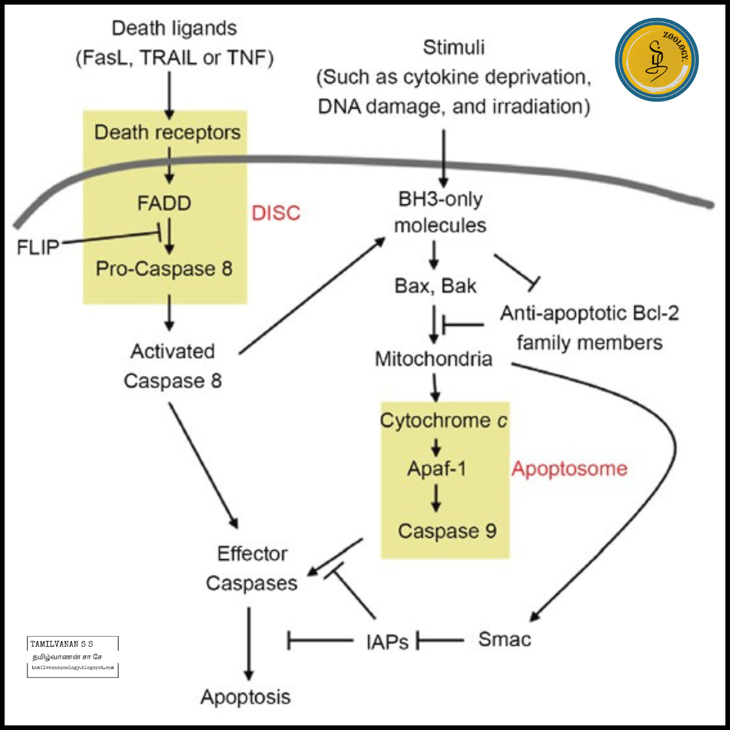
Apoptosis can be initiated through one of three pathways. In the intrinsic or mitochondrial pathway, the cell kills itself because it senses cell stress, while in the extrinsic pathway the cell is instructed to kill itself through signal transduction stimulators from other cells. 

The Perforin/Granzyme pathway is mediated by cytotoxic T cells. In this third pathway, apoptosis is induced via either Granzyme B or Granzyme A. All three initiation pathways (apart from Granzyme A) induce cell death through the Execution pathway that involves the activation of caspase-3.

Intrinsic Pathway

The stimuli that initiate the intrinsic pathway produce intracellular signals that may act in either a positive or negative fashion. Negative signals involve the absence of certain growth factors, hormones and cytokines that can interfere with the suppression of cell death programs, thereby triggering apoptosis. 

In other words, there is the withdrawal of factors, loss of apoptotic suppression, and subsequent activation of apoptosis. Positive signals include, but are not limited to, radiation, toxins, hypoxia, hyperthermia, viral infections, and free radicals. 

These stimuli cause changes in the inner mitochondrial membrane that results in an opening of the mitochondrial permeability transition (MPT) pore, loss of the mitochondrial transmembrane potential and release of two main groups of normally sequestered pro-apoptotic proteins from the intermembrane space into the cytosol.

The first group consists of cytochrome c, Smac/DIABLO, and the serine protease HtrA2/Omi. Cytochrome c binds and activates Apaf-1 as well as procaspase-9, forming an “apoptosome”. The clustering of procaspase-9 in this manner leads to caspase-9 activation. Smac/DIABLO and HtrA2/Omi are reported to promote apoptosis by inhibiting IAP (Inhibitors of Apoptosis Proteins) activity. IAP supresses caspases.

The second group consists of AIF (Apoptosis-Inducing Factor), endonuclease G and CAD (Caspase-Activated DNAse). AIF transfers to the nucleus and causes DNA fragmentation into ∼50–300 kb pieces and condensation of peripheral nuclear chromatin. 

This early form of nuclear condensation is referred to as Stage I condensation. Endonuclease G also transfers to the nucleus where it cleaves nuclear chromatin to produce oligo-nucleosomal DNA fragments.

AIF and endonuclease G both function in a caspase-independent manner. CAD is subsequently released from the mitochondria and transfers to the nucleus where, after cleavage by caspase-3, it leads to oligo-nucleosomal DNA fragmentation and a more pronounced and advanced chromatin condensation. This later and more pronounced chromatin condensation is referred to as Stage II condensation.

Extrinsic Pathway

The Extrinsic initiation pathway involves receptors of the TNFR (Tumour Necrosis Factor Receptor) family. In the extrinsic, death receptor pathway of apoptosis, ligation of death receptors on the cell surface leads to caspase activation. This pathway relies on the formation of a Death-Inducing Signalling Complex (DISC), which always includes FADD and caspase-8. 

The death receptors known so far are TNFR1 (ligand TNF-alpha), CD95 (ligand FasL), and TRAILR1 plus TRAILR2 (ligand TRAIL). In the case of ligated TNFR1, adapter TRADD (Tnf Receptor type 1-Associated Death Domain) is first engaged, which in turn recruits FADD. TRADD is only required for apoptosis when induced by TNF-alpha. 

The other ligated death receptors engage FADD directly. Fas associated via death domain (FADD) protein is responsible for the recruitment of caspase-8 to form the DISC. 

The presence of the FLICE-like inhibitory protein (cFLIP) in the FADD-caspase-8-cFLIP complex determines if and how cells die. As such, cFLIP is a switch that determines cell fate. cFLIP comes in two major isoforms, the long isoform cFLIPL and the short isoform cFLIPS. The cFLIPS protein is an inhibitor of caspase-8 and blocks DISC-dependent procaspase-8 activation. 

The cFLIPL protein regulates the extent of activation and possibly substrate specificity of procaspase-8. Low levels of cFLIPL can enhance apoptotic signaling, whereas apoptosis is inhibited when cFLIPL levels are high.

CD95 and TRAIL-R ligation induces apoptosis by direct recruitment of FADD-caspase-8 in a complex called the DISC. As described above, isoform levels of cFLIP then determine whether apoptosis is blocked or engaged. 

Ligation of TNFR1 can both induce caspase-8-mediated apoptosis as well as block apoptosis via the NF-κB-induced expression of cFLIP in a feedback loop. Receptor-interacting serine/threonine kinases 1 (RIPK1) is key in regulating TNFR1-induced FADD-caspase-8-mediated apoptosis. 

TNFR1 ligation leads to the recruitment of TRADD, TRAF2, cIAP1/2, and RIPK1 (complex I). RIPK1 ubiquitination by cIAP1/2 mediates activation of NF-κB and the production of pro-inflammatory and pro-survival gene expression. RIPK1 is deubiquitinated by CYLD and leaves complex I to recruit FADD to form the ripoptosome (complex IIa) involving caspase-8 and cFLIPL. 

Homodimerization and activation of caspase-8 on FADD induces apoptosis. One of the expressed pro-survival genes, cFLIPL, heterodimerizes with caspase-8, resulting in inhibition of caspase-8 activation and apoptosis. Alternatively, RIPK1 interacts with RIPK3 to either stimulate RIPK3 oligomerization in the necrosome (complex IIb), or to suppress it. 

Oligomerized RIPK3 is a prerequisite to trigger phosphorylation of the downstream mediator Mixed-Lineage Kinase-Like (MLKL) that triggers necrosis. Thus, death receptor TNFR1 provides two separate pathways downstream the TRADD recruitment: deubiquitinated RIPK1 either allows the activation of DISC in the ripoptosome (complex IIa) igniting apoptosis, or it interacts with RIPK3 to activate MLKL in the necrosome (complex IIb). 

MLKL is the effector protein that, once activated, transfers to the plasma membrane where it induces rupture and subsequent cell death. This regulated form of necrosis is also known as necroptosis. The release of cellular components this way results in an inflammatory response.

Perforin/Granzyme Pathway

One aspect of the adaptive immune system is recognizing and eliminating target cells through the induction of apoptosis, involving CD8+ cytotoxic T lymphocytes (CTLs) and natural killer (NK) cells. CTLs recognize target cells by the presentation of major histocompatibility complex class I (MHC I) molecules on surface of the target cell, while NK cells recognize target cells by the loss of class I molecules, of killer cell immunoglobulin receptors (KIRs), and of killer cell lectin like receptors (KLRs). 

Although there is a difference in the process of target cell recognition, once identified, both CTLs and NK cells execute similar effector function to remove the target cell. This is achieved through the release of cytotoxic granules containing perforin, granzymes, and granulysin, which work together to induce apoptosis in the target cells. This effector-killing of target cells enables eradication of intracellular pathogens and malignancies.

A key protein in the cytotoxic granule is perforin. Classically, perforin is known to form a pore in cell membranes, allowing passage of granzymes into the cell inducing apoptosis. Granzyme B can either directly, or through caspase-10 activation, convert pro-form caspase-3 into active caspase-3 to initiate the Apoptosis Execution pathway. 

The Granzyme A pathway evades the Execution pathway, leading to DNA cleavage through the SET complex. Once gaining entry to the cell, granzyme A activates a DNA nicking process via DNAse NM23-H1, a tumour suppressor gene product. This DNAse has an important role in immune surveillance by prevention of cancer cell metastasis through induction of tumour cell apoptosis. 

The nucleosome assembly protein SET normally inhibits the NM23-H1 gene. Granzyme A protease cleaves the SET complex thus releasing inhibition of NM23-H1, resulting in apoptosis inducing DNA degradation. 

Execution Pathways

Both extrinsic and intrinsic apoptosis induction pathways lead to an end-point execution phase, considered the final pathway of apoptosis. Execution caspases activate cytoplasmic endonuclease, which degrades nuclear material, and proteases that degrade the nuclear and cytoskeletal proteins. Caspase-3, caspase-6, and caspase-7 function as effector or “executioner” caspases, cleaving various substrates including cytokeratins, PARP, the plasma membrane cytoskeletal protein Fodrin alpha, the nuclear protein NuMA and others, that ultimately cause the morphological and biochemical changes seen in apoptotic cells. 

Caspase-3 is the most important of the executioner caspases and is activated by any of the initiator caspases (caspase-8, caspase-9, or caspase-10). Caspase-3 specifically activates the endonuclease CAD. In proliferating cells CAD is complexed with its inhibitor, ICAD. In apoptotic cells, activated caspase-3 cleaves ICAD to release CAD.

CAD then degrades chromosomal DNA within the nuclei causing chromatin condensation. Caspase-3 also induces cytoskeletal reorganization and disintegration of the cell into apoptotic bodies through the cleavage of Gelsolin. The cleaved fragments of Gelsolin, in turn, cleave actin filaments in a calcium independent manner. This results in disruption of the cytoskeleton, intracellular transport, cell division, and signal transduction.

Phagocytic uptake of apoptotic cells is the last component of apoptosis. Phospholipid asymmetry and externalization of phosphatidylserine on the surface of apoptotic cells and their fragments is the hallmark of this phase. 

The appearance of phosphatidylserine on the outer leaflet of apoptotic cells facilitates noninflammatory phagocytic recognition, followed up quickly by their early uptake and disposal. This process of early and efficient uptake, with no release of intracellular constituents, essentially eliminates the probability of potentially catastrophic inflammatory events.

Friday, September 29, 2023

Lysosome - Structure and Function

Lysosome - Structure and Function 

Introduction

Lysosomes are membrane-bound, dense granular structures containing hydrolytic enzymes responsible mainly for intracellular and extracellular digestion. The word “lysosome” is made up of two words “lysis” meaning breakdown and “soma” meaning body. It is an important cell organelle responsible for the inter and extracellular breakdown of substances. 

They are more commonly found in animal cells while only in some lower plant groups ( slime molds and saprophytic fungi). Lysosomes occur freely in the cytoplasm. In animals, found in almost all cells except in the RBCs. They are found in most abundant numbers in cells related to enzymatic reactions such as liver cells, pancreatic cells, kidney cells, spleen cells, leucocytes, macrophages, etc. 

Structure of Lysosomes

Lysosomes are acidic membrane-bound organelles found within cells, usually around 1 micrometre in length. Lysosomes contain numerous hydrolytic enzymes which catalyse hydrolysis reactions.

The membrane surrounding the lysosome is vital to ensure these enzymes do not leak out into the cytoplasm and damage the cell from within. In order to maintain the acidic pH of the lysosome, protons are actively transported into the organelle across the lysosomal membrane.

Lysosomes are without any characteristic shape or structure i.e. they are pleomorphic They are mostly globular or granular in appearance. It is 0.2-0.5 μm in size and is surrounded by a single lipoprotein membrane unique in composition. 

Diagram of Lysosomes

The membrane contains highly glycosylated lysosomal associated membrane proteins (LAMP) and Lysosomal integral membrane proteins (LIMP).
LAMPs and LIMPs form a coat on the inner surface of the membrane. They protect the membrane from attack by the numerous hydrolytic enzymes retained inside. The lysosomal membrane has a hydrogen proton pump which is responsible for maintaining pH conditions of the enzyme The acidic medium maintained by the proton pump that pumps H+ inside the lumen, ensures the functionality of the lysosomal enzymes. Inside the membrane, the organelle contains enzymes in the crystalline form.

Lysosomal Enzymes

For degradation of extra and intracellular material, lysosomes filled with enzymes called hydrolases. It contains about 40 varieties of enzymes which are classified into the following main types, namely:

Proteases, which digest proteins
Lipases, which digests lipids
Amylase, which digests carbohydrates
Nucleases, which digest nucleic acids
Phosphoric acid monoesters

Collectively the group of enzymes is called hydrolases which cause cleavage of substrates by the addition of water molecules. Most of the lysosomal enzymes function in the acidic medium.

Types of Lysosomes

Primary Lysosomes

Small sac-like structures enclosing enzymes synthesized by the rough endoplasmic reticulum.
Simply called as storage granules storing enzymes.

Secondary Lysosomes

Formed by the fusion of primary lysosome with phagosomes.
Contain engulfed material plus enzymes.
Materials are progressively digested.

Synthesis

The lysosome and the enzymes within it are synthesised separately. Lysosomal proteins are formed in the same way as any other protein. The first step is the initiation of mRNA strand production from relevant DNA segments. The mRNA strands proceed to the rough endoplasmic reticulum, where ribosomes construct the hydrolytic enzymes.

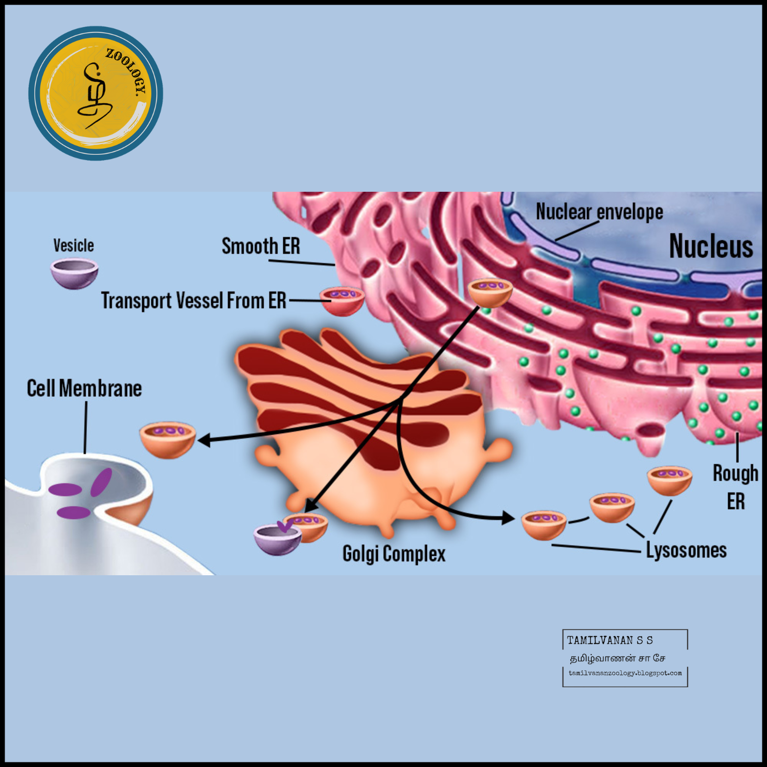
Importantly, these are tagged with mannose-6-phosphate within the golgi apparatus to target them to the lysosome. As a result, vesicles containing these enzymes bud off from the golgi apparatus. Two enzymes are responsible for the attachment of the mannose-6-phosphate tag: N-acetylglucosamine phosphotransferase and N-acetylglucosamine phosphoglycosidase.

This vesicle, now in the cytoplasm, then binds with a late endosome which is another acidic, membrane-bound organelle. The late endosome has proton pumps within its membrane that keep its internal environment acidic. The low pH causes dissociation of the protein from the mannose-6-phosphate receptor. This receptor can then be recycled back to the golgi apparatus.

The phosphate group is also removed from the mannose-6-phosphate tag, to prevent the whole protein returning to the golgi apparatus. The late endosome can eventually mature into a lysosome, after it has received the enzymes from the golgi apparatus.

Functions of Lysosomes

Lysosomes serve two major functions:

Intracellular Digestion

To digest food, the lysosome membrane fuses with the membrane of food vacuole and squirts the enzymes inside.

The digested food then diffuses through the vacuole membrane and enters the cell to be used for energy and growth.

Autolytic Action

Cell organelles that need to be ridden are covered by vesicles or vacuoles by the process of autophagy to form autophagosome.

The autophagosome is then destroyed by the action of lysosomal enzymes.

Processes in which lysosomes play crucial roles include:

a. Heterophagy

The taking into the cell of exogenous material by phagocytosis or pinocytosis and the digestion of the ingested material after fusion of the newly formed vacuole with a lysosome.

b. Autophagy

A normal physiological process that deals with the destruction of cells in the body. It is essential for maintaining homeostasis, for normal functioning by protein degradation, turnover of destroyed cell organelles for new cell formation

c. Extracellular Digestion

Primary lysosomes secrete hydrolases outside by exocytosis resulting in degradation of extracellular materials.

Eg. Saprophytic fungi

d. Autolysis

It refers to the killing of an entire set of cells by the breakdown of the lysosomal membrane. It occurs during amphibian and insect metamorphosis.

e. Fertilization

The acrosome of the sperm head is a giant lysosome that ruptures and releases enzymes on the surface of the egg. This provides the way for sperm entry into the egg by digesting the egg membrane.

f. As Janitors of the Cell

Lysosomes remove ‘junk’ that may accumulate in the cell helping to prevent diseases.

Lysosomal Disease

Nuclear genes organize the production of enzymes in the lysosomes. Nuclear genes are those genes that are being located within the nucleus of a cell, especially in eukaryotes. If there are any mutations in these genes, it will result in the emergence of a lot of human genetic ailments which are collectively called LSD (lysosomal storage diseases). 

Lysosomes as the Therapeutic Targets

There are a lot of lysosomal pathways and their components that can represent the potential pharmacological targets for a huge number of diseases. When we consider lysosomes as the targets, it is very important to note down the need for specificity, i.eThe agents will not target all lysosomes, but they will especially target those lysosomes which are defective in a few organs, cells, or tissues. For several reasons, it is very important to target lysosomes and not to target the whole autophagy process. Firstly, regarding safety, the important role of lysosomes in many key physiological processes means that the therapeutic windows for the intervention of pharmacology with unacceptable side effects may be limited.

Summary of Lysosomes

Lysosomes are cell organelles almost exclusively found in eukaryotic animal cells

Lysosomes are membrane-bound spherical sacs crammed with hydrolytic enzymes

These enzymes can break down many sorts of biomolecules like proteins and fats

Lysosomes are known as the ‘Suicidal Bags’ as they have the capacity to destroy the cell wall with its digestive enzymes, causing autolysis of the cell

Lysosomal enzymes are synthesized within the rough endoplasmic reticulum, where they've brought in by the Golgi body through tiny vesicles. It eventually merges with bigger acidic vesicles

Mutations of the nuclear gene may end in the emergence of rare, diverse human genetic ailments, which are called lysosomal storage diseases or LSD

Golgi Apparatus - Structure and Function


Golgi Apparatus - Structure and Function

Introduction

Golgi Apparatus is a membrane-bound cell organelle present in cells of all the eukaryotic organisms. It is also known as Golgi body, Golgi Complex or just Golgi. Due to its role in a cell, the Golgi Apparatus is called a packaging area of a cell because it is responsible for modifying, packaging (into vesicles) and transport of all the secretory proteins to their respective location inside or outside the cell. Golgi Apparatus is a part of the Endomembrane system which includes the Nucleus, Endoplasmic Reticulum, Golgi, Vesicles and Plasma Membrane. This whole endomembrane system work as a team inside a cell and is responsible for secretory protein pathway.

Golgi apparatus was discovered by Camillo Golgi, in the year 1950. It is also called a Golgi Complex or Golgi body, the membrane-bound organelle of eukaryotic cells (cells with clearly defined nuclei) that is made up of a series of flattened, stacked pouches called cisternae. The Golgi apparatus functions to transport, modify, and pack proteins and lipids into vesicles for delivery to targeted destinations. It is placed in the cytoplasm next to the endoplasmic reticulum and near the cell nucleus. While many types of cells contain only one or several Golgi apparatus, plant cells can contain hundreds.

Structure of Golgi Apparatus

Golgi Apparatus is a membrane-bound cell organelle which is present near the Endoplasmic Reticulum, which is present near the outer membrane of the Nucleus.

It appears like many flattened pouches present close to one another. This flat-tube/pouch-like structure is called as cisternae.

The cisternae of the Golgi apparatus is divided into 3 compartments, these are, Cis (or cis face which is present near the endoplasmic reticulum), Medial (centre part of the cisternae) and trans (or trans face, which is present near the plasma membrane).

Generally, the number of cisternae present in a Golgi Apparatus is only six to eight but in several single-celled organisms, these number can rise up to 60 cisternae per Golgi Apparatus.

The two parts Cis Golgi network and Trans Golgi network are the outmost pouches of the cis and trans face respectively. Cis Golgi network and trans Golgi network, both perform a different and important function in the Golgi Apparatus.

The network of Cisternae is stabilized in the cell via cytoplasmic microtubules which are the cytoskeleton of a cell.

The lumen of the Golgi Apparatus is filled with a matrix which is a fluid protein. The matrix contains many enzymes which play a major role in the modification of the proteins arriving at the Golgi Apparatus.

Animal cells contain one or few Golgi per cell but plant cells contain hundreds of Golgi Apparatus per cell.

Models of Golgi Apparatus 

Endomembrane System of Golgi Apparatus

Two models of the Golgi Complex were proposed by the scientist.

Cisternae Maturation Model

Here it was said that the cisternae mature gradually from cis to trans, that is, each cisterna matures into next cisternae along with the stack and then finally disperse at the trans Golgi network.

The vesicles may move in the opposite direction. For example, the enzymes that must be moved back to Endoplasmic Reticulum, that is the enzymes resident to either Endoplasmic Reticulum or to Golgi itself must be moved back from the trans face to cis face, called Retrograde movement.

Vesicle Transport Model

In this model, it is proposed that the cargo is carried in the forward or anterograde direction by vesicle and cisternae remains stable, that is, they are non-dynamic, unlike in the Cisternae Maturation Model.

Here the cisternae remain at the same place in stable compartments and transport of the vesicles take place instead.

Functions of Golgi Apparatus 

Golgi Apparatus is responsible for some of the most important function inside our cell. Without the Golgi Apparatus, the proteins that are formed in the cytoplasm will have no use.

The proteins, after they are formed, undergoes into a series of modification which is done inside the Endoplasmic Reticulum first and then inside the Golgi Apparatus. This modification works as a tag on the proteins and tells them where to go in the cell. Just like an address on the package makes the package to get successfully delivered, similarly, the modification on the proteins helps them to get delivered to their address inside the cell or outside, depending upon the type.

The Golgi apparatus also modifies the lipids which are to be inserted into the Plasma Membrane.

The proteins coming from the Endoplasmic Reticulum is protected inside a vesicle, that vesicle is received by the cis face of the Golgi Apparatus which is near the Endoplasmic Reticulum.

This vesicle gets fused with the membrane of the Golgi Apparatus and the protein is delivered inside the Golgi. This process of sending of vesicles packed with proteins from Endoplasmic Reticulum to Cis face of the Golgi Apparatus is called as the Anterograde movement. And opposite to this, when the vesicles move form cis face of the Golgi Apparatus to the Endoplasmic Reticulum, it is called a Retrograde movement.

The proteins inside the matrix of the Golgi Apparatus which are embedded on the wall of the cisternae then modify the proteins and lipids by several steps.

When the protein or lipid finally reach the Trans end of the Golgi Apparatus, it buds off along the membrane of the trans-Golgi face. The membrane protects the proteins and is called a vesicle.

The modification on the proteins directs them to go to their particular location. For example, Mannose-6-phosphate is a modification done on their proteins which is destined to go to lysosomes. So any vesicle containing protein tagged with Mannose-6-phosphate inside them will automatically fuse with the membrane of the lysosomes.

Similarly, the proteins or lipids of the plasma membrane with go and fuse with the plasma membrane and those tagged to go outside of the cell also called as secretory proteins like enzymes, hormones, etc, will be secreted out of the cell.

Therefore the exocytosis process of the secretory proteins is regulated by the Golgi Apparatus.

O-linked Glycosylation occurs in Golgi Complex where the synthesis of most of the cell’s saccharides (complex form) are synthesised including GAG’s of proteoglycans.

Thursday, September 28, 2023

Ribosomes - Structure and Function

Ribosomes 

Introduction:

Ribosomes are tiny, granular organelles found in both eukaryotic and prokaryotic cells. They are found inside the cytosol of the cell and play. an important role in protein synthesis by translating the genetic information conveyed by messenger RNA (mRNA) into functional proteins. Ribosomes are composed of two subunits, one larger and one smaller, each of which is made up of proteins and RNA molecules.

"A ribosome is a cellular structure that assembles proteins by linking together amino acids based on genetic instructions from messenger RNA (mRNA)."

Ribosomes were first observed by George Palade (1953) under the electron microscope. It is a kind of complex molecular machine present inside the living cells that produce proteins from amino acids during a process of protein synthesis also called translation. Ribosome translates genetic information stored in messenger RNA into proteins.

The process occur in three stages: initiation, elongation and termination. Within ribosomes, ribosomal RNA (rRNA) catalyzes the peptidyl transferase reaction, to form peptide bonds between amino acids, enabling them to form proteins. After protein is formed in the ribosome, they move to different areas of the cell for various cellular functions.

Ribosomes Location:

Ribosomes are present in the cytosol or attached to the endoplasmic reticulum in both plant and animal cells. They play an important role in translating DNA into proteins. While some ribosomes are permanently associated with the rough endoplasmic reticulum, their association depends on the specific proteins they help to produce. In animal or human cells, there can be as many as 10 million ribosomes. Multiple ribosomes can be linked to the same mRNA strand, a structure known as a Polysome.

Occurrence of Ribosomes:

Ribosomes are distributed universally all throughout the kingdom of animals and plants. They're also found in prokaryotes. The mammalian RBC is the only cell type devoid of ribosomes. For any given type, the density of ribosomes per unit area is rather constant. It is high in active m protein synthesis cells and low in cells where synthesis of protein is low.

Distribution of Ribosomes:

The ribosomes frequently occur freely in the cytoplasm in prokaryotic cells. The ribosomes occur freely in the cytoplasm in eukaryotic cells or remain attached to the outer surface of the endoplasmic reticulum (ER) membrane. They are called free ribosomes if they are not attached to the ER. Free ribosomes represent protein synthesis sites needed to maintain the cytoplasmic matrix's enzyme composition.

Number and Concentration of Ribosomes:

Ribosomes can be observed in all cells containing endoplasmic reticulum. Nearly 100 ribosomes per μ3 are found in rabbit reticulocytes, which corresponds to 1x10⁵ particles per reticulocyte and approximately. 5% of the total cell mass, or approximately 20,000 to 30,000 cell mass. But, if unfavorable nutritional conditions slow the rate of protein synthesis, the number of ribosomes can drop significantly in protein synthesizing cells and bacteria.

Chemical Composition of Ribosomes:

RNA and proteins are the main constituents of ribosomes. The lipids are completely absent or traceable. E. Coli's ribosomes possess almost 60-65% of RNA and 35-40% of their weight protein. Ribosomal RNA differs from tRNA and other RNA classes of most cells in size and base content. In all ribosomes, two types of RNAs are found. They are an essential component and can't be easily removed. 

Ribosome Structure:

The structure of the ribosome is described as follows

Ribosomes consist of both ribonucleic acid (RNA) and protein part. The RNA component is called ribosomal RNA (rRNA), and the protein component consists of various ribosomal proteins.

Ribosomes consists of two subunits – a small subunit and a large subunit and these subunits work together for the process of protein synthesis.

The small subunit reads the genetic information and binds to mRNA. The large subunit catalyzes peptide bond formation and binds to the aminoacylated tRNAs. 

Ribosome has specific binding sites for different molecules involved in protein synthesis. These include: A (aminoacyl) site: The site where aminoacylated tRNA molecules are accepted. P (peptidyl) site: contains the tRNA which carries the growing peptide chain. E (exit) site: the site where deacylated tRNA molecules remains before leaving the ribosome.

The ribosomes when attached to the endoplasmic reticulum, it is called the rough endoplasmic reticulum.

Bound and free ribosomes are similar in structure, and they are involved in protein synthesis.

Characteristics of Ribosomes:

Ribosomes is a cellular structures that take part in the protein synthesis in all living organisms. The characteristics of ribosomes is as follows

Ribosomes are found in both prokaryotic or eukaryotic organisms.

Ribosomes are composed of ribonucleic acid (RNA) and proteins. The RNA component is called ribosomal RNA (rRNA).

Ribosomes consist of two subunits, a small subunit and a large subunit. Both work together during protein synthesis.

Ribosomes have specific binding sites for molecules carrying out protein synthesis.

Ribosomes are found in two regions of the cell: scattered throughout the cytoplasm and attached to the endoplasmic reticulum in some cases, and form the rough endoplasmic reticulum.

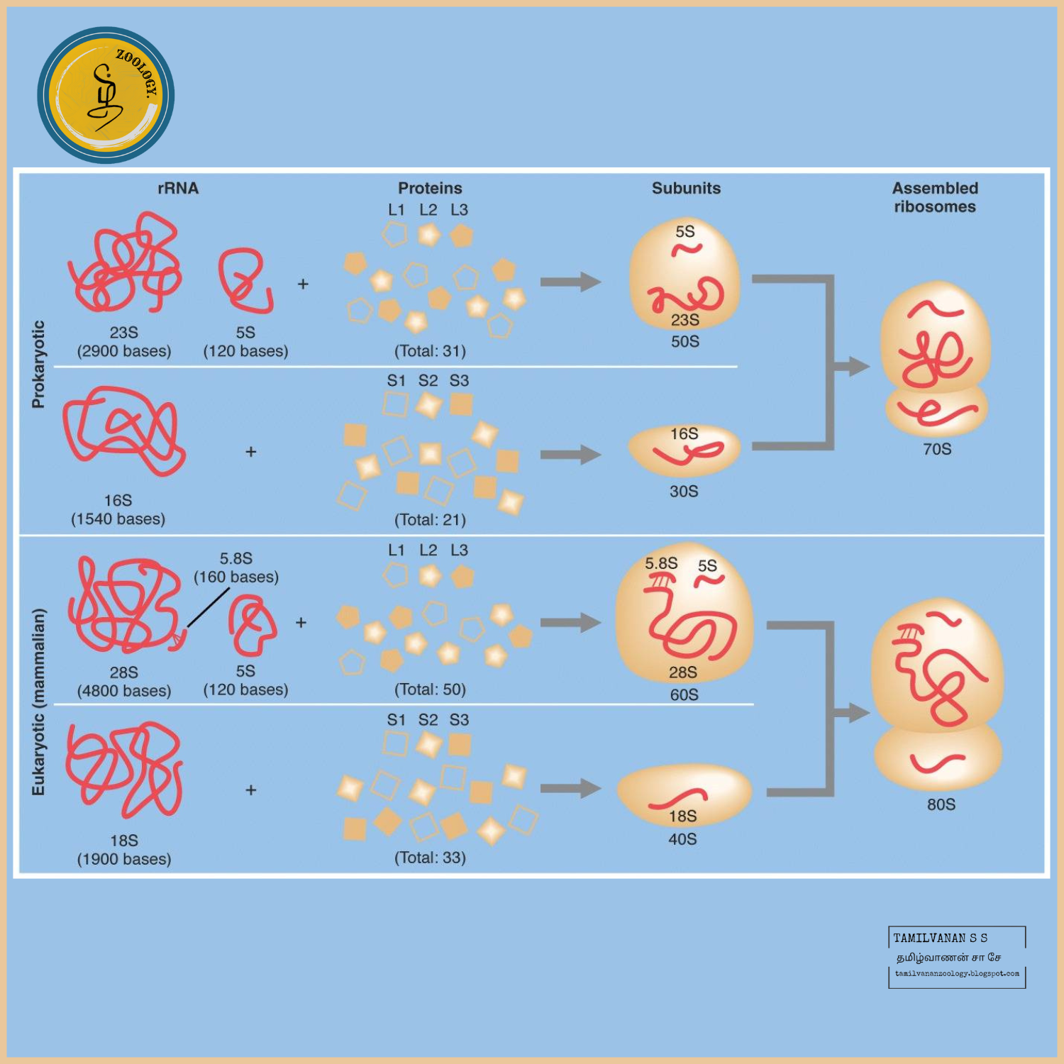
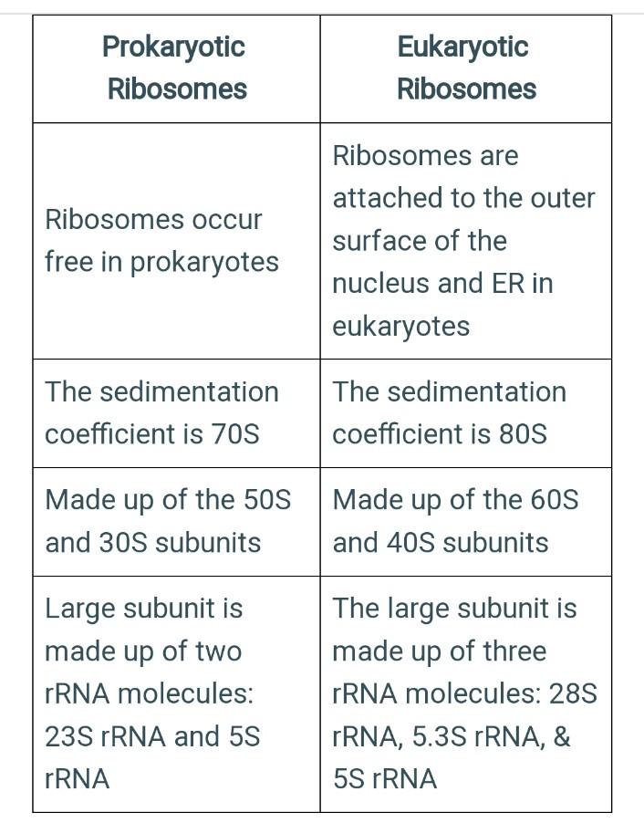
Prokaryotes possesses 70S ribosomes, consisting of a small subunit (30S) and a large subunit (50S). Eukaryotes possess 80S ribosomes, with a small subunit (40S) and a large subunit (60S).

They read the genetic code carried by messenger RNA (mRNA) and use it to assemble amino acids into a specific sequence, ultimately forming proteins.

Prokaryotic Ribosomes:

As compared to eukaryotic ribosomes, the prokaryotic ribosomes are smaller. This is attributed to the association of eukaryotic cell ribosomes with the cytoplasmic or endoplasmic reticulum. 

Prokaryotic ribosomes are called 70S ribosomes and have physical dimensions of approximately 14 to 15 nm by 20 nm, with a molecular weight of roughly 2.7 million, and are made of 50S and 30S subunits. 

The S stands for Svedberg unit in the measurement of ribosome sedimentation coefficient unit. This is a measure of the velocity of sedimentation in a centrifuge; the faster a particle travels when centrifuged, the higher its Svedberg value or the coefficient of sedimentation. 

The coefficient of sedimentation is a function of the molecular weight, volume, and shape of a particle. Normally, heavier and more compact particles have greater numbers of Svedberg or faster sediments. 

There are smaller bacterial ribosomes than eukaryotic ribosomes. A prokaryotic cell usually only has a few thousand ribosomes, while there are several million in a metabolically active eukaryotic cell, such as a human liver cell. 

Proteins that work in the cytoplasm are produced by free ribosomes that are suspended there, while proteins that are bound within membranes or intended for export from the cell are assembled by ribosomes that are bound to rough ER.

The large subunit, called the 50S, is spherical in the prokaryotic cell with a prominent "stalk" and a "central protuberance." It contains the center of peptidyltransferase which catalyzes the formation of peptide bonds between the incoming amino acid and the growing chain of peptides. 

The 50S particle is thick and homogenous. The thin and flexible small subunit of ribosomes in prokaryotes is called the 30S. The 30S houses the decoding center for mRNA and is divided into three domains i.e. head, body, and platform. 

Each one of these domains includes one of the 16S rRNA's main secondary structure domains, namely the 3' major, 5' and central domains. In 16S rRNA, 3' minor domain forms an extended helix running down the 30S subunit surface's long axis, which interacts with the 50S subunit. 

All four 30S particle domains join in a relatively narrow region of the neck. The two "active sites'' face each other around the interface of the subunit and are physiologically linked by the molecule's two ends.

Eukaryotic Ribosomes:

The eukaryotic ribosome (i.e., one not found in mitochondria and chloroplasts) is larger than the prokaryotic 70S ribosome. It is a dimer of the 60S and the 40S subunit, about 22 nm in diameter, and has the sedimentation coefficient of 80S and a molecular weight of 4 million. 

Eukaryotic ribosomes can be either associated with the endoplasmic reticulum or free in the cytoplasmic matrix. When bound to the endoplasmic reticulum to form rough ER, they are attached through their 60S subunits. 

Both free and ER-bound ribosomes synthesize proteins. Proteins made on the ribosomes of the RER are often secreted or are inserted into the ER membrane as integral membrane proteins. 

Free ribosomes are the sites of synthesis for non secretory and non membrane proteins. Some proteins synthesized by free ribosomes are inserted into organelles such as the nucleus, mitochondrion, and chloroplast. They also assist the transport of proteins into eukaryotic organelles such as mitochondria

Types of rRNA

In Prokaryotes

In prokaryotes, the 16S ribosomal RNA is housed in a compact 30S ribosomal subunit. Two rRNA species are present in the large 50S ribosomal subunit (the 23S and 5S ribosomal RNAs). Therefore, it may be concluded that a single rRNA gene in archaea and bacteria codes for the three rRNA types: 16S, 23S, and 5S.

The bacterial 23S, 16S, and 5S rRNA genes are commonly arranged as a co-transcribed operon.

In Eukaryotes

The eukaryotic ribosome is composed of the 40S and a 60S subunit. Eukaryotes, in contrast, usually have several variants of the rRNA genes arranged in repetitive sequences. About 300-400 repeats are found in five clusters on human chromosomes: 13 (RNR1), 14 (RNR2), 15 (RNR3), 21 (RNR4) and 22 (RNR5).

Furthermore, chloroplasts and mitochondria in eukaryotic cells both contain rRNA. Ribosomes can be found in the cytoplasm as free-floating complexes or connected to the endoplasmic reticulum.

Functions of rRNA:

Protein synthesis is the primary function of rRNA. The A, P, and E sites are created within the ribosome by the unusual three-dimensional structure of rRNA, which has internal helices and loops. By attaching to messenger RNA and transfer RNA, these molecules assure that the codon sequence of the mRNA is appropriately translated into the amino acid sequence of proteins.

The A site anchors an entering tRNA that has been charged with an amino acid, while the P site is for binding a developing polypeptide. The tRNA temporarily attaches to the E site following the creation of a peptide bond before exiting the ribosome.

In addition, some ribosomal proteins can bind to rRNA at specific residues, which have been identified after detailed investigation for both the RNA and protein.

Antibiotics like streptomycin and tetracycline have recently been identified as having binding sites on bacterial rRNA. For example, a mutation in the 16S rRNA sequence is the tolerance of Euglena and Escherichia coli to streptomycin.

The 30S rRNA appears to be the source of tetracycline resistance. Similar findings were discovered for Streptomyces to Spectinomycin resistance.

Preribosomal RNA, one of rRNA’s predecessors, has been linked to the production of microRNA, which mediates inflammation and heart illness concerning mechanical stress. This finding adds a new dimension to the role of rRNA.

Ribosomes Functions:

Ribosomes have two principal functions, which involve decoding the messages and the formation of peptide bonds.

Ribosomes participate in the creation of proteins, the DNA makes RNA by DNA transcription.

The mRNA is converted into proteins by the process of translation.

The mRNA is organized in the nucleus and is moved to the cytoplasm for the process of protein synthesis.

The ribosomal subunits in the cytoplasm are bound around mRNA polymers. The tRNA then integrates proteins.

The proteins organized in the cytoplasm are used in the actual cytoplasm, and the proteins synthesized by bound ribosomes are moved to external cells. 

Ribosome Associated Diseases:

Disorders caused by the improper functioning of ribosomes are called ribosomopathies. Mutations that occur in some of the proteins made by ribosomes can cause disorders that are characterized by like bone marrow failure and anemia.

Many congenital syndromes are caused by defective ribosome biogenesis, which includes Diamond-Blackfan anemia (DBA), X-linked dyskeratosis congenita (DKC), cartilage hair hypoplasia (CHH), and Treacher Collins syndrome (TCS).

Diamond-Blackfan anemia (DBA)

It is a rare blood disorder that affects the bone marrow. The bone marrow can make fresh blood cells, including red blood cells, white blood cells, and platelets. In DBA, the bone marrow can’t make sufficient RBC to address the body’s issues. DBA is described as a deficiency of RBC that causes anaemia.

DBA causes abnormal pre-rRNA maturation and shows mutations in one of several ribosomal protein genes that encode structural components of the ribosome.

Summary:

All prokaryotes have 70S ribosomes whereas eukaryotes in their cytosol contain larger 80S ribosomes.

The 70S ribosome consists of subunits 50S and 30S. In catalyzing two biological processes, ribosomes play a key role in the transfer of peptidyl and hydrolysis of peptidyl.

The eukaryotic ribosome (i.e., one not found in mitochondria and chloroplasts) is larger than the prokaryotic 70S ribosome. 

At any given moment, many rRNA molecules dangle from the chromosome at the sites of these clusters of genes that encode rRNA. 

The ribosome reading of each codon results in the incorporation of one amino acid into a progressively longer protein chain. The transfer of RNA (tRNA) molecules, which are the adapter molecules in the translation mechanism, brings the amino acids to the ribosome. 


Female orgasms

Female orgasms A female orgasm can be a highly pleasurable experience during masturbation or sexual activity. While orgasms may ...