Wednesday, August 30, 2023

Spermatogenesis

Spermatogenesis 

Introduction

While the reductive divisions of meiosis are conserved in every eukaryotic kingdom of life, the regulation of meiosis in mammals differs dramatically between males and females. The differences between oogenesis, the production of eggs, and spermatogenesis.


Spermatogenesis

Spermatogenesis is the production of sperm from the primordial germ cells. Once the vertebrate PGCs arrive at the genital ridge of a male embryo, they become incorporated into the sex cords. They remain there until maturity, at which time the sex cords hollow out to form the seminiferous tubules, and the epithelium of the tubules differentiates into the Sertoli cells. The initiation of spermatogenesis during puberty is probably regulated by the synthesis of BMP8B by the spermatogenic germ cells, the spermatogonia. When BMP8B reaches a critical concentration, the germ cells begin to differentiate. The differentiating cells produce high levels of BMP8B, which can then further stimulate their differentiation. Mice lacking BMP8B do not initiate spermatogenesis at puberty (Zhao et al. 1996).

Process 


The spermatogenic germ cells are bound to the Sertoli cells by N-cadherin molecules on both cell surfaces and by galactosyltransferase molecules on the spermatogenic cells that bind a carbohydrate receptor on the Sertoli cells (Newton et al. 1993; Pratt et al. 1993). The Sertoli cells nourish and protect the developing sperm cells, and spermatogenesis—the developmental pathway from germ cell to mature sperm—occurs in the recesses of the Sertoli cells. The processes by which the PGCs generate sperm have been studied in detail in several organisms, but we will focus here on spermatogenesis in mammals.

After reaching the gonad, the PGCs divide to form type A1 spermatogonia. These cells are smaller than the PGCs and are characterized by an ovoid nucleus that contains chromatin associated with the nuclear membrane. The A1 spermatogonia are found adjacent to the outer basement membrane of the sex cords. They are stem cells, and at maturity, they are thought to divide so as to make another type A1 spermatogonium as well as a second, paler type of cell, the type A2 spermatogonium. Thus, each type A1 spermatogonium is a stem cell capable of regenerating itself as well as producing a new cell type. The A2 spermatogonia divide to produce the A3 spermatogonia, which then beget the type A4 spermatogonia. It is possible that each of the type A spermatogonia are stem cells, capable of self-renewal. The A4 spermatogonium has three options: it can form another A4 spermatogonium (self-renewal); it can undergo cell death (apoptosis); or it can differentiate into the first committed stem cell type, the intermediate spermatogonium. Intermediate spermatogonia are committed to becoming spermatozoa, and they divide mitotically once to form the type B spermatogonia. These cells are the precursors of the spermatocytes and are the last cells of the line that undergo mitosis. They divide once to generate the primary spermatocytes—the cells that enter meiosis. It is not known what causes the spermatogonia to take the path toward differentiation rather than self-renewal; nor is it known what stimulates the cells to enter meiotic rather than mitotic division (Dym 1994).

We find that during the spermatogonial divisions, cytokinesis is not complete. Rather, the cells form a syncytium whereby each cell communicates with the others via cytoplasmic bridges about 1 μm in diameter (Dym and Fawcett 1971). The successive divisions produce clones of interconnected cells, and because ions and molecules readily pass through these intercellular bridges, each cohort matures synchronously. During this time, the spermatocyte nucleus often transcribes genes whose products will be used later to form the axoneme and acrosome.

Each primary spermatocyte undergoes the first meiotic division to yield a pair of secondary spermatocytes, which complete the second division of meiosis. The haploid cells thus formed are called spermatids, and they are still connected to one another through their cytoplasmic bridges. The spermatids that are connected in this manner have haploid nuclei, but are functionally diploid, since a gene product made in one cell can readily diffuse into the cytoplasm of its neighbors (Braun et al. 1989). During the divisions from type A1 spermatogonium to spermatid, the cells move farther and farther away from the basement membrane of the seminiferous tubule and closer to its lumen. Thus, each type of cell can be found in a particular layer of the tubule. The spermatids are located at the border of the lumen, and here they lose their cytoplasmic connections and differentiate into sperm cells. In humans, the progression from spermatogonial stem cell to mature sperm takes 65 days (Dym 1994).

Spermiogenesis


The mammalian haploid spermatid is a round, unflagellated cell that looks nothing like the mature vertebrate sperm. The next step in sperm maturation, then, is spermiogenesis (or spermateliosis), the differentiation of the sperm cell. For fertilization to occur, the sperm has to meet and bind with the egg, and spermiogenesis prepares the sperm for these functions of motility and interaction. The first steps involve the construction of the acrosomal vesicle from the Golgi apparatus. The acrosome forms a cap that covers the sperm nucleus. As the acrosomal cap is formed, the nucleus rotates so that the cap will be facing the basal membrane of the seminiferous tubule. This rotation is necessary because the flagellum is beginning to form from the centriole on the other side of the nucleus, and this flagellum will extend into the lumen. During the last stage of spermiogenesis, the nucleus flattens and condenses, the remaining cytoplasm (the “cytoplasmic droplet”) is jettisoned, and the mitochondria form a ring around the base of the flagellum.

One of the major changes in the nucleus is the replacement of the histones by protamines. Transcription of the gene for protamine is seen in the early haploid cells (spermatids), although translation is delayed for several days (Peschon et al. 1987). Protamines are relatively small proteins that are over 60% arginine. During spermiogenesis, the nucleosomes dissociate, and the histones of the haploid nucleus are eventually replaced by protamines. This causes the complete shutdown of transcription in the nucleus and facilitates its assuming an almost crystalline structure. The resulting sperm then enter the lumen of the tubule.

In the mouse, the entire development process from stem cell to spermatozoon takes 34.5 days. The spermatogonial stages last 8 days, meiosis lasts 13 days, and spermiogenesis takes up another 13.5 days. In humans, spermatic development takes nearly twice as long to complete. Because the type A1 spermatogonia are stem cells, spermatogenesis can occur continuously. Each day, some 100 million sperm are made in each human testicle, and each ejaculation releases 200 million sperm. Unused sperm are either resorbed or passed out of the body in urine. During his lifetime, a human male can produce 1012 to 1013 sperm (Reijo et al. 1995).


Kind of Abnormalities in Sperm

The abnormalities found in the sperm include:

Aspermia

This is a condition where a man experiences dry orgasms or orgasms without releasing any semen. This may be the result of retrograde ejaculation, genetic disorders like cystic fibrosis or Klinefelter syndrome, hormonal imbalances or congenital abnormalities. This condition may affect male fertility.

Hypospermia

In such conditions, the total ejaculate is less than 1.5 millilitres. Retrograde ejaculation is the most common cause of this condition. However, it may also be caused by genetic disorders, hormonal imbalances or congenital abnormalities.

Azoospermia

This is a condition wherein a man releases semen that contains no sperm during an orgasm. It is a severe type of male infertility. It may be caused by genetic disorders, hormonal imbalances, congenital abnormalities, untreated STDs and because of post-testicular cancer treatment.

Oligozoospermia

This is a condition wherein the man may have a low sperm count as well as problems with sperm shape and sperm movement. This may be caused by a varicocele vein, hormonal imbalances, undescended testicles, infections of the reproductive tract, environmental conditions and lifestyle choices. In some cases, making a few lifestyle changes may help improve the condition.

Asthenozoospermia

This is a condition where a large percentage of sperm moves abnormally i.e., it doesn’t move in a straight line or does not move at all. This condition may be accompanied by a low sperm count. Some of the causes of this include exposure to toxins, nutritional problems, excessive alcohol consumption, infections and side effects of certain medications.

Teratozoospermia

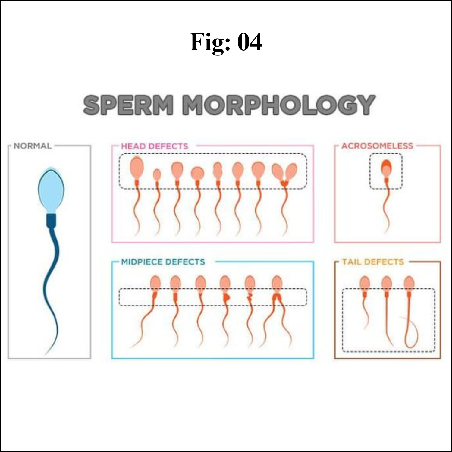
In his condition, the majority of the sperm in the semen are shaped abnormally. The sperms may have more than a single head or tail or an oddly shaped head. This keeps them from moving normally and affects their ability to fertilise an egg.

Oligoasthenoteratozoospermia (OAT)

In such cases the shape, size and movement of the sperm are abnormal. The sperm count may also be lower than normal. This is the most common cause of male infertility.

Necrozoospermia

In such cases all the sperm in the semen is dead. It is a rare cause of infertility.

Leukocytospermia

This is a condition where there is a high amount of white blood cells present in the semen. It is more of a semen abnormality than a sperm abnormality.

Abnormal Sperm Morphology

Teratozoospermia, teratospermia or abnormal sperm morphology is a semen alteration in which a large percentage of sperm have an abnormal shape.


Abnormal Sperm Morphology and Male Infertility

In order for a man to be fertile, only 4 to 14% of normal sperm is needed. Just having abnormal sperm won’t have any impact on fertility; there are other factors. Also, they are sperm numbers, sperm concentration, semen volume, the percentage of sperm that are alive (viability) and the ability to move (motility). An abnormally shaped sperm doesn’t mean that the genetic material it carries will be damaged; it is usually healthy. According to a 2017 study in Asian journal of andrology, men with 0% normally formed sperm had near-normal fertility rates, which indicates there are other significant factors besides sperm morphology normal.


And men with abnormal sperm morphology can impregnate a female. It means that he is not infertile, but he may need a longer time to make the female pregnant. Which means abnormal sperm morphology and pregnancy is possible. If natural conception doesn’t occur, he will have the option of assisted reproductive technology like In vitro fertilisation with ICSI. ICSI is a process, where his sperm can be directly injected into the cytoplasm of an egg, ICSI stands for Intracytoplasmic sperm injection. The advent of ICSI has furthered reduced the significance and perceived need for sperm quality tests because ICSI requires only one sperm to fertilise the egg. ICSI can be done when sperm morphology is less than 2%.


If sperm concentration and sperm motility are normal or high, it may be advisable and reasonable to consider IUI before ICSI-IVF.


Fig & Fig Interactions


Fig (Fruit) & Fig (Wasp) Interactions

Introduction

Everything is connected to everything else. That’s one of the themes that runs through A Perfect Planet. The Earth is a set of interconnected systems where changes in one small thing can have knock-on effects to many others. One of the stories in the series is about tropical fig trees, the birds and primates that feed on their fruit, and the tiny wasps that are fundamental to their life cycles. In my mind the tale of fig trees and fig wasps is one of the most incredible in all of nature. It shows just how interconnected the natural world is and how little things can make big differences.

Interaction of Fig & Fig


" Earth is a set of interconnected systems where changes in one small thing can have knock-on effects to many others. "

To understand how figs and fig wasps interact, it’s important to realize that a fig isn’t a fruit in the way that apples, cherries or plums are. Instead it starts out as a complicated and unique flower head. 

Think of a dandelion flower. Each of those lovely yellow petals is in fact a tiny individual flower with all the petals joined into a narrow tube. 

Now imagine that same dandelion flower never opened; that it was fastened at the top, and instead swelled up as the flowers grew inside. A fig is a bit like that. The round, outer body of the fig is like the base of the dandelion flower (the white ‘pin cushion’ left after you’ve blown the seeds from a dandelion clock), turned inside out, protecting the flowers inside. 

Access to the flowers is through a tiny hole that is blocked by several layers of plant tissue. The only way to pollinate the fig is to squeeze through this tiny gap and get stuck inside. 

So why would a plant evolve a flower that is so difficult to pollinate? Aren’t flowers supposed to be showy and attractive? The reason is that if a plant can ensure that very few things, in extreme cases a single species, can pollinate it, it can evolve specific, targeted and efficient strategies to attract and support its pollinator. 

The difficulty of a pollinator accessing the flowers is balanced by the fact that that pollinator will only take pollen to another flower from the same species – it won’t risk being wasted going to another type of flower. In turn the pollinator evolves structures or strategies to maximize the benefits it gets from its target plant species. 

Many types of plants have evolved close relationships with individual pollinator species, but few quite so intricate as figs and fig wasps.

Pollinating a fig begins when a female fig wasp, which may be just a few millimeters long and laden with pollen, crawls through that tiny hole in the base of the fig. 

To find its way through to the inside of the fig it may need to bite off its own limbs. Once inside, the pollen pollinates some of the tiny individual flowers and the wasp lays eggs in the bases of some others, where the seed should develop. 

The fig sacrifices some of its seed production to provide nursery beds for the wasp’s eggs and larvae to develop. As these larvae develop into adults, the wingless males, which develop first, visit the growing females, fertilize them and, as their dying act, tunnel out of the fig. 

When the females grow to maturity, they work their way out of their growth chamber, picking up pollen from other flowers, and exit through the holes left by their male siblings. 

Seeds develop in the pollinated flowers, the fig matures and becomes sweet and attractive to birds and mammals that eat it and disperse the seeds in their faeces. The emerged females fly to another fig tree, dismember themselves to crawl into a fig and the cycle starts again. Pity the males who know no life outside the confines of a fig. 

Pity also the females who must endure self-mutilation to complete their life cycle. Such is the hand that evolution has dealt these tiny insects. Fig wasps can’t complete their life cycle without figs; fig trees can’t complete theirs without fig wasps. 

This strange marriage has led to figs being one of the most diverse groups of trees in the tropics. Small, chance variations that affect the characteristics of a fig tree, or of its pollinator get hard-wired in as the two co-evolve over time, creating more and more splits in the fig family tree. 

Fig wasps too are extremely diverse with each species of fig being pollinated by only one or a very few species of wasps. The figs we eat are just one species, with several varieties bred to grow fruit without needing to be pollinated at all.

" Fig wasps can’t complete their lifecycle without figs; fig trees can’t complete theirs without fig wasps. "

So much for figs and their pollinators. What is remarkable is how this unique relationship has made fig trees ‘keystone’ elements in tropical forests. The new female wasps need new figs to pollinate and lay their eggs in, even as their original host’s fruits are maturing. 

So at any one time there must be trees with figs at different stages of their development in the forest. Larger animals that use figs as food then have a year-round supply, even through lean seasons where other sources of food are scarce. 

Without figs their ability to survive would be compromised. The forests would not be able to support the diversity and abundance of birds or mammals that they do. In turn these mobile consumers spread the seeds of figs and other fruit trees far and wide, maintaining the diversity of the forest. 

Some figs grow as independent trees but many are ‘stranglers’ that begin their life germinating in the crook of another tree’s branches, high above the forest floor. They send down roots that become trunks and grow to smother their hosts, often spreading to become giant, elephantine ents. 

Some of the world’s biggest trees are figs that owe their existence to a tiny wasp that pollinated a hidden flower to create a seed no bigger than a pinhead which found its way through an animal’s gut and was deposited to germinate, sheltered in the branches of an unsuspecting host.

" With more than 750 species of figs there are bound to be many unknown species and many more intricate relationships to be unearthed."

But this strange story does not end there. Back inside the figs, something else is happening. If nature abhors a vacuum, it also adores an opportunity, and the developing wasp larvae and the other flowers are lots of little opportunities to be exploited. 

Alongside the pollinating fig wasps, whole communities of other wasps have evolved to take advantage of them. There are parasites with improbably long ‘ovipositors’ – the syringe-like extension at the tip of their abdomen – sometimes many times longer than their body, that pierce through the outside of the fig and deposit eggs in the flowers. 

The larvae of some develop and eat the pollinator larvae, while others consume the fig flowers themselves without playing their part and pollinating the fig. Others are parasitoids: species that lay eggs in the pollinator larvae that hatch and eat their host from the inside out. 

In fact the interactions between fig wasps and between wasps and fig trees have many more subtleties and complexities than we can cover here. With more than 750 species of figs there are bound to be many unknown species and many more intricate relationships to be unearthed.

These remarkable natural stories play out in tropical forests around the world, but they simply reflect the way that species interact and ecosystems function anywhere on Earth. 

Our planet and its nature is complex, sometimes fascinatingly so, sometimes unfathomably so. There are many similarly amazing connections happening around you, in your garden, maybe in the soil under your lawn, some of which may have profound implications for how whole ecosystems function, and many of which are yet to be discovered.

Tuesday, August 29, 2023

Reduction Cell Division (Meiosis)


Meiosis (Reduction Division)

Introduction

Meiosis is the process in eukaryotic, sexually-reproducing animals that reduces the number of chromosomes in a cell before reproduction. Many organisms package these cells into gametes, such as egg and sperm. The gametes can then meet, during reproduction, and fuse to create a new zygote. Because the number of alleles was reduced during meiosis, the combination of two gametes will yield a zygote with the same number of alleles as the parents. In diploid organisms, this is two copies of each gene.

Phases of Meiosis

Before meiosis, the DNA is replicated, as in mitosis. Meiosis then consists of two cell divisions, known as meiosis I and meiosis II. In the first division, which consists of different phases, the duplicated DNA is separated into daughter cells. In the next division, which immediately follows the first, the two alleles of each gene are separated into individual cells.

The following are descriptions of the two divisions, and the various phases, or stages of each meiosis. Remember, before meiosis starts the normally diploid DNA has been duplicated. This means there are 4 copies of each gene, present in 2 full sets of DNA, each set having 2 alleles. In the diagram below, the red chromosomes are the ones inherited from the mother, the blue from the father.

At the start of the following diagram, the DNA has already been replicated, which is why the red and blue chromosomes look like the letter “X”. Each one of these “X” chromosomes consists of two sister chromatids – cloned DNA from replication. They are connected at the centromere for storage but can separate into individual chromosomes.


Phases of Meiosis I

Prophase I

Prophase I, the first step in meiosis I, is similar to prophase in mitosis in that the chromosomes condense and move towards the middle of the cell. The nuclear envelope degrades, which allows the microtubules originating from the centrioles on either side of the cell to attach to the kinetochores in the centromeres of each chromosome. Unlike in mitosis, the chromosomes pair with their homologous partner. This can be seen in the red and blue chromosomes that pair together in the diagram. This step does not take place in mitosis. At the end of prophase I and the beginning of metaphase I, homologous chromosomes are primed for crossing-over.

Between prophase I and metaphase I, homologous chromosomes can swap parts of themselves that house the same genes. This is called crossing-over and is responsible for the other law of genetics, the law of independent assortment. This law states that traits are inherited independently of each other. For traits on different chromosomes, this is certainly true all of the time. For traits on the same chromosome, crossing-over makes it possible for the maternal and paternal DNA to recombine, allowing traits to be inherited in an almost infinite number of ways.

Metaphase I

In metaphase I of meiosis I, the homologous pairs of chromosomes line up on the metaphase plate, near the center of the cell. This step is referred to as a reductional division. The homologous chromosomes that contain the two different alleles for each gene are lined up to be separated. As seen in the diagram above, while the chromosomes line up on the metaphase plate with their homologous pair, there is no order upon which side the maternal or paternal chromosomes line up. This process is the molecular reason behind the law of segregation.


The law of segregation tells us that each allele has the same chance of being passed on to offspring. In metaphase I of meiosis, the alleles are separated, allowing for this phenomenon to happen. In meiosis II, they will be separated into individual gametes. In mitosis, all the chromosomes line up on their centromeres, and the sister chromatids of each chromosome separate into new cells. The homologous pairs do not pair up in mitosis, and each is split in half to leave the new cells with 2 different alleles for each gene. Even if these alleles are the same allele, they came from a maternal and paternal source. In meiosis, the lining up of homologous chromosomes leaves 2 alleles in the final cells, but they are on sister chromatids and are clones of the same source of DNA.

Anaphase I

Much like anaphase of mitosis, the chromosomes are now pulled towards the centrioles at each side of the cell. However, the centrosomes holding the sister chromatids together do not dissolve in anaphase I of meiosis, meaning that only homologous chromosomes are separated, not sister chromatids.

Telophase I

In telophase I, the chromosomes are pulled completely apart and new nuclear envelopes form. The plasm membrane is separated by cytokinesis and two new cells are effectively formed.

Results of Meiosis I

Two new cells, each haploid in their DNA, but with 2 copies, are the result of meiosis I. Again, although there are 2 alleles for each gene, they are on sister chromatid copies of each other. These are therefore considered haploid cells. These cells take a short rest before entering the second division of meiosis, meiosis II.

Phases of Meiosis II

Prophase II

Prophase II resembles prophase I. The nuclear envelopes disappear and centrioles are formed. Microtubules extend across the cell to connect to the kinetochores of individual chromatids, connected by centromeres. The chromosomes begin to get pulled toward the metaphase plate.

Metaphase II

Now resembling mitosis, the chromosomes line up with their centromeres on the metaphase plate. One sister chromatid is on each side of the metaphase plate. At this stage, the centromeres are still attached by the protein cohesin.

Anaphase II

The sister chromatids separate. They are now called sister chromosomes and are pulled toward the centrioles. This separation marks the final division of the DNA. Unlike the first division, this division is known as an equational division, because each cell ends up with the same quantity of chromosomes as when the division started, but with no copies.

Telophase II

As in the previous telophase I, the cell is now divided into two and the chromosomes are on opposite ends of the cell. Cytokinesis or plasma division occurs, and new nuclear envelopes are formed around the chromosomes.

Results of Meiosis II

At the end of meiosis II, there are 4 cells, each haploid, and each with only 1 copy of the genome. These cells can now be developed into gametes, eggs in females and sperm in males.

Examples of Meiosis

Human Meiosis

Human meiosis occurs in the sex organs. Male testis produce sperm and female ovaries produce eggs. Before these gametes are made, however, the DNA must be reduced. Humans have 23 distinct chromosomes, existing in homologous pairs between maternal and paternal DNA, meaning 46 chromosomes. Before meiosis, the DNA in the cell is replicated, producing 46 chromosomes in 92 sister chromatids. Each pair of sister chromatids has a corresponding (either maternal or paternal) set of sister chromosomes. These pairs are known as homologous chromosomes. During meiosis I, these homologous chromosomes line up and divide. This leaves 23 chromosomes in each cell, each chromosome consisting of sister chromatids. These chromatids may no longer be identical, as crossing-over may have occurred during metaphase I of meiosis I. Finally, meiosis II takes place, and the sister chromatids are separated into individual cells. This leaves 4 cells, each with 23 chromosomes, or 4 haploid cells.

Fruit Flies

Fruit flies have 4 pairs of chromosomes or 8 chromosomes in regular cells. Before meiosis takes place, each chromosome is replicated, leaving 8 chromosomes and 16 sister chromatids. Meiosis I takes place, and there are 2 cells, each with only 4 chromosomes. Each chromosome is still made of sister chromatids, and some crossing-over may have occurred during metaphase I. Meiosis II now takes place on those two cells. In total, 4 cells are created, again. However, these cells have 4 chromosomes. When two gametes meet to create a new fruit fly, the resulting zygote will have 8 chromosomes of 4 pairs of sister chromosomes, 4 coming from each parent.

Function of Meiosis

Meiosis is necessary for many sexually-reproducing animals to ensure the same number of chromosomes in the offspring as in the parents. The act of fertilization includes two cells fusing together to become a new zygote. If the number of alleles of each gene is not reduced to 1 in the gametes that produce the zygote, there will be 4 copies of each gene in the offspring. In many animals, this would lead to many developmental defects.

In other organisms, polyploidy is common and they can exist with many copies of the same gene. However, if the organism cannot survive if they are polyploidy, meiosis must occur before reproduction. Meiosis occurs in two distinct divisions, with different phases in each.

Sunday, August 27, 2023

Bandura's Observational Learning

Observational Learning

Introduction 

As humans, we start learning on our first day of existence.

Most of this initial learning happens through observation. Observing friends, family members, and the surrounding environment. That’s how we make sense of the world.

In this article, we’ll take an in-depth look at observational learning theory: the definition, four processes, examples, and importance.

We’ll also explore observational learning in the corporate workplace. And how you can use observational learning to improve your corporate training and development programs.

Definition 

Observational learning is the process of learning by watching the behaviors of others. The targeted behavior is watched, memorized, and then mimicked.

Also known as shaping and modeling, observational learning is most common in children as they imitate behaviors of adults.

While at times, we intentionally observe experts to learn new information, observational learning isn’t always intentional. Especially in young children.

A child may learn to swear or smoke cigarettes by watching adults. They are continually learning through observation, whether the target behavior is desirable or not.

Model 

A model is the person performing the task being imitated. In the example of a child learning to swear, the model is the parent that said the swear word. The child is using their parent as a model that they observe performing a behavior.

Good model

Humans don’t just imitate anyone. Most often, we mimic people that:

Are similar to us
Are in high-status positions
Are experts or knowledgeable
Are rewarded for their behaviors
Provide us with nurturing (parents or guardian-figures)

Processes of Observational Learning

Canadian/American psychologist, Albert Bandura, was one of the first psychologists to recognize the phenomenon of observational learning. His theory, Social Learning Theory, stresses the importance of observation and modeling of behaviors, attitudes and emotional reactions of others.

He found that, as social animals, humans naturally gravitate toward observational learning. Children watch their family members and mimic their behaviors. Even infants, at just 3-weeks old, start imitating mouth movements and facial expressions of adults around them.

According to Bandura’s research, there are four processes that influence observational learning:

1.Attention
2.Retention
3.Reproduction
4.Motivation


Let’s take a look at each in more depth:

1. Attention

To learn, an observer must pay attention to something in the environment. They must notice the model and the behavior occurring. Attention levels can vary based on the characteristics of the model and environment – including the model’s degree of likeness, or the observer’s current mood.

In humans, it is likely the observer will pay attention to behaviors of models that are high-status, talented, intelligent, or similar to the observer in any way.

2. Retention

Simple attention is not enough to learn a new behavior. An observer must also retain, or remember, the behavior at a later time.

To increase chances of retention, the observer must structure the information in an easy-to-remember format. Maybe they use a mnemonic device. Or form a daily learning habit.

The behavior must be easily remembered so the action can be performed with little or no effort.

3. Reproduction

The behavior is remembered. But can it be performed in real-life?

Reproduction is the process where the observer must be able to physically perform the behavior in the real-world. Easier said than done.

Often, producing a new behavior requires hours of practice to obtain the skills. You can’t just watch your VP give a brilliant company-wide presentation, then use only the observed tactics in your own presentation 20-minutes later. Those skills take years to craft and perfect.

4. Motivation

All learning requires some degree of personal motivation. For observational learning, the observer must be motivated to produce the desired behavior.

Sometimes this motivation is intrinsic to the observer. Other times, motivation can come in the form of external reinforcement – rewards and punishments.

Bobo Doll Experiment

Bandura’s classic Bobo Doll experiment showed that children would mimic violent behaviors, simply by observing others.

In the experiment, children were shown a video where a model would act aggressively toward an inflatable doll – hitting, punching, kicking, and verbally assaulting the doll. There were three different endings:

1.The model was punished for their behavior
2.The model was rewarded for their behavior
3.There were no consequences

After watching the model, children were given a Bobo doll, identical to that in the video. Their behaviors were observed.

Researchers found that children were more likely to mimic violent behaviors when they observed the model receiving a reward, or when no consequences occurred. On the flip side – children that observed the model being punished for violence showed less actual violence toward the doll.

Examples

Here are a few real-world examples of observational learning:

A child watches their mother eat dinner with a fork. They observe the behavior and quickly learn how to use a fork themselves.
 

A high-school basketball player watches Stephen Curry shoot free-throws. They observe details such as the number of ball dribbles and hand follow through patterns, then try to mimic the behavior themselves.

Influences on Observational Learning

According to Bandura's research, there are a number of factors that increase the likelihood that a behavior will be imitated. We are more likely to imitate:

People we perceive as warm and nurturing

People who receive rewards for their behavior

People who are in an authoritative position in our lives

People who are similar to us in age, sex, and interests

People we admire or who are of a higher social status

When we have been rewarded for imitating the behavior in the past

When we lack confidence in our own knowledge or abilities

When the situation is confusing, ambiguous, or unfamiliar

Pros and Cons of Observational Learning

Observational learning has the potential to teach and reinforce or decrease certain behaviors based on a variety of factors. Particularly prevalent in childhood, observational learning can be a key part of how we learn new skills and learn to avoid consequences.

However, there has also been concern about how this type of learning can lead to negative outcomes and behaviors. Some studies, inspired by Bandura's research, focused on the effects observational learning may have on children and teenagers.

For example, previous research drew a direct connection between playing certain violent video games and an increase in aggression in the short term. However, later research that focused on the short- and long-term impact video games may have on players has shown no direct connections between video game playing and violent behavior.

Similarly, research looking at sexual media exposure and teenagers' sexual behavior found that, in general, there wasn't a connection between watching explicit content and having sex within the following year.

Another study indicated that if teenagers age 14 and 15 of the same sex consumed sexual media together and/or if parents restricted the amount of sexual content watched, the likelihood of having sex was lower. The likelihood of sexual intercourse increased when opposite-sex peers consumed sexual content together.

Uses for Observational Learning

Observational learning can be used in the real world in a number of different ways. Some examples include:

Learning new behaviors: Observational learning is often used as a real-world tool for teaching people new skills. This can include children watching their parents perform a task or students observing a teacher engage in a demonstration.

Strengthening skills: Observational learning is also a key way to reinforce and strengthen behaviors. For example, if a study sees another student getting a reward for raising their hand in class, they will be more likely to also raise their hand the next time they want to ask a question.

Minimizing negative behaviors: Observational learning also plays an important role in reducing undesirable or negative behaviors. For example, if you see a coworker get reprimanded for failing to finish a task on time, it means that you may be more likely to finish your work more quickly.

Friday, August 25, 2023

Skinner's Operant Conditioning


Operant Conditioning
 

Introduction

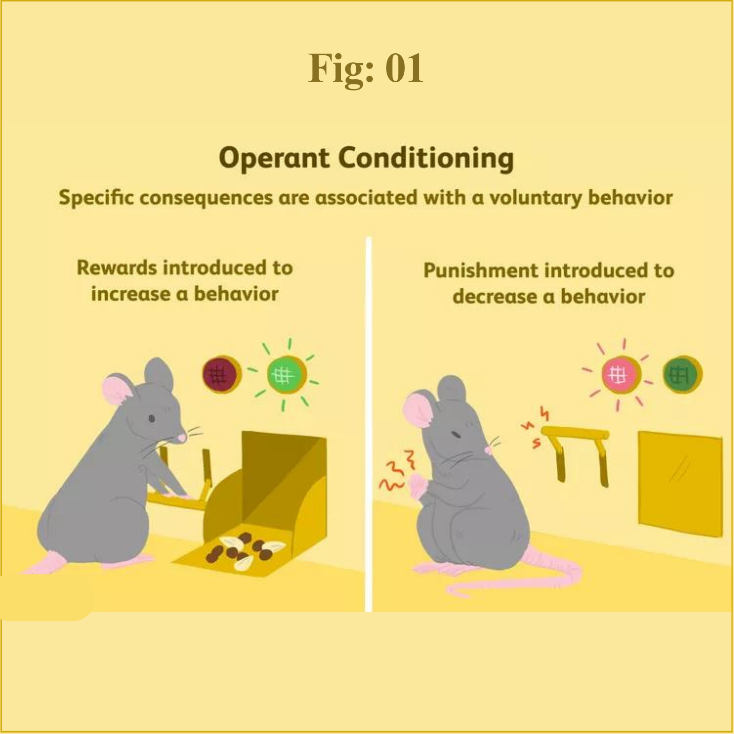
Operant conditioning, sometimes referred to as instrumental conditioning, is a method of learning that employs rewards and punishments for behavior. Through operant conditioning, an association is made between a behavior and a consequence (whether negative or positive) for that behavior.

For example, when lab rats press a lever when a green light is on, they receive a food pellet as a reward. When they press the lever when a red light is on, they receive a mild electric shock. As a result, they learn to press the lever when the green light is on and avoid the red light.

But operant conditioning is not just something that takes place in experimental settings while training lab animals. It also plays a powerful role in everyday learning. Reinforcement and punishment take place in natural settings all the time, as well as in more structured settings such as classrooms or therapy sessions. 

History

Operant conditioning was first described by behaviorist B.F. Skinner, which is why you may occasionally hear it referred to as Skinnerian conditioning. As a behaviorist, Skinner believed that it was not really necessary to look at internal thoughts and motivations in order to explain behavior. Instead, he suggested, we should look only at the external, observable causes of human behavior.

Through the first part of the 20th century, behaviorism became a major force within psychology. The ideas of John B. Watson dominated this school of thought early on. Watson focused on the principles of classical conditioning, once famously suggesting that he could take any person regardless of their background and train them to be anything he chose.

Early behaviorists focused their interests on associative learning. Skinner was more interested in how the consequences of people's actions influenced their behavior.

His theory was heavily influenced by the work of psychologist Edward Thorndike, who had proposed what he called the law of effect. According to this principle, actions that are followed by desirable outcomes are more likely to be repeated while those followed by undesirable outcomes are less likely to be repeated.

Operant conditioning relies on a fairly simple premise: Actions that are followed by reinforcement will be strengthened and more likely to occur again in the future. If you tell a funny story in class and everybody laughs, you will probably be more likely to tell that story again in the future.

If you raise your hand to ask a question and your teacher praises your polite behavior, you will be more likely to raise your hand the next time you have a question or comment. Because the behavior was followed by reinforcement, or a desirable outcome, the preceding action is strengthened.

Conversely, actions that result in punishment or undesirable consequences will be weakened and less likely to occur again in the future. If you tell the same story again in another class but nobody laughs this time, you will be less likely to repeat the story again in the future. If you shout out an answer in class and your teacher scolds you, then you might be less likely to interrupt the class again.

Types of Behaviors

Skinner distinguished between two different types of behaviors

Respondent behaviors are those that occur automatically and reflexively, such as pulling your hand back from a hot stove or jerking your leg when the doctor taps on your knee. You don't have to learn these behaviors. They simply occur automatically and involuntarily.

Operant behaviors, on the other hand, are those under our conscious control. Some may occur spontaneously and others purposely, but it is the consequences of these actions that then influence whether or not they occur again in the future. Our actions on the environment and the consequences of that action make up an important part of the learning process.

While classical conditioning could account for respondent behaviors, Skinner realized that it could not account for a great deal of learning. Instead, Skinner suggested that operant conditioning held far greater importance.

Skinner invented different devices during his boyhood and he put these skills to work during his studies on operant conditioning. He created a device known as an operant conditioning chamber, often referred to today as a Skinner box. The chamber could hold a small animal, such as a rat or pigeon. The box also contained a bar or key that the animal could press in order to receive a reward.

In order to track responses, Skinner also developed a device known as a cumulative recorder. The device recorded responses as an upward movement of a line so that response rates could be read by looking at the slope of the line.


Components of Operant Conditioning

There are several key concepts in operant conditioning. The type of reinforcement or punishment that is used can have an effect on how the individual responds and the effect of conditioning. There are four types of operant conditioning that can be utilized to change behavior: positive reinforcement, negative reinforcement, positive punishment, and negative punishment.

Reinforcement in Operant Conditioning

Reinforcement is any event that strengthens or increases the behavior it follows. There are two kinds of reinforcers. In both of these cases of reinforcement, the behavior increases.

Positive reinforcers are favorable events or outcomes that are presented after the behavior. In positive reinforcement situations, a response or behavior is strengthened by the addition of praise or a direct reward. If you do a good job at work and your manager gives you a bonus, that bonus is a positive reinforcer.

Negative reinforcers involve the removal of an unfavorable events or outcomes after the display of a behavior. In these situations, a response is strengthened by the removal of something considered unpleasant. For example, if your child starts to scream in the middle of a restaurant, but stops once you hand them a treat, your action led to the removal of the unpleasant condition, negatively reinforcing your behavior (not your child's).

Punishment in Operant Conditioning

Punishment is the presentation of an adverse event or outcome that causes a decrease in the behavior it follows. There are two kinds of punishment. In both of these cases, the behavior decreases.

Positive punishment, sometimes referred to as punishment by application, presents an unfavorable event or outcome in order to weaken the response it follows. Spanking for misbehavior is an example of punishment by application.

Negative punishment, also known as punishment by removal, occurs when a favorable event or outcome is removed after a behavior occurs. Taking away a child's video game following misbehavior is an example of negative punishment.

Operant Conditioning Reinforcement Schedules

Reinforcement is not necessarily a straightforward process, and there are a number of factors that can influence how quickly and how well new things are learned. Skinner found that when and how often behaviors were reinforced played a role in the speed and strength of acquisition. In other words, the timing and frequency of reinforcement influenced how new behaviors were learned and how old behaviors were modified.

Skinner identified several different schedules of reinforcement that impact the operant conditioning process:

1.Continuous reinforcement involves delivering a reinforcement every time a response occurs. Learning tends to occur relatively quickly, yet the response rate is quite low. Extinction also occurs very quickly once reinforcement is halted.

2.Fixed-ratio schedules are a type of partial reinforcement. Responses are reinforced only after a specific number of responses have occurred. This typically leads to a fairly steady response rate.

3.Fixed-interval schedules are another form of partial reinforcement. Reinforcement occurs only after a certain interval of time has elapsed. Response rates remain fairly steady and start to increase as the reinforcement time draws near, but slow immediately after the reinforcement has been delivered.

4.Variable-ratio schedules are also a type of partial reinforcement that involve reinforcing behavior after a varied number of responses. This leads to both a high response rate and slow extinction rates.

5.Variable-interval schedules are the final form of partial reinforcement Skinner described. This schedule involves delivering reinforcement after a variable amount of time has elapsed. This also tends to lead to a fast response rate and slow extinction rate.

Examples of Operant Conditioning

We can find examples of operant conditioning at work all around us. Consider the case of children completing homework to earn a reward from a parent or teacher, or employees finishing projects to receive praise or promotions. More examples of operant conditioning in action include:

1.After performing in a community theater play, you receive applause from the audience. This acts as a positive reinforcer, inspiring you to try out for more performance roles.

2.You train your dog to fetch by offering him praise and a pat on the head whenever he performs the behavior correctly. This is another positive reinforcer.

3.A professor tells students that if they have perfect attendance all semester, then they do not have to take the final comprehensive exam. By removing an unpleasant stimulus (the final test), students are negatively reinforced to attend class regularly.

4.If you fail to hand in a project on time, your boss becomes angry and berates your performance in front of your co-workers. This acts as a positive punisher, making it less likely that you will finish projects late in the future.

5.A teen girl does not clean up her room as she was asked, so her parents take away her phone for the rest of the day. This is an example of a negative punishment in which a positive stimulus is taken away.

In some of these examples, the promise or possibility of rewards causes an increase in behavior. Operant conditioning can also be used to decrease a behavior via the removal of a desirable outcome or the application of a negative outcome.

For example, a child may be told they will lose recess privileges if they talk out of turn in class. This potential for punishment may lead to a decrease in disruptive behaviors.

Wednesday, August 23, 2023

Pavlov's Classical Conditioning

Classical Conditioning (Pavlovian Conditioning)

Introduction

Classical conditioning (also known as Pavlovian or respondent conditioning) is learning through association and was discovered by Pavlov, a Russian physiologist. In simple terms, two stimuli are linked together to produce a new learned response in a person or animal.

John Watson proposed that the process of classical conditioning (based on Pavlov’s observations) was able to explain all aspects of human psychology.

Everything from speech to emotional responses was simply patterns of stimulus and response. Watson completely denied the existence of the mind or consciousness. Watson believed that all individual differences in behavior were due to different learning experiences.

Watson (1924, p. 104) famously said:

Give me a dozen healthy infants, well-formed, and my own specified world to bring them up in and I”ll guarantee to take any one at random and train him to become any type of specialist I might select – doctor, lawyer, artist, merchant-chief and, yes, even beggar-man and thief, regardless of his talents, penchants, tendencies, abilities, vocations and the race of his ancestors.

Principles Of Classical Conditioning

Neutral Stimulus

In classical conditioning, a neutral stimulus (NS) is a stimulus that initially does not evoke a response until it is paired with the unconditioned stimulus.

For example, in Pavlov’s experiment, the bell was the neutral stimulus, and only produced a response when it was paired with food.

Unconditioned Stimulus

In classical conditioning, the unconditioned stimulus is a feature of the environment that causes a natural and automatic unconditioned response. In Pavlov’s study, the unconditioned stimulus was food.

Unconditioned Response

In classical conditioning, an unconditioned response is an unlearned response that occurs automatically when the unconditioned stimulus is presented.

Pavlov showed the existence of the unconditioned response by presenting a dog with a bowl of food and measuring its salivary secretions.

Conditioned Stimulus

In classical conditioning, the conditioned stimulus (CS) is a substitute stimulus that triggers the same response in an organism as an unconditioned stimulus.

For example, Pavlov’s dog learned to salivate at the sound of a bell. Simply put, a conditioned stimulus makes an organism react to something because it is associated with something else.

Conditioned Response

In classical conditioning, the conditioned response (CR) is the learned response to the previously neutral stimulus. In Ivan Pavlov’s experiments in classical conditioning, the dog’s salivation was the conditioned response to the sound of a bell.

Acquisition

In the initial learning period, acquisition describes when an organism learns to connect a neutral stimulus and an unconditioned stimulus.

Extinction

In psychology, extinction refers to the gradual weakening of a conditioned response by breaking the association between the conditioned and the unconditioned stimuli.

For example, when the bell repeatedly rang and no food was presented, Pavlov’s dog gradually stopped salivating at the sound of the bell.

Spontaneous Recovery

Spontaneous Recovery is a phenomenon of Pavlovian conditioning that refers to the return of a conditioned response (in a weaker form) after a period of time following extinction.

For example, when Pavlov waited a few days after extinguishing the conditioned response, and then rang the bell once more, the dog salivated again.

Generalization

In psychology, generalization is the tendency to respond in the same way to stimuli that are similar but not identical to the conditioned stimulus.

For example, in Pavlov’s experiment, if a dog is conditioned to salivate to the sound of a bell, it may later salivate to a higher-pitched bell.

Discrimination

In classical conditioning, discrimination is a process through which individuals learn to differentiate among similar stimuli and respond appropriately to each one.

For example, eventually, Pavlov’s dog learns the difference between the sound of the 2 bells and no longer salivates at the sound of the non-food bell.

Working of Classical Conditioning 

There are three stages of classical conditioning. At each stage, the stimuli and responses are given special scientific terms:

Stage 1: Before Conditioning:

In this stage, the unconditioned stimulus (UCS) produces an unconditioned response (UCR) in an organism.

In basic terms, this means that a stimulus in the environment has produced a behavior / response which is unlearned (i.e., unconditioned) and therefore is a natural response which has not been taught. In this respect, no new behavior has been learned yet.

This stage also involves another stimulus which has no effect on a person and is called the neutral stimulus (NS). The NS could be a person, object, place, etc.

The neutral stimulus in classical conditioning does not produce a response until it is paired with the unconditioned stimulus.

Stage 2: During Conditioning:

During this stage, a stimulus which produces no response (i.e., neutral) is associated with the unconditioned stimulus, at which point it now becomes known as the conditioned stimulus (CS).

For classical conditioning to be effective, the conditioned stimulus should occur before the unconditioned stimulus, rather than after it, or during the same time. Thus, the conditioned stimulus acts as a type of signal or cue for the unconditioned stimulus.

In some cases, conditioning may take place if the NS occurs after the UCS (backward conditioning), but this normally disappears quite quickly. The most important aspect of the conditioning stimulus is the it helps the organism predict the coming of the unconditional stimulus.

Often during this stage, the UCS must be associated with the CS on a number of occasions, or trials, for learning to take place.

However, one trial learning can happen on certain occasions when it is not necessary for an association to be strengthened over time (such as being sick after food poisoning or drinking too much alcohol).

Stage 3: After Conditioning:

Now the conditioned stimulus (CS) has been associated with the unconditioned stimulus (UCS) to create a new conditioned response (CR).

Classical Conditioning Examples

Pavlov’s Dogs

The most famous example of classical conditioning was Ivan Pavlov’s experiment with dogs, who salivated in response to a bell tone. Pavlov showed that when a bell was sounded each time the dog was fed, the dog learned to associate the sound with the presentation of the food.


He first presented the dogs with the sound of a bell; they did not salivate so this was a neutral stimulus. Then he presented them with food, they salivated. The food was an unconditioned stimulus and salivation was an unconditioned (innate) response.

He then repeatedly presented the dogs with the sound of the bell first and then the food (pairing) after a few repetitions the dogs salivated when they heard the sound of the bell. The bell had become the conditioned stimulus and salivation had become the conditioned response.

Fear Response

Watson & Rayner (1920) were the first psychologists to apply the principles of classical conditioning to human behavior by looking at how this learning process may explain the development of phobias.

They did this in what is now considered to be one of the most ethically dubious experiments ever conducted – the case of Little Albert. Albert B.’s mother was a wet nurse in a children’s hospital. Albert was described as ‘healthy from birth’ and ‘on the whole stolid and unemotional’.


When he was about nine months old, his reactions to various stimuli (including a white rat, burning newspapers and a hammer striking a four-foot steel bar just behind his head) were tested.

Only the last of these frightened him, so this was designated the unconditioned stimulus (UCS) and fear the unconditioned response (UCR). The other stimuli were neutral because they did not produce fear.

When Albert was just over eleven months old, the rat and the UCSwere presented together: as Albert reached out to stroke the animal, Watson struck the bar behind his head.

This occurred seven times in total over the next seven weeks. By this time the rat, the conditioned stimulus (CS), on its own frightened Albert, and fear was now a conditioned response (CR).

The CR transferred spontaneously to the rabbit, the dog and other stimuli that had been previously neutral. Five days after conditioning, the CR produced by the rat persisted. After ten days it was ‘much less marked’, but it was still evident a month later

Carter and Tiffany, 1999 support the cue reactivity theory, they carried out a meta-analysis reviewing 41 cue-reactivity studies that compared responses of alcoholics, cigarette smokers, cocaine addicts and heroin addicts to drug-related versus neutral stimuli. They found that dependent individuals reacted strongly to the cues presented and reported craving and physiological arousal.

Addiction

Cue reactivity is the theory that people associate situations (e.g. meeting with friends)/ places (e.g. pub) with the rewarding effects of nicotine, and these cues can trigger a feeling of craving.

These factors become smoking-related cues. Prolonged use of nicotine creates an association between these factors and smoking based on classical conditioning.

Nicotine is the unconditioned stimulus (UCS), and the pleasure caused by the sudden increase in dopamine levels is the unconditioned response (UCR). Following this increase, the brain tries to lower the dopamine back to a normal level.

The stimuli that have become associated with nicotine were neutral stimuli (NS) before “learning” took place but they became conditioned stimuli (CS), with repeated pairings. They can produce the conditioned response (CR).

However, if the brain has not received nicotine, the levels of dopamine drop, and the individual experiences withdrawal symptoms therefore is more likely to feel the need to smoke in the presence of the cues that have become associated with the use of nicotine.

Classroom Learning

The implications of classical conditioning in the classroom are less important than those of operant conditioning, but there is a still need for teachers to try to make sure that students associate positive emotional experiences with learning.

If a student associates negative emotional experiences with school, then this can obviously have bad results, such as creating a school phobia.

For example, if a student is bullied at school they may learn to associate the school with fear. It could also explain why some students show a particular dislike of certain subjects that continue throughout their academic career. This could happen if a student is humiliated or punished in class by a teacher.

Monday, August 21, 2023

Pinocytosis


Pinocytosis 

Introduction 

Pinocytosis is the ingestion of extracellular fluids, i.e. the fluid surrounding the cell, together with its contents of small dissolved molecules (solutes). This begins with the cell forming narrow channels through its membrane that pinch off into vesicles, and fuse with endosomes resulting in the hydrolysis or breakdown of the contents. Pinocytosis can be thought of as ‘cell drinking’ as the word comes from the Greek “pino“, meaning ‘to drink’ and “cyto“, meaning ‘cell’. Pinocytosis was discovered by Warren Lewis in 1931 and is also known as fluid-phase endocytosis.

Pinocytosis is an example of endocytosis, a cellular process in which substances are brought inside a cell. Other types of endocytosis include phagocytosis and receptor-mediated endocytosis. All three are about taking in substances into the cell. However, what is the difference between phagocytosis and pinocytosis? Phagocytosis is about “engulfing” a relatively larger substance. Conversely, pinocytosis refers to “cell drinking”.

As for the difference between pinocytosis and receptor-mediated endocytosis, the latter is more specific; substances have to bind to the receptors on the cell surface to initiate endocytosis. Nevertheless, some references classify the latter under the wider, broader, pinocytosis. The uptake of fluid from outside the cell that involves pinosomes (fluid-filled vesicles), irrelevant of the size, is what defines pinocytosis.

Types of Pinocytosis

Pinocytosis can be divided by the size of the molecules to be taken up.

Micropinocytosis refers to the uptake of small molecules with a vesicle size of around 0.1µm. Caveolin-mediated pinocytosis is a common example of micropinocytosis that will be described in more detail below.

Macropinocytosis results in the formation of larger vesicles of around 0.5-5 µm. Macropinocytosis is a non-selective process. It results in the formation of large macropinosomes. The protein actin is largely involved in the formation of protrusions or ruffles in the cell membrane which results in the formation of these large vesicles. Macropinocytosis is used by immune cells such as macrophages to sample bulk extracellular fluid for soluble antigens that can evoke an immune response if necessary.

Pinocytosis can be further divided into 4 sub-types based on the mechanism of action. These are as follows:

Macropinocytosis

Clathrin-mediated endocytosis (also known as receptor-mediated endocytosis)

Caveolae-mediated endocytosis

Clathrin-independent/caveolae independent endocytosis.


Steps of Pinocytosis

What happens during pinocytosis? The membrane surrounding the cell can be described as semi-permeable. This means that it allows some molecules in or out via diffusion. The cell membrane also contains various lipids, fats, and protein channels/carriers.

Only small particles can be taken up during pinocytosis as they are usually dissolved in the extracellular fluid. The resulting vesicle contains this extracellular fluid complete with its solutes.

The vesicle can be described as a membrane-bound organelle; it is made up of the extracellular membrane of the cell enclosing the fluid in a spherical arrangement. Pinocytosis can be initiated by electrostatic interaction between a positively charged substance, such as the charged portion of a peptide or protein, and the negatively charged surface of the cell membrane. This can initiate binding to the cell membrane, altering the shape of the membrane to create a pouch around the fluid containing the charged peptide or protein.

Eventually, the membrane curls around on itself, and the pouch is ‘pinched off ‘ allowing the resulting vesicle to drift into the cytoplasm of the cell.

The pinocytotic vesicles function as carriers of the extracellular fluid into the cell. Let’s take a look at the steps involved in pinocytosis as shown in the diagram below.


Figure 2: the intake of small membrane vesicles from the extracellular fluid is called pinocytosis.

The steps of pinocytosis are shown in the diagram and cited below.

Step 1. A molecule in the extracellular fluid binds to the cell membrane which begins the pinocytosis process.

Step 2. This triggers the cell membrane to create a fold around the fluid containing the molecules to be ingested.

Step 3. The cell membrane invaginates (folds back on itself) to create a pouch.

Step 4. This pouch is then pinched off at the cell membrane and can migrate into the cytosol of the cell.

Function of Pinocytosis

The main function of pinocytosis is to absorb extracellular fluids. It plays an important role in the uptake of nutrients along with the removal of waste products and signal transduction.

Examples of Pinocytosis

What are examples of pinocytosis? In eukaryotic cells, pinocytosis is used widely, from the transport of dissolved fats (e.g. low-density lipoprotein) and vitamins to the removal of waste materials via the kidney cells. It is used by cells of the immune system to check the extracellular fluid for antigens (toxins or foreign substances). It can also be seen in the microvilli of the digestive system. Interestingly, flu viruses can use certain methods of pinocytosis to gain entry to cells as can some bacterial toxins.

Phagocytosis


Phagocytosis 

Introduction 

Phagocytosis, or “cell eating”, is the process by which a cell engulfs a particle and digests it. The word phagocytosis comes from the Greek phago-, meaning “devouring”, and -cyte, meaning “cell”. Cells in the immune systems of organisms use phagocytosis to devour bodily intruders such as bacteria, and they also engulf and get rid of cell debris. Some single-celled organisms like amoebas use phagocytosis in order to eat and acquire nutrients.

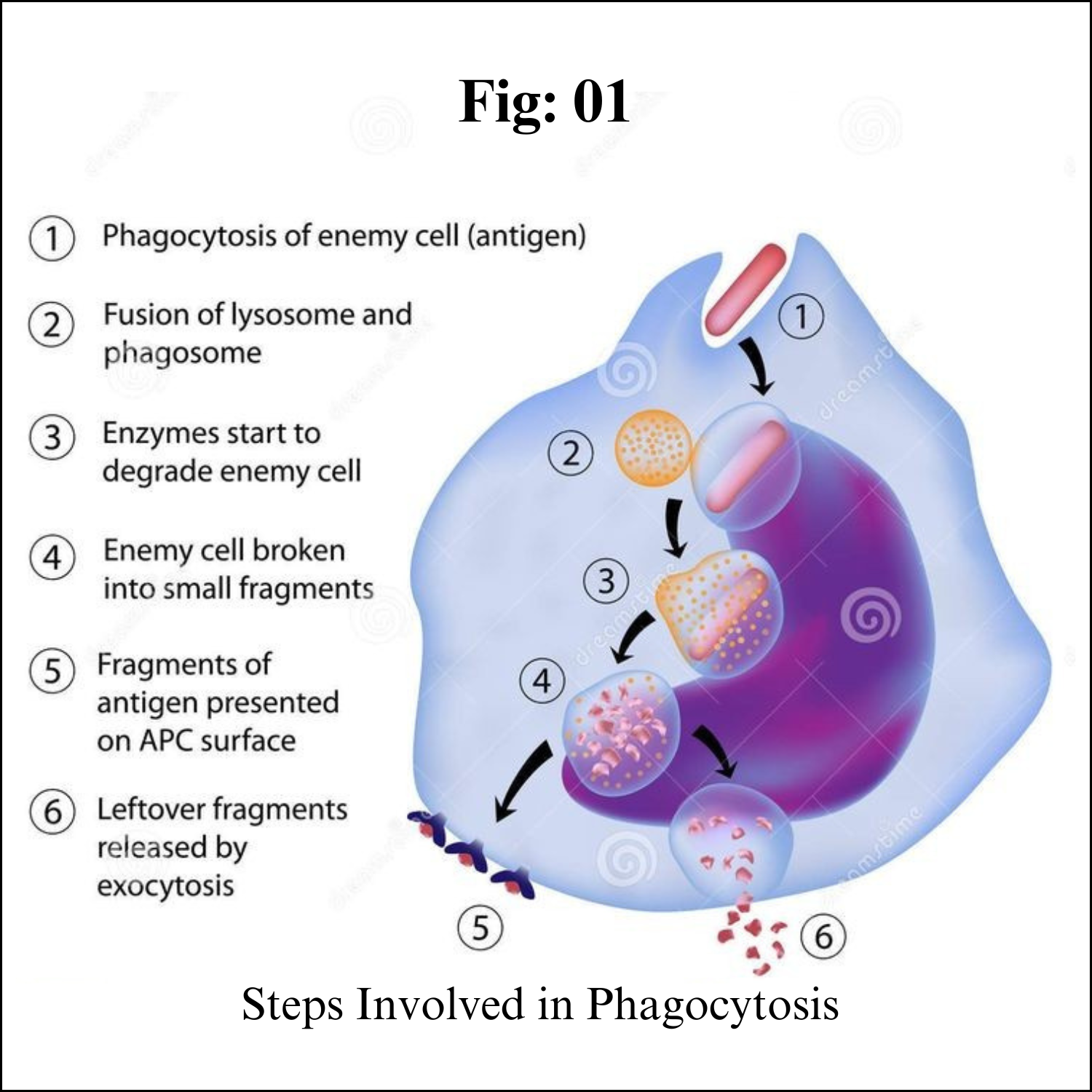
Steps of Phagocytosis

Step 1:

The cell that will perform phagocytosis is activated. This can be a phagocyte, which is a cell in the immune system that performs phagocytosis, or an organism such as an amoeba, which behaves in a similar way to phagocytes when it carries out phagocytosis. In the case of immune cells, activation occurs when the cells are near bacterial cells or parts of bacterial cells. Receptors on the surface of the cells bind to these molecules and cause the cells to respond.

Step 2:

In the immune system, chemotaxis may occur. Chemotaxis is the movement of phagocytes toward a concentration of molecules. Immune cells pick up chemical signals and migrate toward invading bacteria or damaged cells.

Step 3:

The cell attaches to the particle that it will ingest. Attachment is necessary for ingestion to occur. Some bacteria can resist attachment, making it harder for them to be taken into the cell and destroyed.

Step 4:

The cell ingests the particle, and the particle is enclosed in a vesicle (a sphere of cell membrane with fluid in it) called a phagosome. The phagosome transports the particle into the cell.

Step 5:

A lysosome fuses with the phagosome and the particle is digested. Lysosomes are vesicles that contain hydrolytic enzymes that break down molecules. A phagosome fused with a lysosome is called a phagolysosome.

Step 6:

Cellular waste, such as broken down molecules that the cell cannot reuse, is discharged from the cell by the process of exocytosis. Exocytosis is the opposite of endocytosis; it is when cellular waste products travel in vesicles to the surface of the cell membrane and are released, thereby exiting the cell.


This diagram shows the process of phagocytosis. A cell ingests a particle, breaks it down with the enzymes in lysosomes, and expels waste products through exocytosis.

Function of Phagocytosis

The function of phagocytosis is to ingest solid particles into the cell. 

Phagocytosis is a type of endocytosis, which is when cells ingest molecules via active transport as opposed to molecules passively diffusing through a cell membrane. 

Only certain small molecules can pass through the cell membrane easily; larger ones have to go through special channels in the cell or be ingested via endocytosis. 

Other types of endocytosis include pinocytosis, also called “cell drinking”, and receptor-mediated endocytosis, which is when molecules bind to specific receptors on the cell membrane that causes the cell to engulf them.

Phagocytosis is different from pinocytosis because phagocytosis involves the ingestion of solid particles while pinocytosis is the ingestion of liquid droplets. 

Phagocytosis is also used by cells to take in much larger particles than those that are ingested through pinocytosis. Some single-celled protists, such as amoebae, use phagocytosis to ingest food particles; it is literally how they eat food. 

Since their entire body consists of one cell, they can ingest food particles through engulfing them, and then digest these particles by connecting with a lysosome. 

In pinocytosis, the particles that are engulfed do not need to be broken down by a lysosome because they are so small, and instead the vesicle empties its contents directly into the cell.

Examples of Phagocytosis

Phagocytes are found throughout the human body as white blood cells in the blood. One liter of blood contains approximately six billion of them. Many different types of white blood cells are phagocytes, including macrophages, neutrophils, dendritic cells, and mast cells. 

White blood cells are known as “professional” phagocytes because their role in the body is to find and engulf invading bacteria. “Non-professional” phagocytes include other types of cells like epithelial cells, endothelial cells, and fibroblasts. These cells sometimes perform phagocytosis, but it is not their primary function.

As mentioned earlier in the article, amoebae perform phagocytosis in order to consume food particles. 

Amoebae engulf particles by surrounding them with pseudopods, which are temporary armlike projections of the cell that are filled with cytoplasm. Ciliates are another type of organisms that use phagocytosis to eat. 

Ciliates are protozoans that are found in water, and they eat bacteria and algae. Both amoebae and ciliates are protists, organisms that have eukaryotic cells but are not animals, plants, or fungi.

Sunday, August 20, 2023

Coelom (Body Cavity)


Coelom 

Introduction:

The coelom is one of the characteristic features of metazoans. The true coelom is a body cavity formed during embryo development from the three germinal layers. The body cavity meaning a fluid filled space that can accomodate organs. The coelom is lined by mesodermal epithelium cells. Presence or absence of coelom is one of the criteria for classifying animals.

Definition:

" The coelom is the fluid-filled body cavity present between the alimentary canal and the body wall." 

The true coelom has a mesodermal origin. It is lined by mesoderm. The peritoneal cavity present in the abdomen and similar spaces around other organs such as lungs, heart are parts of the coelom. Coelom differs in its structure and formation process. 

Structure, Formation and Types of Coelom:

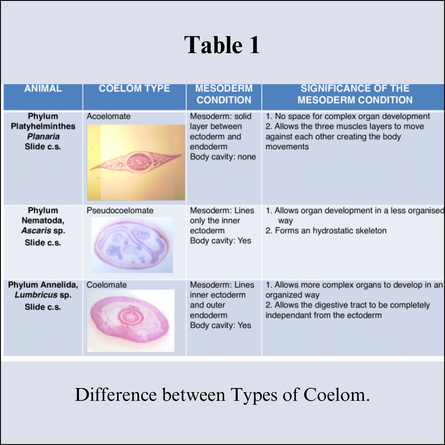
There are three types of structural body formation present in animals related to coelom:

1. Acoelomate:

Coelom is absent. The blastocoel is completely occupied by mesoderm. E.g. Porifera, Coelenterata and Flatworms (Platyhelminthes). There is only spongocoel or coelenteron present.

2. Pseudocoelomate:

True coelom is not present. The blastocoel is partly filled by mesodermal cells. The body cavity is lined by mesoderm only towards the body wall and mesoderm is not present towards the gut. E.g. Roundworms (Aschelminthes)

3. Eucoelomate:

Animals that have a true coelom. The coelom is lined by mesoderm on both the sides, towards the body wall and towards the gut. The blastocoel present in the gastrula gets completely replaced by a true coelom. The body organs are suspended in the coelom by mesenteries. E.g. from the phylum Annelida to Chordata.


Eucoelomates are further divided into Protostomes and Deuterostomes on the basis of different embryonic development. The process of coelom formation in protostomes and deuterostomes is different. 

The coelom is categorized into two types on the basis of formation, namely, Schizocoelom and Enterocoelom.

a. Schizocoelom: It is present in the protostomes. The body cavity or coelom originates from the splitting of the mesoderm. One part attaches to ectoderm and the other surrounds the endoderm. The space between them develops into the coelom. The blastopore forms the mouth. Examples of schizocoelous animals are Annelida, Arthropoda and Mollusca. In Arthropoda and Mollusca the coelom is filled by blood and is known as Haemocoel.

b. Enterocoelom: It is present in the deuterostomes. The coelom is formed from the fusion of the internal outgrowths of the archenteron, that pinches off and fuses together to form coelom lined by mesoderm. Examples of enterocoelous animals are Echinodermates and Chordates.


Coelom Function

Coelom works as a shock absorber and protects from any kind of mechanical shock. It gives more flexibility to the body organs to move and protects from any damage on minor bends by cushioning the internal organs.

The coelomic fluid acts as a hydrostatic skeleton, which helps in the locomotion of soft-bodied animals and gives the body a definite shape. Contracting muscles can push against the coelomic fluid because of the fluid pressure.

The coelomocyte cells, that either float freely in the coelom or attached to the wall, support the immune system. They support the immune system by initiating humoral immune response and phagocytosis.

The coelomic fluid also helps in gaseous transport and transport of nutrients and waste products.

Coelom gives the extra space required by organs to develop and function. E.g. pumping action of the heart, carrying a child in the womb, etc. is possible due to coelom.

Female orgasms

Female orgasms A female orgasm can be a highly pleasurable experience during masturbation or sexual activity. While orgasms may ...