Saturday, October 5, 2024

Female orgasms


Female orgasms

A female orgasm can be a highly pleasurable experience during masturbation or sexual activity. While orgasms may not provide a direct reproductive benefit, the pleasure may help improve mood and relieve stress.

In this article, we look at why female orgasms occur and what happens during an orgasm. We also debunk some common misconceptions.

Causes

Orgasms are a response to sexual stimulation. The female orgasm can result from many types of stimulation, including vaginal, clitoral, and nipple contact.

While vaginal orgasms are less common than those from clitoral stimulation, some women have them — with or without other stimulation.

Not everyone orgasms from the same type of stimulation.

What happens during an orgasm?

During arousal, blood flow to the genitals increases, causing them to become more sensitive.

As arousal increases, a person’s heart rate, blood pressure, and breathing rate may also increase. As orgasm approaches, the muscles may twitch or spasm. Many women experience rhythmic muscle spasms in the vagina during an orgasm.

Orgasms may follow the following stages:

# excitement, during which arousal builds
# plateau, during which arousal increases and levels off
# orgasm, which causes intense feelings of pleasure
# resolution, during which arousal diminishes

Many females can have another orgasm after resolution, whereas males usually require a period of rest before having another orgasm.

What does it feel like?

Orgamsms can feel different for all people, and they may report their experiences in different language.

However, people typically experience intense feelings of pleasure in the genitals and throughout the body. The release of endorphins following an orgasm may cause people to feel happy and relaxed. The skin may also flush.

Women may also experience vaginal contractions during and after an orgasm, and ejaculation is also possible.

Why do females orgasm?

The purpose of the female orgasm is less clear than that of the male orgasm. Researchers have suggested numerous potential benefits, but few have been rigorously tested, and no theory has conclusive scientific support.

A 2016 study argues that the female orgasm may have no obvious evolutionary benefit and that it may be a relic of a time when the hormones associated with orgasm were necessary for a woman to ovulate.

Since there was no evolutionary need to eliminate the female orgasm, it persisted even when it was no longer necessary for fertility.

Orgasm may serve important purposes, however. The pleasure it can cause can encourage females to have sex. This may also promote bonding with a sexual partner, which does have significant evolutionary benefits.

Health benefits

While the internet is filled with articles promising that orgasms improve skin, hair, and overall health, there is little scientific evidence that orgasms offer any specific health benefits.

Scientists have not identified any evolutionary benefits of female orgasms or found that orgasms improve health.
But orgasms are pleasurable, and pleasure can be its own benefit. Pleasurable sex may improve a person’s mood, relieve stress, boost immunity, and foster better relationships.

Common misconceptions

People hold many misconceptions about female orgasms. Some myths include:

Women who cannot orgasm have psychological problems.

While trauma, relationship issues, and poor mental health can make it more difficult to orgasm, many people with healthy sexual attitudes and good relationships still have difficulties.

An orgasm is both a physical and psychological response, and numerous health problems can make it more difficult to enjoy sex in this way.

For example, some women experience vulvodynia, which refers to unexplained pain in the vagina or around the vulva. Treating this and other medical conditions may improve sexual pleasure.

Orgasms from penetrative sex are common or the healthiest form of sexual expression.

Self-appointed experts, mostly men, have long told women that they must orgasm from heterosexual intercourse. However, many women can only orgasm from clitoral stimulation.

Women need to be in love to orgasm.

Orgasm is a complex psychological and biological experience — reaching and experiencing orgasm is not the same for every woman. Some women may need to feel love to orgasm, while others may not.

found people were more likely to orgasm frequently if they:

* received more oral sex
* had longer-lasting sex
* reported higher relationship satisfaction
* asked for what they wanted in bed
* engaged in sexual emails or calls
* expressed love during sex
* acted out sexual fantasies
* tried new sexual positions

A person’s relationship with their partner may or may not influence their ability to orgasm during sex.

A partner can tell if a woman has had an orgasm.

There is no way to tell if a woman has had an orgasm without asking. Some people make noises during an orgasm, while others are silent. Some flush or sweat after an orgasm, but others do not.

A person who wants to know if their partner has had an orgasm can ask without being confrontational.

If the answer is no, avoid judgment, anger, or feelings of inadequacy — these can put pressure on the person to orgasm, which can lead to anxiety and make it more difficult. Instead, discuss whether they would prefer a different approach to sex.

What if you can’t orgasm?

Being unable to orgasm is a common issue, and it can occur for a variety of reasons. Some people may not receive the right kind of stimulation during sex, while others may have experienced trauma linked to sex. Others may simply be uninterested.

Risk factors

A 2018 analysis identified several factors that increase the risk of sexual dysfunction, including:

@ relationship problems
@ stress
@ mental health issues
@ poor physical health
@ genitourinary issues, such as pelvic pain
@ a history of abortion
@ a history of female genital mutilation
@ sexual abuse
@ being religious, perhaps due to sexual shame and stigma

The same study identified several modifiable risk factors that improve sexual experience, including:

# exercise
# daily affection from a partner
# a positive body image
# sex education
# intimate communication with a partner
# Increasing the likelihood

The aforementioned study that compared orgasm frequency among people of various sexual orientations in the United States found that the following behaviors during sex increase the likelihood of women having an orgasm:

1 deep kissing
2 genital stimulation during vaginal intercourse
3 oral sex

If self-help strategies do not work, a doctor who specializes in sexual dysfunction may be able to identify a problem, if there is one.

Many medical issues can make having an orgasm difficult, including:

I    a lack of lubrication
II   hormonal imbalances
III  pelvic pain
IV muscle dysfunction
V  a history of trauma

When trauma or relationship problems make having an orgasm difficult, or when a person feels ashamed of sex or their desires, individual or couples counseling can help.

Summary

Serious scientific research into the female orgasm is relatively recent. Even some doctors may still believe myths about the female orgasm or think that it is unimportant to the female sexual experience.

This means that many people may have trouble accessing reliable information about orgasms.

A competent, compassionate medical professional can help a person understand the process of orgasm and identify potential barriers to sexual satisfaction.

There is no right way to orgasm and no correct way to feel about sex. People should pursue what feels good to them.

Thursday, August 1, 2024

World Breastfeeding week


World Breastfeeding week

August 1 - 7

Benefits of Breastfeeding for Both Mom and Baby

The World Health Organization (WHO) recommends breastfeeding until 2 years old or longer Trusted Source because the benefits continue that long. These agencies recommend starting as early as one hour after birth for the biggest benefits.

Breastfeeding benefits for baby

1. Breast milk provides ideal nutrition for babies

Most healthcare professionals recommend exclusive breastfeeding for at least 6 months or much longer.

Breast milk contains everything baby needs for the first 6 months of life, in all the right proportions. Its composition even changes according to the baby’s changing needs, especially during the first month of life.

During the first days after birth, your breasts produce a thick and yellowish fluid called colostrum. It’s high in protein, low in sugar, and loaded with beneficial compounds. It’s truly a wonder food and not replaceable by formula.

Colostrum is the ideal first milk and helps the newborn’s immature digestive tract develop. After the first few days, the breasts start producing larger amounts of milk as the baby’s stomach grows.

About the only thing that may be lacking from your magical milk supply is vitamin D.

Unless you have a very high intake (and most of us don’t), your breast milk won’t provide enough. Vitamin D drops are usually recommended.

2. Breast milk contains important antibodies

Breast milk is loaded with antibodies that help your baby fight off viruses and bacteria, which is critical in those tender, early months.

This particularly applies to colostrum, the first milk. Colostrum provides high amounts of immunoglobulin A (IgA), as well as several other antibodies.

When you’re exposed to viruses or bacteria, you start producing antibodies that then go into the milk. It’s immunity, baby!

IgA protects the baby from getting sick by forming a protective layer in the baby’s nose, throat, and digestive systemTrusted Source.

Formula doesn’t provide antibody protection for babies. Numerous studiesTrusted Source show that babies who are not breastfed are more vulnerable to health issues like pneumonia, diarrhea, and infection.

3. Breastfeeding may reduce disease risk

Exclusive breastfeedingTrusted Source, meaning that the infant receives only breast milk, is particularly beneficial.

It may reduce your baby’s risk for many illnesses and diseases, including:

Middle ear infections. Breastfeeding, particularly exclusively and as long as possible, may protect against middle ear, throat, and sinus infections well beyond infancyTrusted Source.

Respiratory tract infections. Breastfeeding can protect against multipleTrusted Source respiratory and gastrointestinal acute illnesses.

Colds and infections. Babies exclusively breastfed for 6 months may have a lower risk of getting serious colds and ear or throat infections.

Gut infections. Breastfeeding is linked with a reduction in gut infections.

Intestinal tissue damage. Feeding preterm babies breast milk is linked with a reduction in the incidence of necrotizing enterocolitisTrusted Source.

Sudden infant death syndrome (SIDS). Breastfeeding is linked to a reduced risk of SIDS, especially when breastfeeding exclusively.

Allergic diseases. Breastfeeding is linked to a reduced riskTrusted Source of asthma, atopic dermatitis, and eczema.

Bowel diseases. Babies who are breastfed may be less likelyTrusted Source to develop Crohn’s disease and ulcerative colitis.

Diabetes. Breastfeeding is linked to a reduced riskTrusted Source of developing type 1 diabetes and non-insulin-dependent (type 2) diabetes.

Childhood leukemia. Breastfeeding is linked to a reduction in the risk for childhood leukemiaTrusted Source.

4. Breast milk promotes baby’s healthy weight

Breastfeeding promotes healthy weight gain and helps prevent childhood obesity.

One studyTrusted Source showed that breastfeeding for longer than 4 months had a significant reduction in the chances of a baby developing overweight and obesity.

This may be due to the development of different gut bacteria. Breastfed babies have higher amountsTrusted Source of beneficial gut bacteria, which may affect fat storage.

Babies fed breast milk also have more leptin in their systems than formula-fed babies. Leptin is a key hormone for regulating appetite and fat storage.

Breastfed babies also self-regulate their milk intake. They’re better at eating only until they’ve satisfied their hunger, which helps them develop healthy eating patterns.

5. Breastfeeding may make children smarter

Breastfeeding may help baby ace those tests. Some studiesTrusted Source suggest there may be a difference in brain development between breastfed and formula-fed babies.

This difference may be due to the physical intimacy, touch, and eye contact associated with breastfeeding as well as nutrient content.

StudiesTrusted Source indicate that breastfed babies have higher intelligence scores and are less likely to develop behavioral problems have learning difficulties as they grow older.

However, the most pronounced effects are seen in preterm babies, who have a higher risk for developmental issues.

The researchTrusted Source clearly shows that breastfeeding has significant positive effectsTrusted Source on babies’ long-term brain development.

6. Breastfeeding may help you lose weight

You may have heard this one often. While some women seem to gain weight during breastfeeding, others seem to effortlessly lose weight.

Breastfeeding does burn more caloriesTrusted Source, and after 3 months of lactation, you’ll likely experience an increase in fat burning compared to non-lactating mothers. Though the difference isn’t significant.

7. Breastfeeding helps the uterus contract

During pregnancy, your uterus grows immensely, expanding from the size of a pear to filling almost the entire space of your abdomen.

After delivery, your uterus goes through a process called involution, which helps it return to its previous size. Oxytocin, a hormone that increases throughout pregnancy, helps drive this process.

Your body secretes high amounts of oxytocin during labor to help deliver the baby and reduce bleeding. It can also help you bond with your new little one.

Oxytocin also increases during breastfeeding. It encourages uterine contractions and reduces bleeding, helping the uterus return to its previous size.

Studies have also shown that mothers who breastfeed generally have less blood loss after delivery and faster involution of the uterus.

8. Mothers who breastfeed have a lower risk for depression

Postpartum depression (PPD) is a type of depression that can develop shortly after childbirth.

Women who breastfeed seem less likely to develop postpartum depression, compared to mothers who wean early or do not breastfeed, according to a 2012 studyTrusted Source.

However, those who experience postpartum depression early after delivery are also more likely to have trouble breastfeeding and do so for a shorter duration.

If you have any symptoms of PPD, tell your doctor as soon as possible.

9. Breastfeeding reduces your disease risk

Breastfeeding seems to provide you with long-term protectionTrusted Source against cancer and several diseases.

The total time a woman spends breastfeeding is linked with a reduced risk for breast and ovarian cancer.

Women who breastfeed have a lower risk for:


high blood pressure

arthritis

high blood fats

heart diseaseTrusted Source

type 2 diabetesTrusted Source


10. Breastfeeding may prevent menstruation

Continued breastfeeding also pauses ovulation and menstruation. The suspension of menstrual cycles may actually be nature’s way of ensuring there’s some time between pregnancies.

You may consider this change as an extra benefit. While you’re enjoying precious time with your newborn, it’s just one less thing to worry about.

11. It saves time and money

To top the list, breastfeeding is mostly free, barring expenses for any lactation consulting and breast pumps. By choosing to breastfeed, you won’t have to:


Saturday, May 25, 2024

Reverse Transcription

Reverse Transcription

Introduction

Reverse transcription is the synthesis of DNA from an RNA template. This process is driven by RNA-dependent DNA polymerases, also known as reverse transcriptases. Reverse transcriptases occur naturally in both prokaryotic and eukaryotic organisms, as well as in retroviruses.

Reverse Transcriptase 

Reverse transcriptases (RTs) are RNA-dependent DNA polymerases, a group of enzymes that play a unique role in the flow of genetic information. These enzymes enable the reverse transcription reaction and have been widely used by researchers in a variety of molecular biology applications since their discovery. 

The endogenous properties of reverse transcriptases can be exploited and modulated for successful cDNA-based experiments. In addition to opening up the research into their native roles, including genetic diversity and retroviral replication, reverse transcriptases prove to be important tools for molecular biologists for various applications like gene expression analysis and cDNA sequencing.

The discovery of reverse transcriptase

The original central dogma of molecular biology held that DNA was transcribed to RNA, which in turn was translated into protein. However, this concept was challenged in the 1970s when two scientific teams, one led by Howard Temin at the University of Wisconsin and the other led by David Baltimore at MIT, independently identified new enzymes associated with replication of RNA viruses called retroviruses]. 

These enzymes, called reverse transcriptases, convert the viral RNA into a complementary DNA (cDNA) molecule, which then integrates into the host’s genome. In 1975, Temin and Baltimore received the Nobel Prize in Physiology or Medicine (shared with Renato Dulbecco for related work on tumor-inducing viruses) for their pioneering work in identifying reverse transcriptases.

The prevalence of reverse transcriptase in nature

Reverse transcriptases have been identified in many organisms, including bacteria, animals, and plants, as well as viruses. The natural role of reverse transcriptase is to convert RNA sequences to cDNA sequences that are capable of being inserted into different areas of the genome. In this manner, reverse transcription contributes to:

#Propagation of retroviruses—e.g., human immunodeficiency virus (HIV), Moloney murine leukemia virus (M-MuLV), and avian myeloblastosis virus (AMV).

#Genetic diversity in eukaryotes via mobile transposable elements called retrotransposons.

#Replication of chromosomal ends called telomeres.

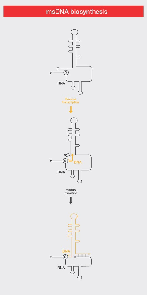
#Synthesis of extrachromosomal DNA/RNA chimeric elements called multicopy single-stranded DNA (msDNA) in bacteria.

Applications of Reverse transcription 

While reverse transcriptases have functional roles in biological systems, they also serve as important tools for studying RNA populations. In molecular biology, reverse transcriptases were first used to produce cDNA to build libraries. cDNA libraries contain DNA copies of mRNA from cells and tissues and are used to gain an understanding of actively expressed genes and their functions at a specific time point.

Although the creation of cDNA libraries was an important step forward in characterizing expressed genes, challenges remained for the study of low-abundance RNAs. 

These were subsequently addressed with the development of the polymerase chain reaction (PCR), a technique to amplify small amounts of genetic material. Reverse transcription combined with PCR, or reverse transcription PCR (RT-PCR), allows detection of RNA even at very low levels of gene expression and paves the way for detection of circulating RNA, RNA viruses, and cancerous gene fusions in molecular diagnostics.

In addition, cDNAs serve as templates in applications such as microarray and RNA sequencing to characterize unknown RNAs in a high-throughput manner.

Monday, April 8, 2024

Erythroblastosis Fetalis

Erythroblastosis Fetalis

Introduction

The adult human body is home to trillions of red blood cells, also known as RBCs or erythrocytes. These blood cells carry oxygen, iron, and many other nutrients to the appropriate places in the body.

When a woman is pregnant, it’s possible that her baby’s blood type will be incompatible with her own. This can cause a condition known as erythroblastosis fetalis, where the mother’s white blood cells (WBCs) attack the baby’s RBCs as they would any foreign invaders.

This condition is highly preventable and the typical, severe form is now very rare in developed countries. Catching it early can ensure a successful pregnancy for mother and child. If left untreated, however, it can be life threatening for the baby.

Erythroblastosis fetalis is now known as hemolytic disease of the newborn.

Symptoms of Erythroblastosis Fetalis

Babies who experience erythroblastosis fetalis symptoms may appear swollen, pale, or jaundiced after birth. A doctor may find that the baby has a larger-than-normal liver or spleen.

Babies who experience erythroblastosis fetalis symptoms may appear swollen, pale, or jaundiced after birth. A doctor may find that the baby has a larger-than-normal liver or spleen.

Blood tests can also reveal that the baby has anemia or a low RBC count. Babies can also experience a condition known as hydrops fetalis, where fluid starts to accumulate in spaces where fluid is normally not present. This includes spaces in the:

Abdomen
Heart
Lungs

Causes

There are two main causes of erythroblastosis fetalis: Rh incompatibility and ABO incompatibility. Both causes are associated with blood type. There are four blood types:

A
B
AB
O

In addition, blood can be either Rh positive or Rh negative. For example, if you’re type A and Rh positive, you have A antigens and Rh factor antigens on the surface of your RBCs. Antigens are substances that trigger an immune response in your body. If you have AB negative blood, then you have both A and B antigens without the Rh factor antigen.

Rh incompatibility

Rh incompatibility occurs when a Rh-negative mother is impregnated by a Rh-positive father. The result can be a Rh-positive baby. In such a case, your baby’s Rh antigens will be perceived as foreign invaders, the way viruses or bacteria are perceived.

Your blood cells attack the baby’s as a protective mechanism that can end up harming the child. If you’re pregnant with your first baby, Rh incompatibility isn’t as much of a concern.

However, when the Rh-positive child is born, your body will create antibodies against the Rh factor. These antibodies will attack the blood cells if you ever become pregnant with another Rh-positive baby.

ABO incompatibility

Another type of blood type mismatch that can cause maternal antibodies against her baby’s blood cells is ABO incompatibility.

This occurs when the mother’s blood type of A, B, or O isn’t compatible with the baby’s. This condition is almost always less harmful or threatening to the baby than Rh incompatibility.

However, babies can carry rare antigens that can put them at risk for erythroblastosis fetalis. These antigens include:

Kell
Duffy
Kidd
Lutheran
Diego
Xg
P
Ee
Cc
MNSs

Diagnosed

To diagnose erythroblastosis fetalis, a doctor will order a routine blood test during your first prenatal visit. They’ll test for your blood type.

The test will also help them determine whether you have anti-Rh antibodies in your blood from a previous pregnancy.

The fetus’s blood type is rarely tested. It’s difficult to test for a fetus’s blood type and doing so can increase the risk for complications.

Frequency of testing

If initial testing shows your baby may be at risk for erythroblastosis fetalis, your blood will be continually tested for antibodies throughout your pregnancy — approximately every two to four weeks.

If your antibody levels start to rise, a doctor may recommend a test to detect fetal cerebral artery blood flow, which isn’t invasive to the baby. Erythroblastosis fetalis is suspected if the baby’s blood flow is affected.

Rh incompatibility

If you have Rh-negative blood, the father’s blood will be tested. If the father’s blood type is Rh negative, no further testing is needed. However, if the father’s blood type is Rh positive or their blood type isn’t known, your blood may be tested again between 18 to 20 weeks of pregnancy, and again at 26 to 27 weeks.

You’ll also receive treatment to prevent erythroblastosis fetalis.

ABO incompatibility

If your baby is jaundiced after birth, but Rh incompatibility isn’t a concern, the baby may be experiencing problems due to ABO incompatibility. ABO incompatibility occurs most frequently when a mother with an O blood type gives birth to a baby who has an A, B, or AB blood type.

Because O blood types may produce both A and B antibodies, the mother’s blood can attack the baby’s. However, these symptoms are generally much milder than a Rh incompatibility.

ABO incompatibility can be detected via a blood test known as a Coombs test. This test, along with a test to determine the baby’s blood type, is performed after the baby is born. It can indicate why the baby may appear jaundiced or anemic.

These tests are usually done for all babies whose mothers have type O blood.

Treatment 

If a baby experiences erythroblastosis fetalis in the womb, they may be given intrauterine blood transfusions to reduce anemia. When the baby’s lungs and heart mature enough for delivery, a doctor may recommend delivering the baby early.

After a baby is born, further blood transfusions may be necessary. Giving the baby fluids intravenously can improve low blood pressure. The baby may also need temporary breathing support from a ventilator or mechanical breathing machine.

Long-term outlook

Babies born with erythroblastosis fetalis should be monitored for at least three to four months for signs of anemia. They may require additional blood transfusions.

However, if proper prenatal care and postpartum care are delivered, erythroblastosis fetalis should be prevented and the baby shouldn’t experience long-term complications.

Prevention

A preventive treatment known as RhoGAM, or Rh immunoglobulin, can reduce a mother’s reaction to their baby’s Rh-positive blood cells. This is administered as a shot at around the 28th week of pregnancy.

The shot is administered again at least 72 hours after birth if the baby is Rh positive. This prevents adverse reactions for the mother if any of the baby’s placenta remains in the womb.

Carbon-14 Dating

Carbon-14 Dating 

Introduction

Radiocarbon dating, or carbon-14 dating, is a scientific method that can accurately determine the age of organic materials as old as approximately 60,000 years. First developed in the late 1940s at the University of Chicago by Willard Libby, the technique is based on the decay of the carbon-14 isotope. Radiocarbon dating has been used for historical studies and atmospheric science, and triggered archaeology’s “Radiocarbon Revolution.”

Carbon-14 dating

The invention of radiocarbon dating elegantly merged chemistry and physics to develop a scientific method that can accurately determine the age of organic materials as old as approximately 60,000 years.

It is based on the fact that living organisms—like trees, plants, people, and animals—absorb carbon-14 into their tissue. When they die, the carbon-14 starts to change into other atoms over time. Scientists can estimate how long the organism has been dead by counting the remaining carbon-14 atoms. The technique was developed in the late 1940s at the University of Chicago by chemistry professor Willard Libby, who would later receive the Nobel Prize for the work.

The breakthrough introduced a new scientific rigor to archaeology, allowing archaeologists to put together a history of humans across the world, but it had a significant effect in other fields, too. 

Carbon dating has helped us reveal how our bodies work, to understand the climate of the Earth and reconstruct its history, and to track the sun’s activity and the Earth’s magnetic fields. Radiocarbon dating was also instrumental in the discovery of human-caused climate change, as scientists used it to track the sources of carbon in the atmosphere over time.

Radiocarbon dating working principle


It starts with cosmic rays—subatomic particles of matter that continuously rain upon Earth from all directions. When cosmic rays reach Earth’s upper atmosphere, physical and chemical interactions form the radioactive isotope carbon-14.

Living organisms absorb this carbon-14 into their tissue. Once they die, the absorption stops, and the carbon-14 begins very slowly to change into other atoms at a predictable rate. By measuring how much carbon-14 remains, scientists can estimate how long a particular organic object has been dead.

From there, the problem becomes how to measure the carbon-14. Libby and fellow chemists at the University of Chicago and other institutions developed techniques to purify a sample so that it emits no other type of radiation except for carbon-14, and then run it through a detector sensitive enough to accurately count the pings emitted by the decay of single atoms. A newer, faster method developed in the 1970s works by using a particle accelerator to count the atoms of carbon-14.

Radiocarbon dating can be used on any object that used to be alive. That includes pieces of animals, people, and plants, but also paper that was made from reeds, leather made from animal hides, logs that were used to build houses, and so forth.

Carbon Dating Invention

Carbon dating was invented in the late 1940s by Willard Libby, a chemistry professor at the University of Chicago and former Manhattan Project scientist.

Libby built upon the work of Martin Kamen (PhD’36) and Sam Ruben, who discovered the carbon-14 isotope in 1940. Carbon-14 has a half-life of about 5,730 years. That means half the atoms in a sample will change into other atoms, a process known as “decay,” in that amount of time.

Libby proposed the idea of carbon dating in the journal Physical Review in 1946. He further developed the concept with members of his research group and published more in Science in 1947 and 1949. 

In a crucial step, Libby’s first graduate student, Ernest C. Anderson, established that organic materials contained essentially the same natural abundance of radiocarbon at all measured latitudes reaching nearly from pole to pole.

Libby worked with colleagues, including anthropologist Robert Braidwood of UChicago’s Oriental Institute (now known as the Institute for the Study of Ancient Cultures), to develop the carbon-14 method. 

Samples taken from artifacts in the museum collections were used to test the accuracy of radiocarbon dating, since archaeologists already knew their ages by tree-ring dating and other evidence. 

The many materials Libby tested while developing the method included a rope sandal found in an Oregon cave, the dung of an extinct ground sloth, linen wrappings from the Dead Sea Scrolls, and part of a funeral ship deck placed in the tomb of Sesostris III of Egypt.

News of the technique spread rapidly. By 1960, more than 30 radiocarbon labs had been established worldwide. (One of the first was led by physicist Hilde Levi, who spent several months at UChicago working with Libby on radiocarbon-related problems in 1947 and 1948).

“Libby’s method remained the only way to measure carbon-14 in samples for several decades and was long considered the most accurate means of dating carbon decay,” said David Mazziotti, a UChicago professor in chemistry. (Today, scientists also use a different way to measure carbon-14 called accelerator mass spectrometry, which can get more precise results from a far smaller amount of sample but is more expensive).

A plaque in the foyer of UChicago’s Kent Laboratory building commemorates the discovery, as a National Historic Chemical Landmark designated by the American Chemical Society. Libby’s invention earned him the 1960 Nobel Prize in chemistry “for determinations in archaeology, geology, geophysics, and other branches of science.”

UChicago science historian Emily Kern has documented how radiocarbon dating developed in an unusual Cold War context.  She described how the technique developed into a wide-ranging, global network from a technology that had roots in World War II’s Manhattan Project to build the atomic bomb. The technology, unbound by national security concerns, meant that carbon-14 laboratories could arise in Australia, Denmark, New Zealand and elsewhere.

Limitations of Carbon-14 Dating

The various dating techniques all have limitations. Each works best for different types of problems. Radiocarbon dating works on organic materials up to about 60,000 years of age.

Conventional radiocarbon dating requires samples of 10 to 100 grams (0.35 to 3.5 ounces) of an object, depending on the material in question. Newer forms of dating can use much smaller amounts, down to 20 to 50 milligrams or 0.0007 to 0.0018 ounces. In both cases, the material is destroyed during the test.

Radiocarbon samples are also easily contaminated, so to provide accurate dates, they must be clean and well-preserved. Dirt and other matter must be washed off with water, but chemical treatments and other cleaning procedures are also often needed. 

This is because there are so few atoms to count; even a little extra carbon from contamination will throw off the results significantly. A million-year-old sample contaminated by only a tiny amount of carbon could yield an invalid age of 40,000 years, for example.

Other dating methods have different strengths. Dendrochronology, also known as tree-ring dating, depends upon the preservation of certain tree species; it can extend to about 12,500 years ago for oak trees and to 8,500 years for bristlecone pine.

Potassium-argon dating can date volcanic materials ranging from less than 100,000 to more than 4 billion years old. Rubidium-strontium dating can be used to determine the ages of items ranging from a few million to a few billions of years old; it is widely used to understand how the Earth and solar system formed and to trace human migration and trade in archaeology.

Improved version

Technological and analytical advances have made radiocarbon dating faster and much more precise—and expanded its range of uses by reducing the size of the sample needed. The latest form of radiocarbon dating, called accelerator mass spectrometry, needs samples of only 20 to 50 milligrams (0.0007 to 0.0018 ounces); however, it is also more expensive.

Another newer development is Bayesian statistical modeling, which applies probability analytics to radiocarbon dates, which always involve an error margin. Bayesian modeling hones the final date range by considering factors such as which layer of sediments the samples come from or their relationship to artifacts of known age.

Discoveries that carbon-14 testing Revealed

Since its discovery, carbon-14 testing has had a major impact on our understanding of fields from archaeology to history to geology.

Dual Process Theory - Psychology

Dual Process Theory

Introduction

Dual process theory is a framework used to explain how people think. It traces its roots back to William James (an early American philosopher and psychologist). At its core is the idea that humans have two different streams or means of thinking. These dual means of thinking give rise to the name dual process theory.

“Dual process theory says that humans have two systems for thinking. System 1 is unconscious, quick, makes use of shortcuts, is a bit sloppy but is relied upon most of the time. System 2 is intentional, calculated and often more accurate, but it takes effort and is slow”.

These dual processes are sometimes referred to as “systems” and known as “system 1” and ”system 2”. System 1 is evolutionarily older, more automatic, instinctive, implicit and unconscious. System 2 is evolutionarily newer, intentional, effortful, explicit and conscious.

Dual process theory continues to evolve. It remains a popular framework in the field of cognitive psychology. It also has some applications in learning theory and in relation to how humans process and store information. More recently it has sprung up in behavioral economics as well. Danny Kahneman’s interpretations in his excellent book “Thinking, Fast and Slow”, helped bring these concepts to the mainstream.

Dual process theory also has a key role to play in understanding how we make decisions.

System 1: Our Automatic Processor

Humans constantly function. The majority of time we do so without really thinking about it. We know what our senses are telling us and we know what they mean we should do.

If we’re hungry, we should eat. And, if we’re a bit tired, we should sleep. If we see some information we dislike, we should ignore it… or perhaps not. We don’t think about walking. And we don’t calculate the trajectory of our steps. We don’t use our knowledge of physics to help us throw a ball. All of these things come naturally.

We’ve developed rules, internal processes and shortcuts in our thinking and decision making that help us survive without conscious effort. And it’s this system of automatic processing that’s known as System 1. We use it to get along in our daily lives without really needing to try too hard or think too much. We also find that the more tired we are, the more we use System 1.

This is economic in many ways. It’s fast too, allowing us to respond almost instantly in many situations. It’s also often reasonably accurate and effective. It also reserves our mental energy for draining thoughtful effort when it’s really required. It does though, rely on generalities and is prone to some sloppy errors.

System 2: Our Controlled Thinking

Sometimes we, as humans, find ourselves in situations where we either don’t have mental shortcuts that we can use, or where we need to be more than just reasonably accurate.

In these circumstances we need to focus on our thoughts. We need to consciously think our way through key factors and reach logical, calculated, informed decisions. To do this we need to slow our thinking down. We ignore our mental shortcuts, we start from the building blocks of information that we have and use logic to reach decisions and conclusions.

This way of thinking is known as System 2 thinking. It often produces better (or at least more reasoned) answers for us, but it’s effortful and it’s slow. This process is excellent in some environments and situations, but dreadful in others. If you rely on system 2 to calculate the moment when a leaping tiger will reach you and plot your escape, then you’ll never finish your calculations.

System 2: Characteristics

System 2 has lots of different characteristics. Some of the most important ones are as follow:

System 2 thinking requires focus and energy

It’s conscious,
Mostly voluntary,
Mostly detached from emotions,
Explicit,
Controlled,
High effort,
Small capacity,
Slow,
More objective (and fact / rule based),
Evolutionarily recent,
Logical and rational.

Dual Process Theory in the World of Work

Many of the challenges that individuals and leaders face in the world of work stem from the very natural tendency for individuals to predominantly use System 1 thinking as opposed to System 2 thinking.

In fact, most cases of sloppy thinking by otherwise capable individuals probably result from their use of System 1 thinking. And this is entirely natural. System 2 thinking requires a lot more effort, and a lot more focus. And this means that to use System 2, individuals normally need to be more motivated.

From a leadership perspective it’s helpful to be aware of these two different types of thinking. The more you can use system 2 thinking yourself, the better the decisions that you make will probably be. And similarly, the more you can help your team use system 2 thinking, the better their decisions will probably be.

Bipolar Disorder

Bipolar Disorder

Introduction

Bipolar disorder, formerly called manic depression, is a mental health condition that causes extreme mood swings that include emotional highs (mania or hypomania) and lows (depression).

When you become depressed, you may feel sad or hopeless and lose interest or pleasure in most activities. When your mood shifts to mania or hypomania (less extreme than mania), you may feel euphoric, full of energy or unusually irritable. These mood swings can affect sleep, energy, activity, judgment, behavior and the ability to think clearly.

Episodes of mood swings may occur rarely or multiple times a year. While most people will experience some emotional symptoms between episodes, some may not experience any.

Although bipolar disorder is a lifelong condition, you can manage your mood swings and other symptoms by following a treatment plan. In most cases, bipolar disorder is treated with medications and psychological counseling (psychotherapy).

Symptoms

There are several types of bipolar and related disorders. They may include mania or hypomania and depression. Symptoms can cause unpredictable changes in mood and behavior, resulting in significant distress and difficulty in life.

*Bipolar I disorder. You've had at least one manic episode that may be preceded or followed by hypomanic or major depressive episodes. In some cases, mania may trigger a break from reality (psychosis).

*Bipolar II disorder. You've had at least one major depressive episode and at least one hypomanic episode, but you've never had a manic episode.

*Cyclothymic disorder. You've had at least two years — or one year in children and teenagers — of many periods of hypomania symptoms and periods of depressive symptoms (though less severe than major depression).

*Other types. These include, for example, bipolar and related disorders induced by certain drugs or alcohol or due to a medical condition, such as Cushing's disease, multiple sclerosis or stroke.

Bipolar II disorder is not a milder form of bipolar I disorder, but a separate diagnosis. While the manic episodes of bipolar I disorder can be severe and dangerous, individuals with bipolar II disorder can be depressed for longer periods, which can cause significant impairment.

Although bipolar disorder can occur at any age, typically it's diagnosed in the teenage years or early 20s. Symptoms can vary from person to person, and symptoms may vary over time.

Mania and hypomania

Mania and hypomania are two distinct types of episodes, but they have the same symptoms. Mania is more severe than hypomania and causes more noticeable problems at work, school and social activities, as well as relationship difficulties. Mania may also trigger a break from reality (psychosis) and require hospitalization.

Both a manic and a hypomanic episode include three or more of these symptoms:

*Abnormally upbeat, jumpy or wired.

*Increased activity, energy or agitation.

*Exaggerated sense of well-being and self-confidence (euphoria).

*Decreased need for sleep.

*Unusual talkativeness.

*Racing thoughts.

*Distractibility.

*Poor decision-making — for example, going on buying sprees, taking sexual risks or making foolish investments.

Major depressive episode

A major depressive episode includes symptoms that are severe enough to cause noticeable difficulty in day-to-day activities, such as work, school, social activities or relationships. An episode includes five or more of these symptoms:

*Depressed mood, such as feeling sad, empty, hopeless or tearful (in children and teens, depressed mood can appear as irritability).

*Marked loss of interest or feeling no pleasure in all — or almost all — activities.

*Significant weight loss when not dieting, weight gain, or decrease or increase in appetite (in children, failure to gain weight as expected can be a sign of depression).

*Either insomnia or sleeping too much.

*Either restlessness or slowed behavior.

*Fatigue or loss of energy.

*Feelings of worthlessness or excessive or inappropriate guilt.

*Decreased ability to think or concentrate, or indecisiveness.

*Thinking about, planning or attempting suicide.

Other features of bipolar disorder

Signs and symptoms of bipolar I and bipolar II disorders may include other features, such as anxious distress, melancholy, psychosis or others. The timing of symptoms may include diagnostic labels such as mixed or rapid cycling. In addition, bipolar symptoms may occur during pregnancy or change with the seasons.

Symptoms in children and teens

Symptoms of bipolar disorder can be difficult to identify in children and teens. It's often hard to tell whether these are normal ups and downs, the results of stress or trauma, or signs of a mental health problem other than bipolar disorder.

Children and teens may have distinct major depressive or manic or hypomanic episodes, but the pattern can vary from that of adults with bipolar disorder. And moods can rapidly shift during episodes. Some children may have periods without mood symptoms between episodes.

The most prominent signs of bipolar disorder in children and teenagers may include severe mood swings that are different from their usual mood swings.

Causes

The exact cause of bipolar disorder is unknown, but several factors may be involved, such as:

*Biological differences. People with bipolar disorder appear to have physical changes in their brains. The significance of these changes is still uncertain but may eventually help pinpoint causes.

*Genetics. Bipolar disorder is more common in people who have a first-degree relative, such as a sibling or parent, with the condition. Researchers are trying to find genes that may be involved in causing bipolar disorder.

Risk factors

Factors that may increase the risk of developing bipolar disorder or act as a trigger for the first episode include:

#Having a first-degree relative, such as a parent or sibling, with bipolar disorder.

#Periods of high stress, such as the death of a loved one or other traumatic event.

#Drug or alcohol abuse.

Complications

Left untreated, bipolar disorder can result in serious problems that affect every area of your life, such as:

Problems related to drug and alcohol use.

Suicide or suicide attempts.

Legal or financial problems.

Damaged relationships.

Poor work or school performance.

Co-occurring conditions

If you have bipolar disorder, you may also have another health condition that needs to be treated along with bipolar disorder. Some conditions can worsen bipolar disorder symptoms or make treatment less successful. Examples include:

Anxiety disorders.

Eating disorders.

Attention-deficit/hyperactivity disorder (ADHD)
Alcohol or drug problems.

Physical health problems, such as heart disease, thyroid problems, headaches or obesity.

Prevention

There's no sure way to prevent bipolar disorder. However, getting treatment at the earliest sign of a mental health disorder can help prevent bipolar disorder or other mental health conditions from worsening.

If you've been diagnosed with bipolar disorder, some strategies can help prevent minor symptoms from becoming full-blown episodes of mania or depression:

#Pay attention to warning signs. Addressing symptoms early on can prevent episodes from getting worse. You may have identified a pattern to your bipolar episodes and what triggers them. Call your doctor if you feel you're falling into an episode of depression or mania. Involve family members or friends in watching for warning signs.

#Avoid drugs and alcohol. Using alcohol or recreational drugs can worsen your symptoms and make them more likely to come back.

#Take your medications exactly as directed. You may be tempted to stop treatment — but don't. Stopping your medication or reducing your dose on your own may cause withdrawal effects or your symptoms may worsen or return.

Fecal Microbiota Transplantation (FMT)

Fecal Microbiota Transplantation (FMT)

Introduction

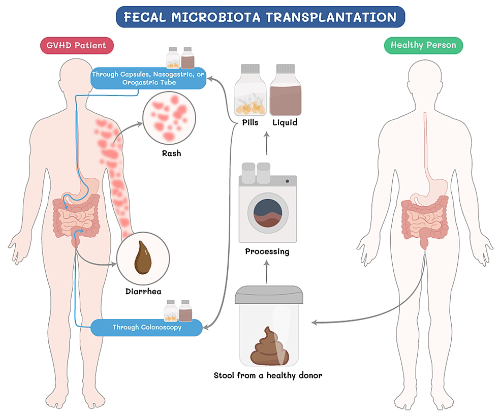
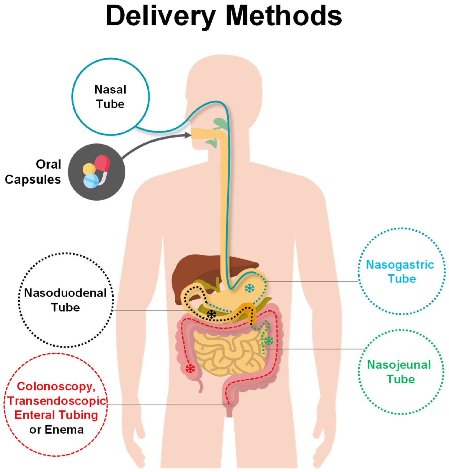
Fecal microbiota transplantation (FMT) is a procedure that delivers healthy human donor stool to a child via colonoscopy, enema, nasogastric (NG) tube, or in capsule form (popularly called “poop pills”). It may be prescribed for debilitating gasterointestinal infections, such as Clostridium difficile (C. diff), that keep recurring despite antibiotic therapy.

C. diff is a serious infection — one that causes debilitating diarrhea and in the U.S., in 2011 alone, C. diff was responsible for 29,000 deaths in adults. Treating C. diff begins with administering antibiotics; however, recurrence is common. By the third episode of severe C. diff, it is unlikely antibiotics will cure the infection; so, your child’s doctor may consider FMT as an option.

Based on successful treatment of C. diff infections, FMT is now being looked at for a wide range of conditions. However, it’s still considered an experimental treatment, and shouldn’t be attempted without medical supervision.

Not all children are good candidates for FMT. The procedure carries some risk, particularly if your child is taking immunosuppressant medication or has had a recent bone marrow transplant.

Working principle

The GI tract has an “ecosystem” of thousands of bacteria and other organisms that help keep the body healthy. When your child is given an antibiotic, this ecosystem is disrupted, allowing the growth of disease-causing bacteria, such as C. diff.

With a fecal transplant, “good” microorganisms from the donor stool are infused into the patient. Healthy bacteria begin to grow and prevent C. diff from recurring.

Stool donors are rigorously screened and stool samples are extensively tested before being used for FMT.

Preparation

The long-term results of FMT are unknown, so you’ll need to sign an informed-consent form on behalf of your child.

To prepare for the transplant, your child should have an empty GI tract. This often means drinking only clear fluids and abstaining from eating for 24 hours before the procedure.

If your child has mild or moderate C. diff, they may also be asked to drink a liquid to encourage a bowel movement; however, children with more severe infections will not follow this same protocol.

Procedure

There are several different FMT techniques:

#Colonoscopy: A thin, hollow tube with an attached camera is placed up the colon, and a catheter-tipped syringe is used to inject donor stool through the channel.

#Enema: Although less invasive than a colonoscopy, a fecal enema often needs to be performed more than once, because the donor stool doesn’t reach the colon.

#Nasogastric (NG) tube: Using a thin, flexible feeding tube, doctors insert donor stool through a patient’s nostril, down the throat, and into the stomach.

#Oral capsules, known as “poop pills.”

Future

Recent studies show how FMT can be beneficial to children who have specific GI conditions, such as Crohn’s disease or ulcerative colitis. We are also starting to test FMT for peanut allergy, based on evidence that transferring “good” bacteria can rebalance the immune system.

FMT is a hot topic among researchers and clinicians. ClinicalTrials.gov reveals some 170 studies on FMT, with applications as diverse as type 2 diabetes, cirrhosis, and HIV. And, in a clinical trial at Boston Children’s Hospital, researchers are looking at whether giving peanut-allergic people the “good” bacteria from a non-allergic person (in the form of “poop pills”) prevents allergic reactions.

Blood– Brain Barrier (BBB)

Blood– Brain Barrier (BBB) 

Introduction

The brain is the epicentre of an eclectic array of physiological activity. It integrates information from the external environment with signals from the internal environment in order to execute specific activities.

With all the special activities that occur at the neuronal level, it is paramount that the chemical environment in which these cells operate is strictly regulated. This is the primary function of the blood-brain barrier.

This article will look at the overall structure of the blood-brain, blood-nerve, and blood-cerebrospinal fluid barriers found throughout the nervous system. Additionally, special attention will be paid to those areas within the brain that lack a blood-brain barrier and to clinically relevant points with respect to this membrane.

Definition

Blood-Brain Barrier (BBB) is a selectively permeable membrane regulates the passage of a multitude of large and small molecules into the microenvironment of the neurons. It achieves this feat by with the aid of multiple cellular transport channels scattered along the membrane. 

These include:

*amino acid transporters.

*glucose transporter 1 (GLUT1).

*nucleouside & nucleotide transporters.

*monocarboxylate transporters (MCT1 and MCT2).

*ion transporters (Na+/K+-ATPase pumps) that facilitate the transport of essential molecules into the brain.

In addition to facilitating the uptake of amino acids, the amino acid transporters may inadvertently transport undesirable heavy metals into the brain’s immediate environment. 

Consequently, at high enough concentrations, this will result in neurotoxicity. GLUT1 and the MCT transporters carry glucose, and lactate and ketones, respectively.

Blood–brain barrier structure

The brain has a large network of arterial and venous vessels taking blood to and from (respectively) brain tissue. However, most of the action occurs at the level of the capillaries. Both the luminal and abluminal (outer surface of the vessel) sides are lined by key structures that contribute to the integrity of the cells. 

Firstly, squamous epithelial cells form the endothelial wall of the capillaries; the luminal surface of these cells comes into contact with circulating blood and its constituents. The abluminal surface is in contact with a circumferentially continuous basement membrane.

The endothelial cells are anchored to each other by zonula occludens or tight junctions, as well as zonulae adherens. The former provide structural support to the endothelial wall, while the latter physically connects adjacent cells. 

Additionally, the tight junctions circumscribe the cells and provide a seal with all adjacent cells. Therefore, the endothelium functions as an impermeable barrier between the capillary lumen and brain tissue.

Studies conducted on other mammals have implicated pericytes as integral components in the formation of the blood-brain barrier. These cells encircle endothelial cells of capillaries and are able to contract in order to regulate capillary blood flow. 

Consequently, the contractility also regulates the amount of blood flowing through the capillaries, thus enhancing the blood-brain barrier. Furthermore, some theories suggest that pericytes not only promote the formation of tight junctions, but they also inhibit the production of chemicals that promote vascular permeability.

Astrocytes are highly branched cells with small bodies found both in white matter (fibrous astrocytes) as well as in grey matter (protoplasmic astrocytes). 

The podocytes of both fibrous and protoplasmic astrocytes not only encircle nerve fibres and neuronal somas (respectively), but they also surround the abluminal surface of the capillaries. At this point, the processes are referred to as perivascular endfeet.

Blood–nerve barrier

A similar architectural construct exists in the peripheral nervous system that also limits the interaction between the peripheral nerves and circulating blood. This system is informally referred to as the blood-nerve barrier.

Blood–cerebrospinal fluid barrier

There is a similar barrier system in place that acts as an interface between blood and cerebrospinal fluid. This is known as the blood-cerebrospinal fluid barrier. The main similarities between the blood-cerebrospinal fluid barrier and the blood-brain barrier are:

Endothelial cells found in the capillary beds.

Circumferential basement membrane around the abluminal surface of the capillary.

And perivascular endfeet of the astrocytes also on the abluminal surface of the capillaries.

The blood-cerebrospinal fluid barrier, however, also has fenestrated endothelial cells which allow easier passage of water, gases and lipophilic substances from the blood to the cerebrospinal fluid. 

Additionally, there are choroidal epithelial cells that are integral to the production of cerebrospinal fluid.

Choroid epithelium consists of ciliated cuboidal cells, equipped with microvilli, encompassing capillary tufts. Although the choroid epithelium is continuous with the ependymal layer (simple ciliated columnar cells) of the ventricle, it contains more tight junctions and consequently act as an effective barrier between blood and cerebrospinal fluid.

Circumventricular organs

There are regions of the brain where the blood-brain barrier is absent. This anatomical adaptation allows areas of the brain to monitor homeostatic changes within the systemic circulation. As a result, the brain is able to detect these changes and effect necessary protective physiological processes to mitigate these activities. There are seven such areas that are collectively referred to as the circumventricular organs. They can be further subdivided in secretory and sensory organs:

Secretory organs

Secretory organs, as the name suggests, are structures that release their products directly into the bloodstream or cerebrospinal fluid. The products may either be neurohormonal or other proteins. 

The secretory circumventricular organs include the neurohypophysis (posterior pituitary gland), pineal gland, subcommissural organ and median eminence.

1.Neurohypophysis – is also known as the posterior pituitary gland. It is the region of the hypophysis cerebri that originates from neuroectoderm and stores hypothalamic hormones (namely oxytocin and vasopressin). Hormones are delivered to the posterior pituitary gland by way of nerve fibres travelling from the paraventricular and supraoptic hypothalamic nuclei.

2.Pineal gland – is a pine-shaped organ situated in the posterior aspect of the third ventricle. It lies just superior to, and in the midline of, the corpora quadrigemina (superior and inferior colliculi). The gland is encapsulated and lobulated (internally). Its constituent cells – pinealocytes – produce and cyclically release melatonin with the oscillating circadian rhythm (which is regulated by the suprachiasmatic nucleus). The absence of a blood-brain barrier here allows melatonin to be secreted into the rich blood supply coming from the posterior choroidal artery (branch of the posterior communicating artery) and internal cerebral veins (tributary to the great vein of Galen).

3.Subcommissural organ – is situated near to the caudal limit of the pineal recess (in the third ventricle) at the opening of the cerebral aqueduct of Sylvius. Here, it is caudally and ventrally related to the posterior commissure. The ependymal cells here, unlike those covering the other circumventricular organs, are tall ciliated columnar cells. The capillaries at this level are not as abundant or significantly fenestrated as those in other circumventricular organs. The subcommissural organ releases SCO-spondin, which is a glycoprotein that aggregates within the third ventricle and forms Reissner’s fibres. These fibres have been implicated in maintaining the patency of the aqueduct of Sylvius and their absence results in congenital hydrocephalus.

4.Median Eminence – has an intricate communication with the hypophyseal portal system that permits communication between systemic circulation and cerebrospinal fluid by way of the large number of fenestrated capillary beds it contains. The median eminence, which is located at the floor of the hypothalamus, is anterior to the tuber cinereum (ventral extent of the third ventricle). The median eminence also contains specialized cells known as tanycytes, which assist in modifying the permeability of the membrane to allow macromolecules to enter the peripheral circulation.

Sensory organs

Sensory organs are responsible for monitoring the peripheral circulation and responding appropriately to reverse these changes or eliminate toxins. These organs include the subfornical organ, the organum vasculosum of the lamina terminalis and the area postrema.

1.Subfornical organ – is a small region at the interventricular foramen of Monro that is comprised of glial cells, neurons and a densely packed tuft of fenestrated capillaries. Like other circumventricular organs, the subfornical organ is covered by flattened ependymal cells. The subfornical organ has multiple homeostatic functions, including cardiovascular regulation, osmoregulation and energy regulation, among others. This concept is supported by its efferent projections to the lateral hypothalamus, the median preoptic area and organum vasculosum.

2.Organum vasculosum – also known as the organum vasculosum of the lamina terminalis (OVLT) or simply, the vascular organ, it is cranial to the optic chiasm and caudal to the anterior commissure. The vascular bed of this organ is highly fenestrated and the ependyma contains flattened cells with very sparsely distributed cilia. The primary role of the vascular organ is to regulate fluid balance. As such, it receives afferent fibres from the subfornical organ, several hypothalamic nuclei and the locus coeruleus. It then projects to the supraoptic and median preoptic nuclei.

3.Area postrema – is probably the most commonly known circumventricular organ. It is a paired structure that is located at the caudal extent of the floor of the fourth ventricle. This bilateral structure is an important component of what is commonly termed the vomiting center. The absence of a blood-brain barrier in this location allows the area postrema to identify chemical irritants and stimulate a vomiting response.

Gut - Brain Axis (GBA)

Gut - Brain Axis (GBA) 

Introduction

The gut-brain connection is no joke; it can link anxiety to stomach problems and vice versa. Have you ever had a "gut-wrenching" experience? Do certain situations make you "feel nauseous"? Have you ever felt "butterflies" in your stomach? We use these expressions for a reason. The gastrointestinal tract is sensitive to emotion. Anger, anxiety, sadness, elation — all of these feelings (and others) can trigger symptoms in the gut.

The brain has a direct effect on the stomach and intestines. For example, the very thought of eating can release the stomach's juices before food gets there. This connection goes both ways. 

A troubled intestine can send signals to the brain, just as a troubled brain can send signals to the gut. Therefore, a person's stomach or intestinal distress can be the cause or the product of anxiety, stress, or depression. That's because the brain and the gastrointestinal (GI) system are intimately connected.

This is especially true in cases where a person experiences gastrointestinal upset with no obvious physical cause. For such functional GI disorders, it is difficult to try to heal a distressed gut without considering the role of stress and emotion.

Gut health and anxiety

Given how closely the gut and brain interact, it becomes easier to understand why you might feel nauseated before giving a presentation, or feel intestinal pain during times of stress. 

That doesn't mean, however, that functional gastrointestinal conditions are imagined or "all in your head." Psychology combines with physical factors to cause pain and other bowel symptoms. 

Psychosocial factors influence the actual physiology of the gut, as well as symptoms. In other words, stress (or depression or other psychological factors) can affect movement and contractions of the GI tract.

In addition, many people with functional GI disorders perceive pain more acutely than other people do because their brains are more responsive to pain signals from the GI tract. Stress can make the existing pain seem even worse.

Based on these observations, you might expect that at least some patients with functional GI conditions might improve with therapy to reduce stress or treat anxiety or depression. 

Multiple studies have found that psychologically based approaches lead to greater improvement in digestive symptoms compared with only conventional medical treatment.

Gut-brain connection, anxiety and digestion  

Are your stomach or intestinal problems — such as heartburn, abdominal cramps, or loose stools — related to stress? Watch for these and other common symptoms of stress and discuss them with your doctor. 

Together you can come up with strategies to help you deal with the stressors in your life, and also ease your digestive discomforts.

Female orgasms

Female orgasms A female orgasm can be a highly pleasurable experience during masturbation or sexual activity. While orgasms may ...